Intrapartum A&P (see AP for pelvis) Flashcards
Cervical ripening
- Ripening primarily caused by collagen rearrangement
- Before and during labor
- Tight collagen bundles become less dense and more loosely packed at midpregnancy .
- Before and during labor
- Hormonal/inflammatory changes →
- Increased water content in cervix → softening
- Promoted by proteoglycan decorin and reduction in fibronectin.
- Lack of progesterone inhibition (switch to estrogen dominence) → collagen breakdown and elastin remodeling mediated by matrix metalloproteinases
- Increased water content in cervix → softening
- Realignment of elastin and smooth muscle fibers plays a minor role too
4 phases of cervical change: (not related to stages of labor)
- Softening (remodeling)
- Can start as early as 4 weeks post LMP
- Ripening (accelerated softening at the end of pregnancy)
- Dilation
- Repair
Define: labor
- Classically the occurrence of regular painful contractions that promote dilation of the cervix.
- Contractions that occur at regular intervals with increasing frequency, duration, and intensity are the hallmark of labor
Phases of parturition
-
Phase 0 - Quiescence:
- Period in late pregnancy of uterine quiescence.
- Inhibitors of uterine contractions include progesterone, prostacyclin, relaxin, nitric oxide, and other hormones.
-
Phase 1 - Activation:
- Uterotropins (e.g., estrogen) stimulate upregulation of myometrial receptors for oxytocin and prostaglandins, and activation of gap junctions between myometrial cells.
-
Phase 2 - Stimulation:
- Uterotonins (e.g., oxytocin and prostaglandins) promote labor progression.
-
Phase 3 - Involution:
- Uterine involution after birth is mediated by oxytocin
Define: station
- Where the lowermost part of the fetal presenting part resides relative to an imaginary line drawn between the ischial spines of the woman’s pelvis (0 station)
- Measured in cm from 0 station
- Lowermost presenting part above 0 station is -1, -2, -3, -4, or -5
- Lowermost part of the presenting part (the bone, not swelling or soft tissue) lower than 0 station is +1, +2, +3, +4, or +5 station.
- At 0 station, the BPD is most always descended through the inlet (engaged).
- At +5 station, presenting part will be visible at the vaginal introitus.

Define: synclitism and asynclitism
- Relationship of the sagittal suture of the fetal head to the symphysis pubis and the sacrum of the maternal pelvis
- Determined when the AP diameter of the head is in alignment with the transverse diameter of the pelvic inlet (in other words occiput transverse)
- Synclitism - sagittal suture is midway between the symphysis pubis and the sacral promontory
-
Asynclitism - fetal neck is tilted so that the head leans laterally toward the fetal shoulder somewhat → sagittal suture is closer to the symphysis pubis or to the sacral promontory
- Anterior vs posterior asynclitism: based on which parietal bone (based on relation to maternal pelvis) is dominant
- Anterior - anterior parietal bone (closer to maternal symphysis) is lowermost/leading part → sagittal suture will be closer to sacral promintory
- Posterior - posterior parietal bone (closer to sacral promintory) is lowermost/leading part → sagittal suture will be closer to symphysis
- Normal labor - fetal head usually enters the pelvic inlet with a moderate degree of posterior asynclitism and then changes to anterior asynclitism as it descends farther into the pelvis before the mechanism of internal rotation occurs.
- This sequential change from posterior to anterior asynclitism facilitates descent → fetus takes advantage of the roomiest portions of the true pelvis

Define: molding
- Change in the shape of the fetal head as a result of the soft skull bones’ overriding/overlapping one another because they are not yet completely fused, so that movement is possible at the location of the sutures.
- Minor degrees are normal
- The shape of the head depends on the presentation and attitude → which parts of skull are subjected to pressure.
- Most common - occiput cephalic presentation → parietal bones override the occipital bone (may feel bony ridge) → obliterates/minimizes the posterior fontanel.
- When the parietal bones overlap at the sagittal suture (not uncommon), the parietal bone that was anterior in the pelvis overlaps with the “posterior” parietal bone, which was depressed because of pressure from the sacral promontory
- Molding involves the entire skull - overlapping in one area is counterbalanced by movement elsewhere → harmony between the base and the vertex of the skull → prevents destructive tension and possible rupture of the dura mater
Define: caput succedaneum
- Formation of an edematous swelling over the most dependent portion of the presenting fetal head
- Pressure around the presenting part by the cervical opening → congestion and edema
- Worse if membranes are ruptured and the fetal head (rather than the membranes) is functioning as the dilating wedge against the cervical opening
- If the fetal head is unusually molded or significant caput is present, the head may not be engaged in the pelvis at all, so caput and molding can be clinically quite important.
- Differentiated from cephalohematoma (more serious) by the fact that caput succedaneum crosses suture lines as a generalized swelling, whereas a cephalohematoma (bleeding beneath the periosteum) may occur over more than one cranial bone but is limited to each individual bone and does not cross any sutures.
- A few millimeters of caput succedaneum is considered normal.
- may develop during a somewhat prolonged labor resulting from uterine inertia with weak contractions.
- Extensive caput succedaneum, (identification of fetal sutures and fontanels impossible, combined with a more severe degree of molding), is usually seen when the pressure has been great and labor prolonged - cephalopelvic disproportion must be suspected in such a case. A sizable caput may also be seen from positional pressure when the fetus was in an OP position for a relatively prolonged period.
Discuss the uterine preparation that occurs prior to labor and identify the role of prostaglandins, corticotropin releasing hormone (CRH) and oxytocin
- Loss of progesterone (PR) dominence
- Switch in dominant PR receptor type: PR-B (promotes quiescence/suppresses inflamation) → PR-A (pro-inflammatory) → functional PR withdrawal → increased production of corticotropin-releasing hormone (CRH) by the placenta (peaks at time of birth) →
- Placenta makes CRH → crosses to fetus → stimulates fetal HPA axis →
- Fetal adrenal cortisol production → fetal lungs maturation → increased fetal pro-inflammatory surfactant and phospholipid production → induces uterine contractions
- Fetal adrenal DHEA-S (estrogen substrate) → estrogen production/dominence → onset and progression of labor.
- Fetal membrane production of stimulatory prostaglandins
- Estrogen → uterine myometrial cells express receptors for prostaglandins and oxytocin and develop gap junctions (allow direct communication between muscle fibers)
- Prostaglandins facilitate uterine contractions, increase myometrial sensitivity to oxytocin, and stimulate formation of gap junctions.
*
Discuss theories of the onset of labor
- Complex interplay between biochemical and mechanical influences originating in both the maternal and fetal systems and the complex interactions of hormonal systems regulating one another’s activity.
- Cascade of events that include multiple redundant loops (no single factor identified).
- 2 (known) primary mechanisms:
- Change from progesterone dominance and uterine quiescence to estrogen-stimulated uterotropin activation
- Placental production of corticotropin-releasing hormone
Describe the myometrial physiology underlying uterine contractions:
- Trigger (stretch or hormone receptor binging) → action potentials pass through gap junctions → contraction of adjacent uterine smooth muscle bundles → coordinated wave of uterine contraction.
- Prostaglandins produced by activated myocytes work in a paracrine fashion, stimulating nearby myocytes to depolarize in a wave of contraction
- Decreased intracellular Ca++ concentrations + action of myosin light-chain phosphatase enzymatically reverses the actin–myosin linkages → smooth muscle relaxation.
- Uterine smooth muscle contractions are intermittent, which allows for reperfusion of the uterine muscle, placenta, and fetus between contractions.
- Each contraction builds in intensity (“increment”), reaches its peak (“acme”), and then decreases in intensity until the muscle returns to a state of relaxation (“decrement”), which persists until the next contraction begins

Describe the mechanism of cervical effacement and dilatation
- Effacement - shortening of cervix
- Muscle fibers lengthen at the internal os → stretches the endocervix upward into LUS.
- Dilation - the widening of the external os.
- Force of contractions + hydrostatic action of amniotic fluid = force that promotes dilation of the low-resistance cervix
- If ROM, the pressure of presenting part promotes dilation
Describe the mechanisms of labor, cardinal movements of the fetus, including causes for each step, for each of the positions of the vertex
- Engagement
- BPD of fetal head passes into pelvic inlet
- Descent
- Occurs throughout/simultaneously via contractions (1st stage) and pushing (2nd stage)
- Flexion
- Crucial for descent
- Fetal head meets resistance (increases with descent) → flexes so fetal chin rests on the thorax → _smaller suboccipitobregmatic diameter substituted for the larger fetal head diameter_s when the head is not completely flexed (ie military attitude, or some degree of extension)
- Some degree may occur prior to engagement
- Internal rotation
- AP diameter of the fetal head aligns with the AP diameter of the maternal pelvis
- Amount of internal rotation determined by the distance the occiput must travel from its original position entering the pelvis to the OA or OP (expressed in degrees)
- Head is turned 45° from shoulders and shoulders enter pelvic inlet in oblique position
- Extension
- Birth of head in OA positions only (resistance from curve of carus
- Restitution
- Rotation of head 45° to be at right angle with shoulders again
- External rotation
- Fetus’s shoulders rotate 45°, bringing the bisacromial diameter into alignment with the AP diameter of the pelvic outlet.
- Causes the head to rotate externally another 45° into the LOT or ROT position
- Lateral flexion or expulsion
- Anterior shoulder comes into view at the vaginal opening, impinges under the symphysis pubis; the posterior shoulder then distends the perineum and is born by lateral flexion.
- After the shoulders are assisted to exit, the remainder of the body follows the curve of Carus and the fetus is readily born.
Largest pelvic diameter for:
- Pelvic inlet
- Midplane
- Pelvic outlet
- Inlet - transverse
- Midplane - AP
- Outlet - AP
Where does right oblique diameter start?
- Right sacroiliac joint (and moves diagonally to left iliopectineal eminence
- (Vice versa for left oblique diameter)

Describe normal uterine contractions by defining or describing:
Retraction
- Muscle bundles in the uterine fundus progressively shorten with each contraction (do not relax fully) → upper portion of the uterus thickens and gradually becomes a smaller cavity → reduced fundal capacity → descent of the fetus toward the more passive LUS
- LUS muscle bundles become longer in response to contractions in the fundus, creating a distensible structure that is able to accommodate the fetus, a process that promotes fetal descent.
- Physiologic retraction ring - line of demarcation between the active and passive segments
- Bandl’s ring - pathologic retraction ring of obstructed labor seen when the active segment becomes so much thicker and shorter the line between the two uterine segments is visible

Describe normal uterine contractions by defining or describing:
Descending gradient
- Triple descending gradient of fundal dominance - propagation of the contraction wave:
- Contractions:
- Start in the fundus
- Last longer in the fundus
- Progress from the fundus toward the isthmus
- Contractions:
Describe normal uterine contractions by defining or describing:
Effect on uterine shape and position
- Upper uterine segment becomes smaller and thicker (Retraction), firm during contractions
- Lower uterine segment becomes longer, softer/distended, more passive
- During active labor, the uterine divisions between the active and passive segments become increasingly evident. By abdominal palpation, even before ROM, the two segments can sometimes be differentiated.
Describe normal uterine contractions by defining or describing:
Intensity, Frequency, Duration
- Intensity - strength of contraction
- Frequency - beginning of one contraction to the beginning of the next
- Duration - beginning to end of one contraction
Describe normal uterine contractions by defining or describing:
Tonus and intrauterine pressure readings at rest and during contractions
- IUPC - quantitative measurement of uterine contraction intensity in Montevideo units (MVU)
- MVU = Amplitude - baseline resting tone
- Add all contractions in a 10 minute period together
- “Adequate” (according to 1960s study) 200 - 250 MVU
- Patient generally sense contractions when the contractions exert ~15 mm Hg of intrauterine pressure (this is when a provider can feel it abdominally too)
- Latent phase contractions = 10 - 40 mm Hg
Identify the purpose of the mucus plug during pregnancy and its significance in labor
- Protective barrier created by cervical secretions from proliferation of the glands of the cervical mucosa early in pregnancy → seals the cervical canal throughout pregnancy
- Often expelled over the course of several days, and noticed by the woman as mild irregular spotting when wiping after urination or defecation.
- As a predictive sign of labor, bloody show usually signals the onset of labor within the next few days.
- Not accurate if a vaginal examination or sexual intercourse has recently occurred
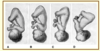
Identify 4 types of cephalic presentations
(A) Vertex
(B) Sinciput (military/forehead)
(C) Brow
(D) Face
Identify 8 variations of cephalic presentation
- LOA
- LOT
- LOP
- OP
- ROP
- ROT
- ROA
- OA
Describe landmarks and state measurement of common diameters of the term fetal skull
- Suboccipitobregmatic: 9.5 cm
- Submentobregmatic (ie face presentation): 9.5 cm
- Biparietal diameter (BPD): 9.5 cm
- Occipitofrontal (essentially AP diameter): 11 cm
- Supraoccipitomental: 13.5 cm


