Infections In the Lung Flashcards
What are the clinical features of pneumonia?
- fever and chills
- unrelenting cough
- sputum production (purulent/yellow)
- chest pain (if pleura inflamed)
- impaired gas exchange resulting in SOB/dyspnoea and tachypnoea, and hypoxemia
Hospital patients are more susceptible to what type of pneumonia-causing agents?
gram negative bacteria (e.g. pseudomonas)
Immunocompromised hosts are more likely to get pneumonia caused by
fungi and protozoa (e.g pneumocystis jirovecii)
What are the 4 routes of infectious pathogens into the lungs?
- inhalation of pathogens in air droplets
- aspiration of infected secretions from URT
- aspiration of infected particles
- gastric contents, food, drink, foreign bodies
- haematogenous spread (via blood)
What are the 3 main causes of pneumonia?
- URT flora
- S. pneumoniae, H. influenzae, S. aureus
- enteric saprophytes, by contaminaiting airways or blood stream
- E. coli, Pseudomonas
- extraneous pathogens
- Legionella pneumophilia, TB
What are the 2 patterns of infective pneumonia?
- alveolar inflammation
- neutrophils in the alveolar spaces = consolidation
- Strep, Staph Haemophilus, G-ves
- neutrophils in the alveolar spaces = consolidation
- interstitial inflammation
- lymphocytes, macrophages, sometimes plasma cells in the connective tissue septa between the alveoli (interstitium)
- viruses, atypical pneumonia viruses (mycoplasma pneumoniae)
- lymphocytes, macrophages, sometimes plasma cells in the connective tissue septa between the alveoli (interstitium)

What are the two types of alveolar pneumonia?
- bronchopneumonia
- consolidation is patchy, multi-focal; very often bilateral (more than 1 lobe)
- lobar pneumonia
- involves an entire lobe and often inflammation of the adjacent pleura

What is the most common cause of lobar pneumonia?
S. pneumoniae (90%)
and H. influenzae
How is lobar pneumonia acquired?
- community acquired in adults 20-50
- commonly following viral URTI
What is the clinical presentation of lobar pneumonia?
- abrupt onset
- fever & chills
- rasied WBC
- cough
- pleuritic chest pain
- haemoptisis
- G+ diplococci in sputum
- bacteraemia
What are the 4 stages of lobar pneumonia?
- congestion of alveolar capillaries
- alveolar spaces filled with proteinaceous exudate containing G+ diplococci (Strep)
- red hepatization (consolidation)
- haemorrhage into air spaces
- grey hepatization
- fibrin, neutrophils, macrophages in alveolar spaces
- resolution
What is a cute bronchopneumonia?
- most common pattern of bacterial pneumonia
- patchy consolidation, often multi-focal and involving more than one lobe or lung
- centered on bronchioles, spreads into surrounding alveolar spaces
Acute bronchopneumonia is common in
- extremes of life
- secondary to pre-existing chronic disease
- COPD, congestive heart failure, malignancy, CF
- v. in hospitalized patients (G- bacteria & staph important causes)
- post-op complications that impair clearance of respiratory secretions
- secondary infection following viral UTI
Histologically, acute bronchopneumonia presents with
bronchioles and alveoli filled with neutrophils

What are the complications of pneumonia?
- pleuritis
- pyothorax (pus in pleural space)
- if becomes walled off by fibrous tissue = empyema
- abscesses
- cavities contaning pus (purulent exudate)
- commonly caused by staph aureus pneumonia, Klebsiella, or Pseudomonas
- chronic complications like bronchiectasis
What causes lung abscess?
- typical complication of pneumonia caused by s. aureus, klebsiella, pseudomonas
- aspiration of infected material from URT or gastric contents
- distal to a bronchial obstruction by tumours
- septic emboli to the lung (eg in infective endocarditis)
What are the causes of pneumonia with interstitial inflammation?
- viruses
- bacteria (atypical pneumonia)
- inflammatory responses to drugs
- immunological diseases
- collagen vascular diseases (lupus, vasculitis)
- radiation
What is the pathology of infective pneumonia with interstitial inflammation caused by bacteria and viruses?
- widened alveolar septa
- infiltrated with lymphocytes, plasma cells, and macrophages
- bronchioloits
What is the histologic presentation of interstitial pneumonia?
- may be oedema fluid, red cells, and fibrin in alvelolar spaces
- there are no alveolar neutrophils or inflammatory cells tf no consolidation
- macroscopically the lung appears wet, dark, and heavy

What are the causes of atypical pneumonia?
- mycoplasma pneumoniae
- coxiella burnetti
- legionella spp
- chlamydia pneumoniae
What is atypical pneumonia?
- community acquired pneumonia lacking clinical and radiological signs of consolidation
What are the symptoms of atypical pneumonia?
- systemic symptoms predominate over respiratory
- malaise
- aches and pains
- headaches
- diarrhoea
- dry/non-productive cough or no cough at all
- often ambulatory despite extensive radiological signs of pneumonia
- clinical presentation follows intersitial pneumonia pathology eg no consolidation
How does atypical pneumonia present on CXR?
- no consolidation pattern
- widespread changes throughout both lung fields
- reticulonodular infiltrate (dots and dashes)

What is tuberculosis?
- chronic granulomatous pneumonia due to infection with Mycobacterium tuberculosis
- tubercle = granuloma
What is primary TB?
- typically in childgood
- pathology is characterised by a Ghon’s complex
- area of inflammation (peripheral mid-zone of lung) called a Ghon focus
- mediastinal or hilar lymphnodes = granulomatous lymphadenopathy
- Ghon focus + enlarged lymph nodes = Ghon’s complex
- granuloma consists of:
- multinucleated giant cells
- epitheliod macrophages
- lymphocytes
- central caseuous (cheesy) necrosis
- usually asymptomatic
- heals, often involving calcification
- remains dormant until secondary infection arises

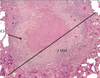
tuberculous granuloma
What are epitheliod macrophages?
- large, rounded pink cells
- form aggregates

What is the large arrow pointing to? The small arrow?
- large: caseous necrosis
- small: multinucleated macrophages/giant cells

What causes the formation of granulomas in TB?
- cell-mediated immune reaction type IV hypersensitivity
- monocytes exit peripheral blood in the area of the infection
- enter the tissue
- stimulated by cytokines (IFNy) to become epitheloid macrophages
- this forms a lump

What is secondary TB?
- reactivation of dormant TB or reinfection
- lobar pneumonia involving upper lobe
- much more extensive caseation than primary due to stronger cell-mediated immune response
- can erode bronchi causing cavitation
What are the complications of secondary pulmonary TB?
- spread of caseation into surrounding lung
- erosion of blood vessels –> haemoptisis
- erosion of the bronchial tree –> cavitation and widespread infection to other parts of the lung via airways
- pleural inflammation and fibrosis
- lung scarring
What are the clinical features of TB?
- variable weight loss
- mailaise
- fevers
- night sweats
- haemoptyisis
- dyspnoea
- chronic cough
- more severe and more acutely developed in pt with miliary and bronchopneumonia TB
How does TB spread within the body?
- via lymphatics
- pleura
- other parts of lung
- other lung
- via bronchial tree (infective caseous material in bronchial tree)
- extensive TB bronchopneumonia
- can be coughed up –> laryngeal TB
- then swallowed –> intestinal and oesophageal TB
- haematogenous spread
- bia bloodstream to other organs
- brain, urogenital tract, bones
- bia bloodstream to other organs
What is miliary TB?
- most important form of extra-pulmonary TB
- occurs in primary and secondary TB
- more common in secondary unles immuno-compromised
- bacteria disseminates via bloodstream
- can involve lung and/or multiple other organs:
- liver, spleen, bone marrow, brain
What is the pattern of miliary TB?
- numerous small granulomas (~2-3mm) in lung and other organs

What is single-organ TB?
- usually caused by secondary TB with caseation
- other organs can be seeded with TB from primary infection
- seen in spine (Potts disease) and urogenital tract
- in kidney, see butterfly-shaped lesions of caseous necrosis in upper pole
- some cavitation occurs due to caseous material entering the collecting system

