Immuno Flashcards
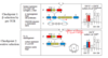
Two signal hypothesis
B cells - need 2 signals to weed out bad and get rid of autoreactive
- agR - trigger –> signal transduction, receptor is crosslinked (if never find ag – die!)
- cytokine - membrane associated signal (if autoreaction - a little activation – death!)
signal 1 only: death and inactivation
both signals = activation

CXCL13
attracts B cell into pimary follicle
When does a cell become double positive? Is this step reversible?
Once a functional beta chain is expressed the T-cell becomes CD4+/CD8+. This is reversible, if the cell is unable to functionally express the alpha chain before g:d, the cell exits the a:b pathway and becomes DN again.
CCL21
chemokine that activates integrins on T cells
so T cells migrate into LN
Th2
provide help to B cells for ab production, especially switching to IgE
parasites
C3 convertase
made by C3+B+D
C3Bb
cleaves C3 into C3a (secreted) and C3b (on pathogen)

burkitt’s lymphoma
mature memory B cell, resembes germinal center B cell
GALT
additional lymphoid tissues
tonsils, adenoids, appendix, peyer’s patches
what if immature B cell binds soluble univalent self ag?
B cell is signaled to make IgD and become unresponsive to ag
anergic
enters peripheral circulation but doesn’t survive for long
Complement Cascade functions
Lyse bacteria (MAC)
opsonize bacteria for phagocytosis - coat w proteins so immune can take up
release vasoactive and chemotactic moleucules that attract more immune cells to the site
PRRs
Pattern recognition receptors
also called PAMPs, and DAMPs,
rec structures that are commonly found in pathogens/molecules released during an infection
T cell from DP?
NKT, treg, Th, Tc

plasma cell
ssecrete abs
IgM and IgG
fight current infection
late-pro B
V-DJ rearrangement on H chain
CTLA-4
restrain T cell activation
binds B7 with really high avidity
if block - boost immune response to cancer
AIRE
on medullary epithelium in thymus
make secreted molecules to see if Tcells respond against them
defect - ab response vs insulin
Autoimmune regulator (AIRE) is a transcription factor that can stimulate the global transcription of tissue specific genes to have a low level of protein expression.
This ‘micro-expression’ is sufficient for MHC presentation and protection of even these unique cell types.
Lack functional AIRE - autoimmune polyendocrinopathy-candidiasis-ectodermal dystrophy (APECED) - broad spectrum autoimmunity.
NK cell regulation
balance of inhibitory and stiulatory signals
MHC I on normal cells - rec by inhibitory receptors and by activating receptors - does not kill! it’s a balance and activating receptors ALWAYS signal
altered/absent MHC I (stress) can not stimulate negative signal - triggered by activating receptor only! triggers apoptosis in target cell
mechanism for T cell migration in HEV
L-selection –> glycam1 - rolling
chemokines activate integrins
integrins (LFA) bind tightly to ICAM
migrate into LN

macrophages in thymus
help clear out dead/non productively arranged apoptotic cells
selectin
adhesion molecules on endothelial - weak adhesion and allows neutrophil to roll along vascular endothelial surface
mobility
cells and effectors travel to sites of infection and bring info back
IgA
mucosal immunity
augment barrier
MHCI/CD8
MHC holds peptides generated in the cytoplasm
expressed by all cells
alerts CD8+ T cells to the presence of infection/mutation and kill infected/transformed cells
CD8 binds alpha 3 domain of MHCI
ZAP70
TCR binds MHC II –> Lck on CD4 P ITAMs
ZAP-70 (kinase) binds ITAMs and is P by Lck
ZAP-70 P PLC - cleaves PIP2 to DAG and IP3, IP3 allows Ca to come in
Mast cells
cover self in IgE – IgE receptor
binding - release of granule contents
allelic exclusion
on B cell
homogenous BCR with high avidity binding
BCR can be membrane bound or secreted
only rearrange 1 chromosome at a time
if no allelic exlusion - 2 H and 2 L, lower affinity, decreased ability to stimulate

memory B cell
IgG
prepare for future infection
neutrophil
kill!
short lived - die within hrs of entering inflammed - pus
most abundant WBC
circulates in blood unless specifically attraced to inflamed site
phagocytose and kill bacteria with highly toxic granule contents
large reserve in bone marrow –> increase in WBC that means a lot of been released (signal)
complememnt: C3b is on the bacteria and C3A is released– chemotactic for nuetrophil
IRAK-4
complex at macrophage surface - TLR
if bind - activated!
phosphorylates TRAF-6 - kinase cascade —> IKK
IKK - binds and activates NFKb –> activates transcription of genes for inflammatory cytokinse!
made in cytoplasm and secreted via IR
if definiciency - 50% chance you will die in first 3 years, then you are fine

t cell dependent B cells
need T cell
B2 cells
- BCR
- from T cell membrane or cytpkine (CD40L!)
negative selection - T cells
central tolerance - elim high affinity to avoid attacking own tissues
high affinity TCR with pMHC –> apoptosis (negative selection) or agonist selection of Treg
self AG is expressed under AIRE
many self ag-reactive T cells that do not encounter ag in the thymus reach periphery - anergic/ignorant of self ag
common erythroid megakarocyte progenitor
megakaryocyte –> platelets
erythroblast –> erythrocyte
When is a lymphoid progenitor committed to the T-cell lineage? Describe the T-cell receptor (TCR) at this stage.
Loss of CD34 stem cell marker, still double negative for CD4/CD8 (DN T-cell progenitor)
TCR is not expressed, TCR gene is
Treg
suppress T cell response
Factor H and Factor I
inactivation C3b - disassemble so no feed forward
binds to sialic acid on human cells

IL-7
stromal cell factor - secreted - binds pre-B cell
required
CAMs still attach

FcRn
recycles IgG across placenta
reactivity
distinugish self vs non self
harmless vs harmful
somatic hypermutation
B cells
affinity maturation
When a B cell recognizes an antigen, it is stimulated to divide (or proliferate). During proliferation, the B cell receptor locus undergoes an extremely high rate of somaticmutation that is at least 105-106 fold greater than the normal rate of mutation across the genome
This directed hypermutation allows for the selection of B cells that express immunoglobulin receptors possessing an enhanced ability to recognize and bind a specific foreign antigen.
systemic inflammation
liver - acute phase proteins (complememt)
bonemarow - nuetrophil mobilization
hypothalamus - increase body temp - decrease pathogen replication
fat, muscle - protein and energy mobilization to increase body temp
Which cells present antigens for negative selection?
Bone marrow derived dendritic cells and macrophages. Thymocytes.
hat is the final product of positive selection?
single positive T cell, CD4+ or CD8+
what are cells that first enter the thymus?
uncommitted lymphoid progenitor cells
CD34
CTL
kill virus infected cells
viruses and some intracellular bacteria
large pre-B
VDJ rearranged, H chain is made
surrogate L chain to partner with
B cell in lymph node
maturation
B cell is guided by chemokines to primary lymphoid follicles
B cells with a netwrd of special stromal cells -FDCs, attract B cells by secretng chemokines and give signals to mature the B cell
pre-b cell leukemia
pre b cell, bone marrow
sepsis
macrophages activated in liver and spleen - secrete TNF-alpha into bloodstream
systemic edema - decreased BV, hypoproteinemia, collapse of blood vessels
disseminated intravascular coagulation - wasting, organ failure, septic shock
MAC
C5b + C6,7,8, lots of 9
make a pore in bacteria!

repeated rearrangement
nonproductive rearrangements - turn RAG back on - occur until positive selection to allow recognition of MHC
interaction w MHC - turn off Rag recombonase

MHC I
hold peptides generated from proteins in the cytoplasm
self or viral
all nucleated cells express MHCI
CD8+ reconize MHC I and directly kill cells
What is peripheral tolerance?
Protection of self by repression of auto-reactive T-cells in the periphery that may have excluded negative selection.
T cell independent B cells
marginal B cells interact w repetative non-protein ag –> crosslink
CR (complement receptor enhances BCR signaling
CR2–> C3d
clustering of ag receptors - allows P of ITAMs
B cell activation by TI-2 ag (LPS, bacterial DNA
CTL priming
CD4 and CD8 cells bound to APC
CD4 get MHC II and B7 signal –> induce to make CD40L and IL-2
CD40L –> CD40 on APC - icreases B7 - co stim CD8 cell
T cell also sends IL-2 to CTL
APC presents MHC I and II

T cell priming
mature dendritic cell primes naive t cell in T-cell zone
MHC2
B7 –> CD28
Nod-like receptors
cytosolic sensors of PAMPs and DAMPs - if something is in the cytosol
reside in inactive form - looks like TLR
binding of bacterial ligand s–> recruit RIPK2, activate NFkappaB
LOF - don’t detect gut microbes fast enough (Crohn’s)
GOF - Blau - sterile inflammation and over activation
CXCL8
chemokine secreted by inflammation - activate integrins and available for WBC to bind via CXCL8R

positive selection - T cells
DPCD4CD8 interact w pMHC on cortical epithelial cells
if intermediate affinity –> cell activation, migration to the medulla, shut off CD4 or CD8 dep on which MHC it recognizes
high affinity interactions –> CD4 that express FoxP3 and become Tregs OR cell death
no/low affinity interactions –> further alpha rearrangement or death by neglect (95%)
primary immune response
first time you see the pathogen/antigen
CD8
recognize MHC I
kill infected cells
inflammation cytokine
TNF-alpha (Remicade blocks it)
induces inflammation
also IL-6, IL-12, IL-1beta, CXCL8
small pre-b
H: VDJ rearranged
L: V-J rearranging
H chain is in the ER
When does rearrangement of the alpha chain occur?
once the cell becomes double positive (i.e. has successfully rearranged the B chain)
antigen activated B lymphoblast
alternative splicing to secrete Ig
isotype swithing
somatic hypermutation
Where does negative selection occur?
Cortex Medulla boundary - where dendritic cells begin to be enriched
CTLA-4
on T cells
competes with CD28 for B7 binding and dampens immune response
if inactivate CTLA-4 –> does not compete to bind B7 - inreased immune response against cancer
IgM
largest
infection
complement
CCR7
immature dendritic cells activated by PAMPS when encounter pathogens
TLR singaling induces CCR7, enhances processing of pathogen derived ags
CCR7 directs migrantion into lymphoid tissues and augments expression of co-stimulatory molecules and MHC molecules
barrier defenses
Skin, GI tract, respiratory tract, urogenital tract, eyes
mechanical, chemical and microbiological defenses
DAF and MCP
disrupt C3bBb covertase on human cell surface

structure of Ab
3D beta barrel with beta strands and beta folds
HV regions are next to each other
CDR loops are beta bends
medullary venules
where progenitors exit and enter
Granzymes
serine proteases which activate apoptosis once in the cytoplasm of the target cell
What signalling pathway prevents T-cells from entering a B-cell, NK cell, or myeloid developmental pathways?
Notch
cytokines for virus infected cells
IFN-alpha, IFN-beta
activate PRK - no DNA replication
increase MHC class I on all cells
activate dendritic, macrophages
activate NK cells to kill virus infected cells
induce chemokinse to recruit lymphocytes
ICAM
induced on endothelial cell by chemokines
mediate tight binding of wbc

MHC II isotypes
HLA-DP, HLA-DQ, HLA-DR
1 from each parent
alpha and beta chain encoded in different genes A and B, each from different parent –> more diversity!!
mom: DPA1 DPB1
dad: DPA2 DPB2
you: A1B1 A1B2 A2B1 A2B2
HLA can have 2 beta chains depending on the allele - more diversity!
thymus cortex
immature thermocytes, branched cortical epithelial cells, macrophages
first phases of development: gene rearrangement and positive selection
from ectoderm epithelial cells

What is central tolerance?
Protection of self by negative selection in the thymus
thymus medulla
mature thermocytes, medullary epithelial cells, macrophages
latter phases of development
less lymphoid cells
negative selection to remove autoreactive - AIRE
from bone marrow derived dedritic cells

L-selectin
selectin for T cell rolling on endothelial lining of LN
binds sugars (glyCAM-1)
T Cell checkpoint 2
positive selection
V(a)-J(a) rearrangement
surface expression of a:b:CD3
selective events begin
DP, low CD3
after alpha chain is rearranged
Where does gene rearrangement occur for T-cells?
Cortex
FOXN1
transcription factor required for development of thymus
nude mouse doesn’t have and won’t reject human tumor
immature b cell
H: VDJ rearranged
L: VJ rearranged
H chain (mu), L chain (l or k)
IgM on surface
What happens when a T-cell has successfully rearranged g:d receptors?
g:d receptor assembles with CD3 and shuts down further rearrangement of a:b. exit thymus to the periphery.
lymphoma
in tissue
Bcl11b
inhibits NK cell fate in T cells
B cell receptor development
in the bone marrow - generate diverse/clonally expressed BCR
circulation of lymphoid cells
one way valves - flow of lymph is always away from peripheal tissues - no pump!
unlike blood, no pump so circulation relies on body’s movement
naive lymphocytes enter LN from blood, ags from infection reach LN via lymphatics, lymphocytes and lymphatics return to blood via thoracic duct

immature B cell
leaves bone marrow, enters peripheral circulation
high IgM, low IgD
alternative splicing to give both d and m
gains access to primary lymphoid follicle and matures
why don’t all viruses downmodulate MHC1?
NK cells kill cells that have downmodulated MHCI and can no longer communicate with CD8+ T cells
SCF
stromal cell factor - binds Kit receptor on pro-B cell

MHC restriction
TCR needs to recognize both MHC and peptide
B1 cells
in fetus
restricted repertoire
self renewing
spontaneous production of Ig
mostly IgM
no t cell help needed
no somatic hypermutation
no memory
IgE
allergy
Tfh
b cell help
isotype switching
antibody production
all type of pathogens
Marginal Zone B cells
anti-polysaccharide
spleeninc marginal zone
secrete in bone marrow
react to things in blood and bind polysaccharide
common lymphoid progenitor
B cell, plasma cell
T cell, effector T cell
NK cell
acute lymphoblastic leukemia
lymphoid progenitor in bone marrow
LFA-1
T cell adhesion integrin for LN
IL-4
B cell s- activation growth, increased MHC II production
T cell s- growht, survival
Perforin
aids in delivering contents of granules into cytoplam of target cells
polymerize in PM of target cells
Treg
mediate peripheral tolerance
regulatory T cells, during negative selection, these cells do not bind either too strongly or too weakly to MHCII molecules.
weak binding → would become effector
strong binding → would undergo apoptosis
clonal selection
each lymphocyte bears a single receptor with unique specificity
engagemnt of that receptor stimulates the cell to divide and expand in clonal expansion
all the progen have identical receptors/specificity
self-reactive receptors are removed through negative selection
What are the three selection steps a T cell progenitor must pass in order to become a mature T cell?
B-chain selection for functionality
Positive selection for MHC reactivity
Negative selection for autoimmunity
What is the first developmental choice a committed T-cell progenitor must make? how does this choice occur?
gamma:delta (g:d) vs. alpha:beta (a:b)
DN T cells are simultaneously rearranging g, d, b genes
the first gene that is able to make a functional receptor g:d or b (NOT a)
the beta and alpha chain are rearranged sequentially, not simultaneously
if at any point the g:d genes successfully rearrange the T cell becomes committed to the g:d
MHCII/CD4
MHCII holds peptides generated from endocytosed roteins
expressed by APCs and alerts CD4 T cells to the presence of extracellular pathogens
helps apcs eradicate the infection
CD4 binds Beta-2 domain of MHC II
pT-alpha
In the ER the beta chain associates with a “training wheel light chain’ pT-alpha (invariant polypeptide)
Successful heterodimerization (beta + pTa) leads recruitment of CD3 and zeta chain
this is a double negative 3 (DN3) cell
beta chain
invariant polypeptide (pTa)
CD3
zeta chain
How does a T cell become single positive?
During positive selection, when the TCR binds to an MHC. The co-binding of either CD4 or CD8 will trigger a signalling cascade that will repress the other receptor and commit the cell to one lineage.
CD3
signal transduction for TCR, nexxt to it on T cell membrane
T Cell costimulation
costiumlation for T cells
B7 (on APCs) interacts with CD28
only professional APCs express B7 like dendritic cells, macrophages, B cells
when APCs are activated through TLR, incrase activation of MHC and B7 and other proteins that help T cell activation
if T cell hgets specific signal without co-stimulation –> becomes anergic!!
refractory period to get rid of auto cells
activated cells no longer need costimulation

T-cell area
in secondary lymphoid tissue
B cell encounters ag, dentained in t cell areas ad activated by ag specific T h cell
link btwn adaptive and innate
activated dendritic cells pick up ag and bring it to LN
IFNg
activate MHC I and II on macrophages
inflammasone
post translationally!
sense cell stress and pathogens
generated in pro-inflammasome - cleave for activity - secrete pro-IL - secrete as active form after 2nd signal
MHC I peptide loading
intracellular antigen –> proteasome –> peptide transport into ER and bind to MHC I –> present on cell surface

systemic inflammation cytokines
IL-1
IL-6
TNF-alpha
B Cell, L chain rearrangement
Kappa and lambda genes on both chromosomes! 4 chances
either m:k or m:l

MHC I structure
1 chain! a 1, 2, 3
beta2 microglobin - binds anything
alpha helpx and beta pleated sheet
peptide fits right in groove

How are progenitors biased toward the generation of a:b over g:d T-cells?
Multiple rearrangements for beta chain can occur on the same allele → more than one try/chromosome
Homologous chromosome provides a second chance at beta chain rearrangement
g:d T-cells must successfully rearrange 2 heavy chains. a:b T-cells must only rearrange 1 heavy chain and 1 light chain. Heavy chain rearrangement (3 parts) is much less efficient than light chain (2 parts)
C5 vonvertase
cut C5 into C5a and C5b
most isotype in the body
IgA - surface immunity
mature naive b cell
low IgM, high IgD
enters circulation and binds specific ag in lymphoid tissue draining infection
hodgkin’s lymphoma
germinal center B cell
mutated Ig
cross-priming
what if virus doesn’t infect APC - virus needs TCR signal + B7 to activate
APC presents on MHC I to CD8 CTL
against tumors and viruses that don’t infect APC
avoids viral immune avoidance
expansion
cells themselves (clonal lymphocytes) and magnitude of response expands until threat is eliminated
C1
complement fragment, binds IgM on pathogen, dock for start of cpmplement cascade
MHC I isotypes
HLA-A, HLA-B, HLA-C
3 different genes encode MHC I!
each person has one from each parent so up to 6 different class 1 molecules - don’t want a virus that can mutate away from all of them
each gene is highly polymorphic
granulysin
has antimicrobial actions and can induce apoptosis
second checkpoint, B cells
selects for functional light chain

Waldenstrom’s macroglobulinemia
IgM secreting B cell
mutated, no variability within clone
Where does positive selection occur?
Cortex - cortical epithelial cells
maternal and fetus immunity
mom passively transfers IgG
baby first make IgM after a little while
then IgG
then IgA
MHC II structure
2 chains
a1, b1
a2, b2
peptide flops outside of the ends
Th17
enhance neutrophil response
promote barrier activity (epithelia)
secondary immune response
relies on the memory of the first response
MHCII peptide loading
extracellular antigen –> endocytosed –> phagolysosome –> vesicle with MHC II from golgi merges with vescile with peptide and binds –> presents at cell surface

Tysabri
antibody that blocks integrin on nainve T cell outside of lymph organs (gut) so can’t bind to guy endothelium
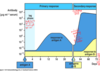
germinal center
in secondary lymphoid tissue
activated B cells proliferate, isotype switching and somatic hypermutation to make highest affinitiy abs
proliferate, differentiate, and mutate their antibody genes (through somatic hypermutation), and switch the class of their antibodies (for example from IgM to IgG) during a normal immune response to an infection
maturing B cells (centroblasts) migrate from the dark zone to the light zone and start to express their antibody on the cell surface and at this stage are referred to as centrocytes. The centrocytes are in a state of activated apoptosis and compete for survival signals derived from follicular dendritic cell and TFH cells. This rescue process, known as germinal center selection, is believed to be dependent on the affinity of their surface antibody to the antigen. Such that, a B cell that has successfully gained mutations that confer a higher affinity surface antibody towards antigen gains a survival advantage over lower affinity B cell clones and those that have gained deleterious mutations. Cyclic re-entry into the dark zone once again as centroblasts allows a chance for otherwise non-selected B cell mutants to gain more mutations in order to improve affinity towards antigen. Interactions with T cells are also believed to prevent the generation of autoreactive germinal center B cells
C3
most important complement protein - in the plasma, starts plasma cascade
H. influenzae (type B)
recurrent bacterial pathogen
capsular polysaccharide
population bottleneck –> athabascan infant dont inherit best Vkgene allele for an effective Hib polysaccharide response - not a protein - can’t make a T cell inependent germinal center resonse
notch
inhibits B Cell fate in T cells
T cell from double negative?
g:d T
DP

T cells help B cells
in B cell follicle- results in germinal center formation
T cell rec MHC II on B cell
T cell CD40L –> B cell CD40
upreg AID - class switching and somatic mutation

B cell negative selection
alteration, elimination or inactivation of B ell receptors that bind to components of the human body
How does a progenitor get the merit badge of “pre-T cell?” What cell processes does this trigger?
successful superdimerization and downstream signalling
stops recombination
signals proliferation (clonal expansion of cells carrying the same beta chain)
begins process of CD4/CD8 expression
B cell positive selection
promotion of a fraction of immature B cells to become mature B cells in the secondary lymphoid tissue
mature B cells circulate in lymph, blood, secondary lymphoid tissues
C3a, C5a
Secreted!
anaphylatoxins: dilate blood vessles, increase vascular permeability, expression of adhesion molecules on endothelium, chemoattraction of neutrophils and monocytes, activation of neutrophils, machrophages, mast cells
functions of primed T cells
help CD8+ T cells prme - cross presentation (CD 4+ Th)
kill target cells (CD8 CTL)
help B cells in germinal center rxn - class switch, somatic hypermutation (Tfh cells)
Differentiate into cytokine-producing Th subsets and migrate
C3b
on pathogen surface - opsonize for phagocytosis - coat pathogen
CLIP
invariant chain blocks binding of peptides to MHC II in ER, in vesicles invariant chain is cleaved leaving CLIP which blocks the binding of peptides to MHC II in vesciles
HLA-DM facilitates the release of CLIP and allows proteins to bind

pIGR
recycles dimeric IgA onto mucosal surfaces
follicular dendritic cells (FDCs)
in primary follicle of secondary lymphoid tissues
interactions of B cell w FDC and chemokines (BAFF and BAFFR) –> maturation of the B cell
depo of ag - immune complexes stick! B cells stick to ag

T helper cells (follicular)
induces activation induced deaminase (AID) for somatic hypermutation
downregulate IL-7 receptor
MHC II
hold peptides generated from endocytosed proteins, often from extracellular bacteria
only specialized APCs express it
CD4+ Tcells rec MHCII and help innate immune cells and clear an infection
first checkpoint, B cells
pre-bcr with surrogate light chain
after H chain rearrangement
selects for functional H chain

receptor editing, b cells
in bone marrow
no rxn with self ag –> immature Bcell moves to the blood and expresses IgD and IgM
rxn with celf ag –> immature B cell retained in bone marrow, self ag ligates immature b cells IgM and immature B cell continues to rearrange light chain genes
if new R is self-reactive keeps rearranging, if not, b cell leaves bone marrow
4 light chain gene rearrangements, apoptosis if nothing
common myeloid progenitor
common granulocyte precursor, neutrophil, eosionphil,basophil
unknown precursor, mast cell and monocyte –> dedritic cell, macrophage
CD8
T cells bind to MHC I
ag made in cell (viral)
if rec type I MHC in positive selection: become CTK
Somatic Recombination
in the germline DNA
splicing at RNA level

CD59
on human cells - binds to C5b678 complex - prevents recruitment of C9 - no MAC pore on human cells!

pre BCR
heavy chain, surrogate light chain (VpreB)
coreceptors - alpha and beta

death by neglect
if TCR doesn’t recognize self-MHC, not productive
95% of T cells
VDJ recombination in T cells
double-negative
Rag!
V and D, also diversity around the junction
many ways to combine and diversity when they join
when rearranged - fixed! don’t evolve
somatic rearranging, no somatic hypermutation
DiGeorge syndrome
no embryonic fomration of thymic epithelium
no t cells

CD4
bind to MHC II
helper T cell
if rec MHC II - become T helper and get CD4
What happens to T cells that are unable to make appreciable contacts with MHC molecules during positive selection?
reactivation of RAG genes to attempt to generate another alpha chain
MHC binding → progress
No MHC binding → apoptosis
TAP
transporter - transports protein fragments into the ER
peptides put on MHCI (attached to chaperones) in the ER and exported
used for a lot of viruses to evade the immune system (cytomgalovirus, herpes simplex - close up TAP)
leukemia
in blood
CCL21
chemokine that attracts immature B cells to HEV and into LN
T cell checkpoint 1
beta-selection by pre-TCR
surface expression of beta chain with surrogate A chain
Beta rearrangement stops and cell proliferates
CD4/CD8 induction, alpha transcroption starts
from double neg to double pos

Th1
activate infected macrophages
provide hel to B cells for ab production
microbes that live in macrophage vesicles
extracellular bacteria
dark zone
t cell help and proliferation
rapid and mutative cellular division in the dark zone
g:d TCR
rec non-classical MGC molecules presenting things like lipids
often hard wired for rapid responses to ag and/or danger signals at barrier surfaces
responses –> damage, breakdown of epithelium
B2 cells
after birth
great diversity
most in secondary lymph organs
replaced from bone marrow
IgG>IgM
somactic hypermutation
memory
Early pro-B cell
D-J rearrangement in H chain
light zone
stimulated by antigen of FDCs
if interact w ag and t cell –> proliferation
migrate to the light zone where they are known as centrocytes, and are subjected to selection by follicular helper T (TFH) cells in the presence of follicular dendritic cells (FDCs).
Cyclic re-entry into the dark zone once again as centroblasts allows a chance for otherwise non-selected B cell mutants to gain more mutations in order to improve affinity towards antigen.
spleen
red pulp - filters blood and gets rid of old RBC
white pulp - captures blood pathogens for elimination
TLR
membrane bound PRRs
on cell surface - recognized LPS (bacterial outermembrane)
rec: differnt from us, essential to microbe survival, common among microbes of a given class
dsRNA, polysaccharide, flagella
CD4
rec MHC II and help innate immune cells clear infection
don’t want to kill APC!
M Cells
in epithelial cells of GALT
full of holes - let ag come in and stimulate directly - direct sampling
B cell differentiation sites
long bone –> blood –> LN, spleen, peyer’s patches
B-Cell CAMs
keep close relation to stromal cells in marrow

how can MHC binding be degenerate?
anchor residues
certain binding sites need to match with the peptide it’s presenting, the rest can be anything
anchor residues are fixed and the rest can flop out
What type of cell do double positive cells interact with to undergo positive selection? What is unique about this cell type?
Cortical epithelial cells, express both MHCI and MHCII
stromal cell
in bone marry - specialized microenvironment for maturing B cell
B cell moves but maintains contact
CDR
on ab
3 regoing on H and L chain with most variability
B Cell, H chain rearrangement process

IL-2
growth cytokines
RIG-I like Receptor
cytosolic RNA sensors (viral RNA)
IgG
blood
extracellular fluid
bind pathogen
complement
structure differs at hinge length
alpha-beta TCR
rec MHC molecules
interact w ag only in the context of cleaved peptide + MHC
