Histology - Exam 3 Flashcards
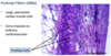
Describe ameloblasts.
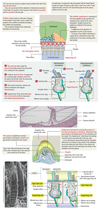
Ameloblasts
- Form enamel which covers only the tooth crown.
- Enamel is the hardest substance in the body.
- Enamel is 96% inorganic salts, about 90% of which is calcium phosphate in the form of apatite crystals and 4% organic matter and water.
- Enamel is laid down in prisms.
- Each prism is formed by one ameloblast.
- Secreting apical domains = Tomes’ processes.
- Increment lines of Retzius are periods of rhythmic growth.
Describe arterioles.
Arterioles
- Small arteries
- Tunica media consists of 1-3 layers of smooth muscle cells.
- Mean arterial pressure depends on proper tone of smooth muscles in arterioles (peripheral resistance arterioles).
- Thickness of smooth muscle layer decreases as diameter becomes smaller.
- Give rise to metarterioles which have a discontinuous layer of smooth muscle tissue.

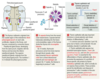
Describe B cell development.
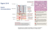
B Cells - Development
- Maturation of B cells involves the appearance of certain cell surface receptors:
- IgM and IgD
- MHC class II proteins
- Complement receptors
- Ig Fc receptors

Describe bile.
Bile
- Hepatic bile is produced and secreted by the liver.
- Bile from the gallbladder is hepatic bile that has been stored and concentrated.
- Components:
- Bile acids:
- Cholic and chenodeoxycholic acids (synthesized in hepatocytes).
- Deoxycholic acid and lithocholic acid (converted by bacteria).
- Water and electrolytes.
- Cholesterol and phospholipids (esp. lecithin).
- Pigments and organic molecules:
- Major pigment is bilirubin.
- Bile acids:
Describe bilinogen.
Billinogen
- Oxidized into excreted products OR:
- Reabsorbed into the blood and carried back to the liver TO BE:
- Re-excreted by the liver OR:
- Excreted in the urine.

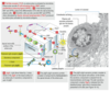
Describe bilirubin formation.
Bilirubin Formation
- Hemoglobin is released from damaged RBCs.
- Hemoglobin is phagocytized by macrophages.
- Split into globin and heme.
- Heme ring is opened to free iron.
- Heme is transported in the blood by transferrin.
- Straight chain of pyrole nuclei is formed.
- Heme is converted by heme oxygenase into biliverdin.
- Biliverdin -> free (unconjugated) bilirubin.
- Free bilirubin is transported attached to plasma albumin to liver hepatocytes.
- Free bilirubin is released from plasma albumin within the liver cells and conjugated:
- With glucuronic acid -> bilirubin glucoronids (80%).
- With sulfate -> bilirubin sulfate (10%).
- With a variety of other substances (10%).
- Conjugated bilirubin is secreted (active transport) into the intestine.
- Conjugated bilirubin in the intestine is converted by bacterial action into urobilinogen.

Describe Clara cells.
Clara Cells
- These cells are found only in bronchioles.
- Number of Clara cells increases as ciliated columnar cells decrease.
- Histologically, these cells can be identified by an apical surface that bulges into the lumen of the airway.
- Secrete surface-active lipoprotein that prevents collapse of terminal bronchioles during exhalation.
- Contain abundant SER.

Describe dermal papillae.
Integument - Dermal Papillae
- Upward projections from each secondary dermal ridge.

Describe dust cells.
Dust Cells
- Macrophages, derived from monocytes.
- Phagocytize particles such as pollutants, bacteria, and surfactant that are not trapped in the mucous and expectorated.
- Relation to CHF:
- In CHF, fluid containing the breakdown products of hemoglobin (iron-containing hemosiderin) leak into alveolar spaces and are phagocytized by the dust cells.
- The iron-containing dust cells are referred to as heart failure cells.
Describe elastic arteries.
Elastic Arteries
- Conducting arteries.
- Stretch during systole and recoil during diastole.
- Tunica media consists of layers of elastic fibers organized into elastic laminae:
- 40 layers in newborn to 70 layers in older adult.
- CT is interspersed between the elastic laminae.
- Include:
- Aorta
- Pulmonary trunk
- Large branches of aorta.

Describe endothelium.
Endothelium

Describe ethanol metabolism.
Ethanol Metabolism

Describe GALT.
Gut-Associated Lymphoid Tissue
- The bulk of the body’s immune defenses is centered in GALT.
- Components:
- Transitory aggregations of lymphocytes, neutrophils, eosinophils.
- Permanent structures:
- Appendix
- Peyer’s patches
- Mesenteric lymph nodes
Describe gastric glands.
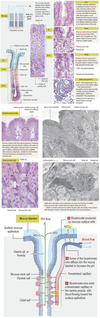
Gastric Glands
- Simple branched tubular glands.
- Narrow isthmus opens into bottom of a gastric pit.
- Fundus (base) of the gland extends into the lamina propria.
- Cells of the gastric glands:
- Mucous neck cells secrete soluble mucous.
- Stem cells in neck replace other cells of the gastric pit.
- Chief cells (zymogenic cells).
- Secrete pepsinogen.
- Parietal cells (oxyntic cells):
- Manufacture HCl.
- Secrete intrinsic factor.
- Enteroendocrine cells (Amine Precursor Uptake and Decarboxylation - APUD cells):
- Diffuse neuroendocrine cells that secrete hormones.
Describe hair follicles.
Hair Follicles
- Develop from epidermis as elastic, keratinized threads.
- Components of follicle:
- Root sheaths (external and internal)
- Hair shaft
- Hair bulb
- Sebaceous glands and arrector pili muscles are associated with hair follicles.
- Hair bulb:
- Expanded lower part of hair follicle.
- Matrix
- Vascularized dermal papilla.
- External root sheath:
- Down growth of epidermis.
- Internal root sheath:
- Generated by bulb matrix.
- Layers:
- Henle’s layer (outermost).
- Huxley’s layer
- Cuticle
- Interlocks with cuticle of hair shaft.
- Hair shaft:
- Layers:
- Medulla (innermost)
- Cortex
- Cuticle
- Layers:

Describe interpapillary peg.
Integument - Interpapillary Peg
- Downward growth of epidermis along crest.

Describe keratinization.
Epidermis - Keratinization

Describe keratinocyte stem cells.
Keratinocyte Stem Cells
- These cells can reestablish epidermis in severely burned patients.
- Migration pathways:
- Bulb-epidermis stem cell pathway.
- Bulb-sebaceous gland stem cell pathway.
- Bulb-hair stem cell pathway.
Describe Kupffer cells.
Liver - Kupffer Cells
- Phagocytic cell.
- Derived from monocytes.
- Lines hepatic sinusoids.

Describe langerhans cells.
Langerhans Cells
- Dendritic cells.
- From monocytes.
- Antigen-presenting cells.
- Primarily in stratum spinosum.
- Migrate from epidermis to lymph nodes.
- Birbeck granules.

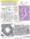
Describe lymph node histology.
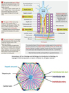
Lymph Node - Histology
- 1-25mm in diameter.
- Hilus - entry and exit point for vessels:
- Efferent lymphatic vessels as well as arteries and veins enter/leave through the hilus.
- Afferent lymphatic vessels enter the convex side of the node.
- Capsule - dense collagen fibers, some elastic fibers and smooth muscle fibers.
- Trabeculae
- Cortex:
- Outer:
- Contains lymph follicles (nodules).
- Follicles:
- Contain B cells, follicular dendritic cells, and migrating dendritic cells.
- Secondary follicles:
- Mantle (dark stained)
- Germinal center (lighter stained)
- Primary:
- Lack mantle and germinal center.
- Deep (inner):
- Contains TH cells, macrophages
-
High endothelial venules (HEVs)
- Port of entry for circulating differentiated lymphocytes to seed lymph node.
- Medulla:
- Irregular arrangement of loose medullary sinuses and dense medullary cords:
- Sinuses are lined with macrophages.
- Cords consist of blood vessels, lymphoblasts and plasma cells.
- Site of lymphocyte reentry into lymph stream.
- Thymic-dependent areas in subcortical and deeper medullary regions.
- Irregular arrangement of loose medullary sinuses and dense medullary cords:
- Outer:

Describe lymphoid tissue.
Lymphoid Tissue
- Appears in body as a gradient from diffuse lymphoid tissue to lymphoid organs.
- Lymphoid organs:
- Primary - where precursor cells mature into immunocompetent cells, and are programmed to recognize a specific antigen.
- Thymus and bone marrow.
- Secondary - trapped antigens stimulate clonal expansion of mature T & B cells.
- Lymph nodes, spleen, tonsils.
- Primary - where precursor cells mature into immunocompetent cells, and are programmed to recognize a specific antigen.
- Lymphocytes mature in primary lymphoid organs and then take up residence in secondary lymphoid organs.
- Lymphoid follicle (nodule)
- Primary vs. secondary (with germinal center).
Describe melanocytes.
Melanocytes
- Derived from melanoblasts.
- Do not form desmosome attachments in epidermis.
- Inject melanin granules into keratinocytes.
- Pathway for melanin formation:
- Tyrosine -> 3,4-dihydroxyphenylalanine (DOPA) -> dopaquinone -> melanin
- Requires tyrosinase.

Describe Merkel cells.
Merkel Cells
- Mechanoreceptors
- May also act as diffuse neuroendocrine cells.
- Usually in stratum germinativum.
- Contains catecholamine-like granules.