Histo: Upper GI Disease Flashcards
What is a key histological feature of the oesophageal mucosa?
Presence of submucosal glands

what is the Z-line?
The point in the oesophagus at which the epithelium transitions from being squamous to being columnar

What does the body and fundus of the stomach have in abundance?
Specialised glands responsible for producing acid and enzymes
Which part of the stomach tends to be affected by H. pylori-associated gastritis?
Pylorus and antrum

What are the three layers of the gastric mucosa?
- Columnar epithelium
- Lamina propria
- Muscularis mucosa

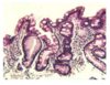
What is the normal villous: crypt ratio?
2:1

What does the presence of goblet cells in the stomach signify?
Intestinal metaplasia
NOTE: goblet cells are NOT normally seen in the stomach
What is the characteristic histological feature of acute oesophagitis?
Presence of lots of neutrophils
This is usually caused by GORD

What are the complications of acute oesophagitis?
- Ulceration
- Fibrosis
- Haemorrhage
- Perforation
- Stricture
- Barrett’s oesophagus
Define Barrett’s oesophagus
Metaplastic process by which the normal sqaumous epithelium of the lower oesophagus is replaced by columnar epithlieum
NOTE: this is also known as columnar-lined epithelium (CLO)

What are the two forms of metaplasia in barrett’s oesophagus?
Gastric metaplasia - without goblet cells
Intestinal metaplasia - with goblet cells

What further degree of metaplasia is associated with an even greater risk of cancer than Barrett’s oesophagus?
Intestinal metaplasia - goblet cells become visible
NOTE: metaplasia is reversible
Define dysplasia.
Changes showing some of the cytological and histological features of malignancy but with no invasion through the basement membrane.

What is the most common oesophageal carcinoma in developed countries?
Adenocarcinoma of the oesophagus - associated with reflux and mainly found int eh lower oesophagus

What is squamous carcinoma of the oesophagus associated with?
- Smoking and alcohol
- It tends to affect the middle/lower oesophagus
- It is the most common type of oesophageal cancer in Africa
What are the main histological features of squamous cell carcinoma of the oesophagus?
Cells produce keratin (normal oesophageal squamous epithelium is non-keratinised)
Intercellular bridges

How is eosinophilis oesophagitis treated?
- Steroids
- Allergen removal
NOTE: this is associated with an allergic reaction (asthma of the oesophagus). It is due to allergy to food causing muscle spasm and dysphagia.
What is this?
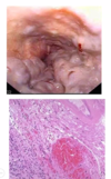
Oesophageal varices
What is the commonest cause of oesophageal varices?
- Cirrhosis of the liver (Most common)
- Portal vein thrombosis
What are the common causes of acute gastritis?
List some causes of chronic gastritis
- Autoimmune (body, auto-antibodies e.g. antiparietal)
- Bacterial (H. pylori)
- Chemical (NSAIDs, bile reflux)
NOTE: the key inflammatory cells in chronic gastritis are lymphocytes
What is the type of pattern seen in H.pylori gastritis?
Chronic gastritis +/- activity
What are the outcomes of H.pylori gastritis?
CLO-IM-dysplasia
Adenocarcinoma
Lymphoma (MALToma)

What is mucosa-associated lymphoid tissue and what is their presence indicative of?
- Chronic gastritis caused by H. pylori infection induces lymphoid tissue in the stomach
- The presence of lymphoid follicles in a stomach biopsy, is highly suggestive of H. pylori infection
- This is important because it is associated with an increased risk of lymphoma


