Histo: Neuro-Oncology Flashcards
How much more common are secondary brain tumours than primary brain tumours?
Brain metastases are 10x more common that primary tumours
Describe the locational classification of brain tumours.
Extra-axial (coverings):
- bone
- meninges
- nerves
- cranial soft tissue
Intra-axial (parenchyma):
- Derived from normal cell populations of the CNS (e.g. glia, neurones, neuroendocrine, vessels)
- Derived from other cell types (e.g. lymphomas, germ cell tumours)
List the different cell types within the CNS that can give rise to brain tumours.
- Neurones
- Astrocytes
- Oligodendrocytes
- Ependyma
- Choroid plexus epithelium
- Meningothelial cells
- Embryonal cells
What is the aetiology of CNS tumours
Largely unknown
- Environmental: radiation associated with meningioma
- Genetic predispostion: familial CNS tumour syndromes
What is the most common genetic syndrome associated with brain tumours?
Neurofibromatosis
What is the inheritance pattern of neurofibromatosis?
Autosomal dominant
Where are the genes that cause neurofibromatosis located?
- NF1 = 17q11
- NF2 = 22q12
What tumours are associated with following Familial CNS Tumour Syndromes:
- NF1
- NF2
- Brain Tumour polyposis syndrome 1
- Gorlin syndrome
- Von Hippel Lindau syndrome
- NF1 - neurofibroma, astrocytoma
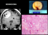
- NF2 - bilateral vestibular schwanoma, meningioma
- BTP1 - malignant glioma
- Gorlin syndrome - medulloblastoma
- VHL - haemangioblastoma
AD inheritance
What are some common signs of CNS tumours
Raised ICP:
- Headache (worse in morning, coughing, lying down)
- Vomiting
- Altered mental status
List some manifestations of brain tumours that are:
- Supratentorial
- Subtentorial
Supratentorial
- Focal neurological deficit
- Seizures
- Personality changes
Subtentorial
- Cerebellar ataxia
- Long tract signs (e.g. hyperreflexia)
- Cranial nerve palsies
What are some neuroimaging modalties
- CT
- MRI
- MR spectroscopy - assess tumour metabolic activity
- Perfusion MRI
- fMRI
- PET
Outline the management options for brain tumours.
Surgery - aim for maximal safe resection with minimal damage to the patient. Debulking may be performed and biopsies may be taken.
Radiotherapy - used for gliomas and metastases
Chemotherapy - mainly for high-grade gliomas and lymphomas
What is the role of histopathology and molecular pathology
- Definitive diagnosis
- Prognostic tests
- Assessement of treatment response
What is the WHO classification of brain tumours based on?
- Tumour type (cell of origin)
- Tumour grade (aggressiveness/degree of malignancy)
- Molecular profile - most tumour types have specific molecular markers
NOTE: metastases are not graded
What are some criteria that tumour grade is based on?
- Mitotic activity
- Degree of cell and tissue differentiation
- Degree of necrosis
Outline the meaning of the different WHO grades for brain tumours.
- Grade I = benign, long-term survival
- Grade II = death in > 5 years
- Grade III = death in < 5 years
- Grade IV = death in < 1 years
NOTE: grades I and II are low
GRADE GUIDES Mx
Which brain tumours are staged?
None
Except medulloblastoma
What is the most common type of primary brain tumour?
Glial tumours, specifically astrocytoma

How are the types of glial tumours seen in children and adults different?
Diffuse gliomas
- Mainly seen in adults
- Supratentorial
- Malignant progression
- Astrocytomas, oligodendrogliomas
Circumscribe gliomas
- Mainly seen in children
- Posterior fossa
- Rarely undergo malignant transformation
- Astrocytomas, ependymomas
Which genetic mutations are associated with gliomas in adults and in children?
- Diffuse infiltration (adults) - IDH1/2
- Circumscribed gliomas (children) - MAPK (BRAF)
List some examples of circumscribe gliomas.
- Pilocytic astrocytoma (MOST COMMON)
- Ependymoma
- Subependymal giant cell astrocytoma
List some key features of pilocytic astrocytomas (WHO grade 1)
- Mainly occurs in children (1st and 2nd decade of life)
- Associated with NF1
- Often located cerebellar, optic hypothalamus, or brainstem
- BRAF mutation in 70% of cases
What is the hallmark histological feature of pilocytic astrocytoma?
- Piloid (hairy) cell
- Often see Rosenthal fibres and granular bodies
- Slow-growing with low mitotic activity

List some key features of astrocytoma (WHO grade 2, 3, and 4)
- Usually affects adults 20-40 years old
- Cerebral hemispheres are the most common site in adults
- Can progress to become a higher grade (malignant progression)
- IDH1/2 mutation in 80% of cases