Hemodynamic disorders Flashcards
what is oedema?
An abnormal increase in interstitial fluid
What does Oedema mean?
- Increased hydrostatic pressure
- Salt and water retention
- Reduced plasma oncotic pressure
- inflammation
- lymphatic obstruction
(HSWOIL)
What are the two types of oedema?
- Localised oedema
- Generalised oedema
Describe generalised oedema
Fluid in serous cavities (pleural, pericardial, peritoneal)
more than 5L
Causes:
- heart failure
- inflammation
- venous hyoertension
- lymphatic obstruction
Describe localised oedema
Pulmonary and cerebral oedema
Causes:
- Heart failure (congestive)
- Hypoproteinaemia (low protein content)
- Nutritional oedema (low albumin)
What is generalised pitting oedema?
(difference between pitting and non-pitting)
When oedema occurs in subcutaneous tissues AND serous cavities.
Pitting oedema- when you apply pressure to swollen area (each a finger press), the impression will stay there, it will leave a pit.
Non-pitting oedema will return back to the swollen shape.
What is pulmonary oedema?
In pulmonary capillaries, the oncotic pressure is greater than the hydrostatic pressure.
left heart failure increases the hydrostatic pressure in the pulmonary capillary bed
Fluid will accumulate first in the the interstitial space and then eventually spills into the alveolar spaces.
What is this xray showing?

Pulmonary oedema.
You should be able to see clear black spaces for the lungs.
In the xray, the pt has an enlarged heart whcih suggests heart failure.
Fluid accumulates in the alveoli leading to almost no oxygen diffusion (which causes shortness of breath)
Breathlessness and pulmonary oedema?
Breathlessness (dyspnoea) is the main symptom.
It is typically worse when lying flat (orthopnoea)
Fluid in the alveolar spaces predisposes to bacterial infection in the lungs (pneumonia).
The microscopic picture is an image of proteinaceous material within the alveoli

What is cerebral oedema?
it is localised oedema. Caused by the following:
Vasogenic- increased permeability of capillaries and venules
Cytotoxic- derangement of sodium-potassium mebrane pumps e.g. ischaemic strokes
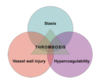
What is thrombosis?
It is an abnormal blood clot formation in the circulatory system
What are the three causes of thrombosis?
(Virchow’s triad)
- endothelial injury (to the vessel wall)
- Stasis or turbulent blood flow
- Blood hypercoagulability- coagulation pathways are activated and thrombosis occurs
what is venous blood flow?
Stasis and hypercoagulability are key factors. Most of the time they form in deep leg veins (venous thrombosis-DVT) Sometimes can occur higher up.
It can be caused genetically or acquired disruption in the lamina blood flow (e.g. long term bed rest)
Pulmonary embolism is the most important potential complication.

What is arterial thrombosis?
Almost always related to vessel wall injury caused by atherosclerotic plaques.
Narrowing of the artery (stenosis) causes ischarmia of the tissue supplied bu the artery.
A complee blockage of the artery by thrombus causes infarction of the tissue supplied by the artery.

Arterial thrombosis can cause real problems with the heart…
You can get red thrombus in a coronary artery leading to myocardial infarction.
There are 4 different fates of thrombi
- Propagation
- Embolisation
- Dissolution (break down)
- Organisation and recanalisation (coronary artery)
(PEDRO!!)
When do thrombi become noticable?
- When they obstruct arteries or veins
- When they embolise (obstruction of artery or vein with a clot)
What is an emboli?
It is abnormal material within the circulatory system that is carried in the blood to a site distant from its point of origin. Most emboli are fragments of dislodged thrombus.
Rare types of emboli include: fat, air, amniotic fluid, tumour.
Emboli can lodge vessels and block them off
What does ischaemia mean?
It describes something that has a lack of oxygen
What does infarct mean?
It is an area of ischaemic necrosis caused by occlusion (blockage) to arterial or venous supply.
What is the difference between a red infarct and white infarct?
Red infarct- venous occlusion happens in tissue with geocirculation like the lungs
White infarct- arterial occlusion- solid organ wedge shape- spleen or kidney.
Infarcts can heal by repair. Although structural integrity is maintained, there is a permenant loss of tissue
What is bowel infarction?
Infarcted segment of small bowel is dark
What is myocardial infarction?
Normal myocardium should not be so thick. the white areas are the areas of ischaemia- these areas have scarred.

DVT and pulmonary embolism
Most venous thromoembolism originate from DVT.
Emboli that block the pulmonary artery is the most significant consequence.
