Hematopoiesis - Strom 03.16.15 Flashcards

1
Q

What are pluripotent stem cells?

A
  • Defined by ability to “salvage” all elements of hematopoiesis in recipient after their bone marrow contents are wiped out by irradiation or chemotherapy
  • Rare (1 in 20 million), and express receptors for key growth factors
  • Morphologically, “blasts” (small round cells with hypodense chromatin and no morphologic features associated w/differentiation), but make up only a tiny fraction of the morphologic blasts in the bone marrow (so they can’t be ID’d clearly by morphology)
  • In research settings and gene therapy protocols they can be purified almost to homogeneity by identifying certain characteristic cell surface markers
2
Q

Can you use a microscope to pick out burst (BFU) and colony forming units (CFU) from the bone marrow? How are they functionally defined?

A
  • NO - although it would be really helpful if you could
  • Functionally defined by the fact that if you dump a mixture of cells from bone marrow into petri dish, a particular cocktail of growth factors causes formation of colonies with morphologic and immunophenotypic features of maturing cells in specific lineages
3
Q

What are the 4 key heme growth factors?

A
  • Thrombopoietin (TPO)
  • Erythropoietin (EPO): this drug is the reason many pts with renal failure are alive now
  • Granulocyte-macrophage colony stimulating factor (GM-CSF)
  • Granulocyte colony stimulating factor (G-CSF)
4
Q

What percentage of cells in the bone marrow are blasts? What is the breakdown within this group of cells?

A
  • Blasts <4%
    1. Erythropoiesis: 20-30% (incl. thrombopoiesis)
    2. Myelopoiesis: 60-70%
    3. Lymphopoiesis: 10-20%
    4. Pluripotent: probably <0.1%
  • NOTE: microscopic view doesn’t quite match this b/c can’t identify the CFU/BFU cells morphologically; most likely have no features associated w/differentiation at all – that is, they are morphologic “blasts”
5
Q

Describe the normal morphologic maturation of granulocyte precursors.

A
  • Continuous process, but represented categorically:

Blast (3-4%) -> Promyelocyte (2-8%) -> Myelocyte (10-13%) -> Metamyelocyte (10-15%) -> Bands and neutrophils (25-40%)

  • More mature species outnumber less mature species b/c granulocyte maturation a process of differentiation and cell division; cells thought to lose ability to divide only after the myelocyte stage
  • Can be affected by # of disease states, e.g., it can INC in overall numbers, be shifted to the left or right, or be blocked part way through (“maturation arrest”)
6
Q

What are the key regulators of granulopoiesis?

A
  • GM-CSF: how its production is tied in to the multiple biological factors that can stimulate increase in neutrophil count not well understood
  • Acts well up the differentiation chain, such that it will also stimulate production of eosinophils (which branch off this process b/t morphologic blasts & promyelocytes)
  • G-CSF: acts more specifically on neutrophil precursors -> exact morphologic stage of the least differentiated cell it acts on is not clear
  • NOTE: both of these factors released via bone marrow stromal cells; aka, myelopoiesis
7
Q

Describe the normal maturation of red cell precursors.

A
  • Epo production stimulated by hypoxia, sensed by renal peritubular cells -> renal failure usually results in anemia (add to differential diagnosis of unexplained anemia)
  • Blast (3-4%) -> Pronomoblast -> Basophilic erythroblast -> Polychromatophilic erythroblast -> Normochromic erythroblast
  • 4 or 5 cell divisions, but red cell precursors seen in bone marrow mostly most mature erythroid precursors, “normochromic erythroblasts”
    1. After 5th division, nuclei extruded and nascent red cells released (polychromasia, reticulocytes)
8
Q

Describe thrombopoiesis (megakaryocytes).

A
  • Platelet production very different than RBC, leukocyte production -> megakaryocytes: polyploid factory cells (nuclei divide multiple times, so instead of normal diploid (2N) state, contain 16-32 haploid genomes)
  • Megakaryocytes extend snakelike tubes called “proplatelets” into highly perforated (fenestrated) blood vessels in bone marrow (sinuses) -> mature platelets lopped off one at a time from ends of the proplatelets
  • Regulated by hepatocytes and other sources (blast, 3-4% -> immature -> mature)
9
Q

Where is TPO made? What does it do?

A
  • Thought to be made at a constant rate, primariily in liver
  • Binds both platelets and megakaryocytes; when bound to megakaryocytes, stimulates their production from immature precursors and platelet production from mature megakaryocytes
  • Low platelet count allows more TPO to bind megakaryocytes, stimulating more thrombopoiesis
10
Q

What is the basic structure of the EPO and TPO receptors? What would you expect if both of these were stuck in the ON position?

A
  • EPO (expressed on erythroid precursors) and TPO (aka, c-MPL) receptors (on mature/immature megakaryocytes) have very similar structures and functions
  • Both transmembrane dimers with two copies of relatively inactive kinase (Jak-2) attached to their cytoplasmic tails
  • If stuck in ON position, you would expect: BLOOD CLOTS
11
Q

What happens when the Type 1 hematopoietic growth factor receptors are activated? Why is it important to understand this?

A
  • Cytokine binding to receptor swings cytoplasmic tails, and their bound kinases (Jak-2), close together -> kinases phosphorylate each other, increasing activity
  • More active kinases phosphorylate other targets further down cytoplasmic tail, initiating series of other signal transduction steps (& altered transcription patterns) w/divergent actions in different lineages (EPO vs. TPO)
  • Both processes result in differentiation, proliferation of precursors in their respective lineages
  • Acquired mutations in receptors, or Jak-2 kinase, are clinically relevant – paticularly if they cause constitutive activation of these signal transduction pathways
12
Q

What happens to bone marrow cellularity with age?

A

It declines. The image shown is a bone marrow biopsy from a 5 y/o.

13
Q

What is this?

A

High power view of bone marrow biopsy. You can see some erythroid (top right), myeloid (middle), and megakaryocyte (middle) cells here.

14
Q

What is this? Is it normal or abnormal? Why?

A
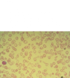
  • Aspirated (sucked out with needle) normal bone marrow material
  • Should be able to identify the neutrophils and bands present, and the smaller cells with dark and very round nuclei (mostly normochromic erythroblasts)
  • More important point is that there are a lot of different cell morphologies present, making this NORMAL -> when you see bone marrow aspirate containing predominantly one cell type, there’s a problem
15
Q

What is heme? What does heme synthesis require?

A
  • Heme is an oxygen carrier
  • Requires iron, B6, succinyl CoA, and glycine (which requires B12 and folate)
16
Q

What does globin production require?

A
  • Requires amino acids of course, and malnutrition definitely causes anemia
  • In this country, problems with globin production more commonly due to problems with the genes for its two component proteins (alpha and beta), also required for synthesis
17
Q

What is required for red cell DNA synthesis?

A
  • Requires deoxynucleoside triphosphates, which require ribonucleotide reductase and thymidine (B12 and folate)
  • Although thrown away at end of process, nucleus is replicated multiple times in red cell production, so the production process kind of counterintuitive – red cells lack nuclei, but you have to make tons of DNA in order to produce red cells (again requiring both adequate nutrition and a couple of key cofactors (vitamins))
18
Q

How is the red cell production process regulated?

A
  • Primarily by erythropoietin
  • Will fail if kidneys fail, or if bone marrow gets filled up by cells that don’t belong there (which can include fibroblasts and the collagen they produce)
19
Q

Is anemia a diagnosis? Why or why not?

A
  • Anemia might have been a diagnosis once, but it is not now - any more than fatigue or pallor or tachycardia are diagnoses
  • Anemia is a laboratory finding; it remains undiagnosed until you find a cause for it
20
Q

What are the 3 possible disease mechanisms for anemia?

A
  1. Losing red cells from the bloodstream
  2. Not making enough red cells
  3. Both
21
Q

What is the morphology of iron deficiency?

A
  • Morphology: small red cells without much hemoglobin, i.e., microcytosis and hypochromia; lots of variation in red cell size & shape, i.e., anisocytosis & poikilocytosis
    1. Hypochromia will be seen a bit earlier than anisocytosis and poikilocytosis, i.e., when pt’s anemia only mild or moderate
  • Area of central pallor in biconcave discs is enlarged; rule of thumb is that if central pallor diameter > 1/3rd of red cell diameter, cell is hypochromic, or lacking color - i.e. lacking hemoglobin
  • Anemia, microcytosis, hypochromia suggest iron def, but other problems can yield a similar picture -> to confirm iron deficiency, you’ll need additional lab tests
22
Q

What are anisocytosis and poikilocytosis? When will these terms show up in your pt’s lab results?

A
  • Anisocytosis: variation in red cell size
  • Poikilocytosis: variation in shape
  • Descriptive terms that will show up in lab results IF a manual differential count is done; an automated CBC will not generate these terms, but hematology analyzer will tell you if red cells size distribution is increased (“red cell distribution width,” or RDW), which correlates well with morphologic anisocytosis.
  • Both are characteristic of (but not specific for) severe iron deficiency
23
Q

Describe the process by which dietary iron gets into circulation from the gut (receptors covered in other card).

A
  • Dietary iron mostly in oxidized (ferric) state; reduced (in duodenum) before being taken up (in rest of sm bowel)
  • Ascorbate (vit C) reducing agent duodenal reductase (duodenal cytochrome b) uses (to reduce one thing you have to oxidize another) -> uptaken iron re-oxidized for transport (via serum oxidase, ceruloplasmin)
  • Free iron in plasma would be bad thing, augmenting bacterial growth and catalyzing formation of superoxide radicals from oxygen -> so, transport handled by specialized circulating protein, transferrin, which only works with Fe+++ (it can’t transport ferrous iron)
24
Q

What transporters are involved in iron absorption by gut enterocytes?

A
  • Multistep process with 2 transporters: 1 on luminal mem (DMT-1) and 1 on basement mem (ferroportin)
  • Both transporters regulated by physiologic conditions
    1. DMT-1: iron-dependent regulation of its mRNA translation and stability
    2. Ferroportin: regulatory peptide hepcidin; also responsible for exporting iron from macros that hold body’s iron storage pool– and hepcidin regulates it in that location as well
25
Q

What are the two basic categories for lab measurement of iron in the body? Name the specific tests in each category.

A
  • Measurement of the iron transport system
    1. Serum iron: transferrin-bound iron
    2. TIBC (total iron binding capacity): total amount of transferrin in circulation
    3. Transferrin saturation: serum iron divided by transferrin
  • Measurement of storage pool iron
    1. Serum ferritin: trace amounts of ferriting from storage pool sites leaks out into serum, and its concentration is proportional to total amount in the pool -> most useful initial measurement of iron metabolism in patients with unexplained anemias.
26
Q

The combo of what four measurements establishes a diagnosis of iron deficiency? What is the next step?

A
  • Iron deficiency a result of either increased red cell loss (usually chronic hemorrhage) or reduced dietary iron
  • In either case, storage pool iron depleted, detected as reduced serum ferritin levels
  • Effort to move remaining storage pool (or dietary) iron to production facility (bone marrow) = transferrin production up -> detected as an increased TIBC
  • Intake usually not keeping up with utilization, so serum iron is reduced, meaning transferrin saturation reduced
  • Next step: find out what caused it!
27
Q

What is an example of a “second line” measurement of storage pool iron?

A
  • Soluble (or serum) transferrin receptor
  • Iron-starved macrophages increase the amount of transferrin receptors on their surfaces
28
Q

What is hepcidin? How is it involved in iron regulation?

A
  • Hepcidin: peptide made in liver that regulates iron uptake and transport -> complicated regulatory mech in hepatocytes links its expression to amt of transferrin-bound iron in plasma
    1. INC levels of transferrin-bound iron INC hepcidin production, DEC ferroportin expression (in macros & enterocytes – internalized & degraded)
    2. Low iron: hepcidin expression DEC, ferroportin expression INC, and more iron taken up in gut, put into transport system to feed red cell production
  • NOTE: macros that store iron primarily in bone marrow, spleen, and liver
29
Q

What are some examples of chronic blood loss causing iron deficiency? What is unique about these types of anemia?

A
  • Iron deficiency often results from chronic blood loss
  • Examples: GI tract, urinary tract, dysfunctional uterine bleeding
  • These anemias are due to BOTH red cell loss AND impaired production
30
Q

What is thalassemia? Describe its morphology.

A
  • Thalassemia: reduced globin production resulting from any of dozens of genetic defects
  • Beta thalassemia morphology:
    1. Microcytosis, hypochromia (like iron deficiency, but maybe even smaller)
    2. Frequent target cells (nonspecific b/c commonly also seen in association with liver disease)
    3. Can be severe if homozygous; can be mild and not clinically obvious if heterozygous
31
Q

What do the lab values look like for beta thalassemia? How are they different from those for iron deficiency, and how do you confirm thalassemia dx?

A
  • Lab values: very microcytic anemia (MCV = 69), and normal or INC number of red cells (5.1 x 10^6); also Hg 8 and Hct 24%
  • Reliable clue pt w/microcytic anemia has thalassemia, not iron deficiency is NUMBER of red cells -> DEC in iron def, but usually normal or INC in thalassemias
  • Confirm thalassemia by ordering Hg electrophoresis test, which will detect abnormal Hg (aka, hemoglobinopathy)
32
Q

Describe the organization of the beta globin gene locus.

A
  • Chromosome 11
  • 1 beta globin gene
  • 4 beta globin homologues (epsilon, g-gamma, a-gamma expressed in utero; delta expressed in the fetus, with low-level expression after birth)
  • 1 pseudogene (psi-beta awaiting evolutionary assignment)
33
Q

What is the composition of normal Hgb? How is this altered if expression of the beta globin allele is impaired?

A
  • Normal: alpha-2 beta-2 (Hgb A) with traces of alpha-2 delta-2 (Hgb A2)
  • Mutation: relative amount of delta globin is increased
    1. Increased Hgb A2: result you will most likely see ojn Hgb electrophoresis in pt with beta thal
    2. In severe beta thal cases, fetal Hgb (alpha-2 gamma-2 = Hgb F) will also be detected
  • The severity of different forms of beta thal is a highly variable function of t mutation type, and whether it is present in one (hetero) or two (homozygous) copies
34
Q

What is the structure of the alpha globin gene locus? What are the implications of this?

A
  • Chromosome 16 (gene replicated 4x)
    1. 2 copies expressed in utero: zeta 1 and 2
    2. 2 copies in adults: alpha 1 and 2
  • 2 copies of chrom 16, so haplotypes w/defect in alpha 1, alpha 2, or both can be present in the population and, for individuals whose other haplotype is normal, not cause any problems -> multiple possible mutations in each of these 4 types found in pop; some mutations completely abrogate expression of the gene, some partially
35
Q

How many possible alpha thal genotypes are there?

A
  • 16 possible combinations (genotypes)
  • 6, however, are duplicates (example: haplotypes (b + c ) vs haplotyptes (c + b)), so # of functional possibilities from this example is 10
36
Q

What is alpha thal 1 trait?

A

1 defective allele -> almost no clinical/lab findings

37
Q

What is alpha thal 2 trait?

A
  • 2 defective alleles
  • Mild microcytic anemia, excess Hgb Bart’s (gamma-4) at birth, but normal Hgb electrophoresis as adults
  • Dx: PCR-based (electrophoresis and/or sequencing)
  • Epi: 3% of AA
38
Q

What is Hgb H disease?

A
  • 3 defective alleles
  • Variable degree of microcytic anemia, electrophoresis: 15-30% Hgb H -> can be misdiagnosed as iron def
  • Excess Hgb beta protein in red cells of individuals with this type of genotype -> beta globin can form a functional tetramer - and it does, and can be detected by Hgb electrophoresis
39
Q

What is Hgb Bart’s

A
  • 4 defective alleles
  • Lethal in utero or shortly after birth - apparently before normal developmental expression of hemoglobin beta can produce significant amounts of Hgb H
  • Common allele in SE Asia
40
Q

What are the biochemical requirements for DNA synthesis?

A
  • Ribonucleoside triphosphates have to be reduced to the deoxy form (via ribonucleotide reductase), and UTP has to be methylated to TTP (thymidine)
  • Methyl groups in both cases donated by folate-derived donors, but also require B12 (cobalamin) cofactor
41
Q

How does folate deficiency result in anemia?

A
  • Folate deficiency impairs nucleotide synthesis, that impairs DNA synthesis, and you need to make DNA to make red cells
  • Two different derivatives of folate take part in nucleotide synthesis – the N-10-formyl form in purine synthesis, and the N5, N10 methenyl form in thymidine synthesis (recall that thymidine is just uracil w/a methyl group added - this is how the methyl group gets there)
42
Q

How does B12 deficiency cause anemia?

A
  • B12 (cobalamin) NOT directly required for DNA syn, but if you become deficient in it, nucleotide (and DNA) syn impaired b/c both B12 and N5-methyl-THF are needed to convert homocysteine to methionine –> need a lot of methionine for other metabolic reasons
  • If B12 is lacking, all your folate will get “trapped” in the N5-methyl form, making it unavailable for nucleotide synthesis -> “methyl trap” hypothesis
43
Q

Why does B12 deficiency cause neurologic symptoms?

A
  • Methionine serves as a methyl donor (after converted to S-adenosyl-methionine) in biosyn of a key component of myelin (sphingomyelin) -> B12 def can lead to anemia and characteristic neurologic condition called subacute combined degeneration of the spinal cord
  • If folate supplementation given to B12-deficient pts, it worsens their neurologic status -> biochem unclear (it could be that N5-methyl-THF has its own toxic side effects), but the clinical implications are not; when in doubt, supplement with both vitamins
44
Q

What is the uptake mechanism for Vit B12?

A
  • Mammals can’t make it, so need ito get it from diet
  • HC (haptocorrin) is first binding agent (saliva protein), then bound by intrinsic factor (IF made by parietal cells in stomach – binds B12 after HC digested away in the jejunum, then shepherds it down to distal ileum, where complex taken up and transported into bloodstream
  • In order to make red cells, you have to make DNA, so impaired DNA synthesis via damaged parietal cells may cause anemia (impairing red cell synthesis)
  • Megaloblastic anemia –> check Vit B12 and folate levels; only other role of B12 is to reduce MMA concentration (also an indicator for B12 deficiency).
45
Q

What happens to blood cells if DNA synthesis is impaired?

A
  • Fewer cells produced
  • Normal/enhanced maturation of cytoplasm
  • Impaired nuclear maturation (b/c impaired DNA syn)
  • If DNA syn impaired, production line held up after one or two cell divisions - stage where nucleus is large, and chromatin not condensed into heterochromatin
  • Cytoplasm continues to mature: RNA, blue color, begins to get degraded, and Hgb’s reddish color begins to predominate -> “megaloblastic
46
Q

Are there storage systems for B12/folate?

A
  • Cells have complex storage system for folate derivatives involving linking the molecule to a series of glutamate residues (“polyglutamate” forms)
  • No similar storage system for B12, but internal recycling good -> takes several years of inadequate dietary intake to become functionally B12 deficient
47
Q

What causes megaloblastic anemia? How do you diagnose it?

A
  • Impaired B12 uptake (pernicious anemia)
  • Impaired folate uptake: malabsorption, malnutrition
  • Drug effect: nucleoside analogues (HAART), ribonucleotide reductase inhibitors (Hydroxyurea)
  • Intrinsic bone marrow dysfunction: myelodysplastic syndrome(s)
  • Most can be diagnosed without a bone marrow biopsy
48
Q

How might renal failure be implicated in changes in red cell production?

A
  • Red cell production regulated by organs outside bone marrow, and can be impaired by their dysfunction
  • Anemia a well known, treatable consequence of renal failure
  • Erythropoietin production regulated by O2 sensors located in peritubular cells in renal cortex, and secrete EPO when they sense reduced availability of O2
  • Urge to breath is regulated by CO2 concentration in the bloodstream – NOT by oxygen concentration
49
Q

How is hepcidin regulated by transferrin-bound iron?

A
  • Hepcidin is negatively regulated by iron in the transport system (transferrin-bound iron), however, other factors can *increase *hepcidin production
50
Q

How can chronic and acute inflammatory conditions affect iron stores in the body?

A
  • Chronic and acute inflammatory conditions can induce hepcidin production, impairing use of bone marrow iron stores and uptake of dietary iron
  • Hepcidin uses same mechanism to do both: it impairs activity of ferroportin (causing it to be internalized and degraded), limiting egress of iron from macros that store it, and limits its uptake via the gut epithelial cells that take it up from dietary iron sources -> if persists long enough, pt devos anemia of chronic inflammation
51
Q

What biological role does IL-6 have in anemia of chronic disease?

A
  • One theory is that bacteria really need iron (may be why several human pathogens secrete proteins that lyse red cells)
  • When you’re fighting off a bacterial infection, probably a good idea to clamp down on the availability of iron in the bloodstream – via IL-6 signalling the liver to release hepcidin
52
Q

Explain the mechanism of anemia of chronic inflammation, and how it may be associated with hemochromatosis.

A
  • Iron not mobilized from storage, and not absorbed well from GI tract; transferrin gets pulled out of circulation by hungry red cell precursors
  • “Transferrin saturation” (serum iron divided by TIBC) is actually increased, but net flux of iron through system is reduced, affecting red cells
  • Hepcidin limits iron uptake via increased storage of ferritin (internalization) -> excess iron storage (aka, hemochromatosis) can cause Diabetes, liver disease, and cardiomyopathy
    1. Patient with all of these, high iron levels, and no relevant history may have a hepcidin problem
53
Q

What is the blue in this bone marrow biopsy from a pt with anemia of chronic inflammation? Explain.

A
  • Loaded with iron (blue granules) -> in general, when you suspect anemia of chronic inflammation you will need to get a bone marrow biopsy to confirm it
  • In most contexts (AIDS, TB, RA, cancer), CBC and iron studies do not rule out other bad things happening in the bone marrow, such as acute leukemia, a lymphoma, metastases, or a large number of granulomas
  • Characteristics: normocytic anemia, INC ferritin, DEC or normal serum iron, and INC bone marrow iron stores