Haematology: Blood Coagulation, Haemostasis & Its Investigation Flashcards
(46 cards)
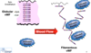
What is haemostasis?
- Protective process designed to stop bleeding and maintain blood flow
The circulatory system of the horseshoe crab contaisn haemolymph. What does haemolymph contain?
- Contains amebocytes which contain:
- Proteins of the coagulation system
- Proteins & peptides of the immune system
How does the fact that the horseshoe crab’s circulatory system is made up of haemolymph affect the way it fights infection?
- It means that in order to fight infection coagulation of that haemolymph would also have to occur
What are some of the functions of haemostasis?
- Respond to tissue injury
- Curtail blood loss
- Restore vascular integrity & promote healing
- Limit infection
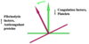
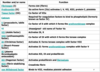
Name the 4 main components of haemostasis
- Endothelium
- Coagulation
- Platelets
- Fibrinolysis
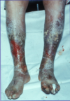
What are the components of a thrombus (blood clot)?
- Fibrin mesh
- Platelets
- Red blood cells

Name the 3 different stages of haemostatsis and state what occurs during each stage
-
Primary haemostasis
- Vasoconstriction
- Platelet adhesion
- Platelet aggregation and contraction
-
Secondary haemostasis
- Activation of coagulation factors
- Formation of fibrin
-
Fibrinolysis
- Activation of fibrinolysis
- Lysis of the plug
What are the different stages of blood clot formation?
- Initiation
- Amplification
- Stable clot
- Lysis

Describe the process of primary haemostasis
- Damage to endothelium exposes sub-endothelium to flowing blood
- This results in sub-endothelial cells releasing Von willebrand factor (VWF)
- Von willebrand factor binds to platelets and causes them to adhere to exposed collagen
- Platelets also become activated as a result of this
- Activated platelets release various pro-thrombotic substances, e.g. thromboxane A2, which help to clump (aggregate) platelets together
- This forms a loose platelet plug
- Loose platelet plug contracts to form a dense, adherent plug
- Platelets in platelet plug form negatively charged phospholipid surface which is required for coagulation
- Allows coagulation factors to interact much better with each other

Why is the phospholipid surface formed by the platelet plug required for secondary haemostasis (coagulation)?
- Allows coagulation factors to interact much better with each other
When released by sub-endothelial cells von willebrand factor goes through a conformational change. Describe what this conformational change is and why it occurs
- As a result of increased shear stress due to endothelium damage when released, von willebrand factor goes from a globular form to a filamentous form
- This filamentous form von willebrand factor then begins to bind to platelets as well as exposed collagen
Where are most of the coagulation factors produced?
- In the liver
Name some of the coagulation factors and state their functions in coagulation (secondary haemostasis)
- Fibrinogen (I) - Forms clot (fibrin)
- Prothrombin (II) - Active form (IIa) activates factors I, V, VII, XIII, protein C and platelets
- Tissue factor (III) - Co-factor of VIIa
- Factor V - Co-factor of X with which it forms the prothrombinase complex
- Factor X - Activates prothrombin by forming prothrombinase complex with factor V
- Von willebrand factor - Binds to VIII, mediates platelet adhesion
Explain how the original waterfall hypothesis describes secondary haemostasis (coagulation)
- States that there are 2 seperate pathways for coagulation - extrinsic and intrinsic pathway which convene to form common pathway
-
Extrinsic pathway
- Tissue damage causes factor VII to activate and become VIIa
- Activated factor VII (VIIa) binds to tissue factor (TF) to form a complex
- This complex then activates factor X to form activated factor X (Xa)
-
Intrinsic pathway
- Blood comes in contact with non-physiological surface
- This causes kallikrein to activate factor XII
- Activated factor XII (XIIa) forms complex with high-molecular weight kininogen (HMWK) which activates factor XI
- Activated XI (XIa) activates factor IX
- Activated factor IX (IXa) forms complex with activated factor VIII (VIIIa) which activates factor X
-
Common pathway
- Activated factor X (Xa) causes factor II (prothrombin) to activate and become IIa (thrombin)
- Thrombin then catalyses conversion of fibrinogen to Fibrin
Deficiencies in particular clotting factors may or may not cause bleeding. Deficiencies in which factors cause bleeding?
- Factor VIII deficiency (Hemophilia A)
- Factor VII deficiency
Deficiencies in which factors don’t cause bleeding?
- Factor XII deficiency
The thrombin produced by the extrinsic pathway isn’t enough for it to catalyse the production of fibrin. How is the extra thrombin needed to produce fibrin produced?
- Extra thrombin produced via a thrombin burst
- This thrombin burst is caused as a result of the thrombin formed via the extrinsic pathway activating factors IX and XI
How does the revised waterfall hypothesis differ in the way it describes the process of secondary haemostasis (coagulation) comapred with the original waterfall hypothesis?
- Revised waterfall hypothesis states that there aren’t seperate pathways that join together in clotting cascade but instead all the reactions are interlinked
- Also states that each reaction needs:
- Ca2+
- Phospholipid surface
- ± Specific co-factors

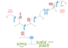
Explain the cell-based model of coagulation
- Initiation of coagulation occurs when sub-endothelial tissue is exposed to the circulation at a site of injury.
- Sub-endothelial cells express tissue factor at their surface, which binds to endogenous activated Factor VII to form a complex
- This complex binds small amounts of Factor X and Factor V to the exposed endothelial surface, which produce small quantities of thrombin
- The thrombin activates platelets that are attracted to the site by the process, as well as other plasma-borne clotting factors
- The activated factors (among them Factor VIII and Factor IX) enable the binding of activated Factor X and Factor V to the surface of activated platelets
- The surface membranes of the activated platelets have gone through conformational changes which expose the ‘reaction sites’ necessary for continuation of the process
- Activated platelets with Va and Xa bound foes on to cause prothrombin to be converted to thrombin which leads to the ‘thrombin burst’
- This thrombin burst is necessary for the large-scale production of fibrin and so the development of an effective clot

What are the main functions of fibrinolysis?
- Limiting blood clots
- Repair and healing
Name the main components of fibrinolysis
- Plasminogen
- Tissue plasminogen activator (t-PA) & urokinase (u-PA)
- Plasminogen activator inhibitor -1 and -2
- α2-plasmin inhibitor
Describe the process of fibrinolysis
- Tissue plasminogen activator (tPA) catalyses the reaction that converts plasminogen into plasmin
- Plasmin then degrades cross-linked fibrin of fibrin clot to produce D dimers
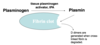
Plasmin can also degrade non-cross linked fibrin or fibrinogen. What is formed as a result?
- Fibrin degradation products (FDP)
How can tPA be used therapuetically?
- tPA and a bacterial activator, streptokinase, can be used in therapeutic thrombolysis for myocardial infarction