Ground Glass Opacities/Interlobal Thickening Flashcards
(14 cards)
Differential diagnosis for ground glass in a central distribution?
- Pulmonary edema.
- Alveolar hemorrhage.
- Pneumocystis jiroveci pneumonia.
- Alveolar proteinosis.
- Mandell, Jacob. Core Radiology (p. 11). Cambridge University Press. Kindle Edition.
DDx for peripheral ground glass or consolidation?
Organizing pneumonia. Chronic eosinophilic pneumonia, typically with an upper lobe predominance. Atypical or viral pneumonia. Pulmonary edema. Peripheral pulmonary edema tends to be noncardiogenic in etiology, such as edema triggered by drug reaction. Peripheral consolidation/ground glass is unusual for cardiogenic pulmonary edema. Mandell, Jacob. Core Radiology (p. 11). Cambridge University Press. Kindle Edition.
Smooth interlobar thickening - what causes it and whats the ddx.
-Conditions that dilate the pulmonary veins cause smooth interlobular septal thickening. -DDx includes Pulmonary edema (by far the most common cause of smooth interlobular septal thickening). Pulmonary alveolar proteinosis. Pulmonary hemorrhage. Atypical pneumonia, especially Pneumocystis jiroveci pneumonia. Mandell, Jacob. Core Radiology (p. 12). Cambridge University Press. Kindle Edition.
Nodular, irregular, or asymmetric interlobar septal thickening - what cuases it and whats the ddx
- Nodular, irregular, or asymmetric septal thickening tends to be caused by processes that infiltrate the peripheral lymphatics, most commonly lymphangitic carcinomatosis and sarcoidosis:
- Lymphangitic carcinomatosis is tumor spread through the lymphatics. Sarcoidosis is an idiopathic, multi-organ disease characterized by noncaseating granulomas, which form nodules and masses primarily in a lymphatic distribution. Mandell, Jacob. Core Radiology (p. 12). Cambridge University Press. Kindle Edition.
What is crazy paving and the ddx?
- Crazy paving describes interlobular septal thickening with superimposed ground glass opacification, which is thought to resemble the appearance of broken pieces of stone.
- DDx:
- Alveolar proteinosis.
- Pneumocystis jiroveci pneumonia.
- Organizing pneumonia.
- Bronchioloalveolar carcinoma, mucinous subtype.
- Lipoid pneumonia, an inflammatory pneumonia caused by a reaction to aspirated lipids.
- Acute respiratory distress syndrome.
- Pulmonary hemorrhage.
- Mandell, Jacob. Core Radiology (p. 13). Cambridge University Press. Kindle Edition.
Centrilobar Nodules - what are they caused by and what is typical of their distribution?
- Opacification of the centrilobar bronchiole (less commonly the centrilobar artery).
- Never extend to the pleural surface.

Centrilobar Nodules - what are the infectious causes?
- Endobronchial spread of tuberculosis or atypical mycobacteria.
- Bronchopneumonia: spread of infectious pneumonia via the airways.
- Atypical pneumonia, especially mycoplasma pneumonia.

Centrilobar Nodules - what are they inflammatory causes?
- HSP is a type III hypersensitivity reaction to an inhaled organic antigen. The subacute phase of HSP is primarily characterized by centrilobular nodules.
- Hot tub lung is a hypersensitivity reaction to inhaled atypical mycobacteria, with similar imaging to HSP.
- RB-ILD is an inflammatory reaction to inhaled cigarette smoke mediated by pigmented macrophages.
- Diffuse panbronchiolitis is a chronic inflammatory disorder characterized by lymphoid hyperplasia in the walls of the respiratory bronchioles resulting in bronchiolectasis. It typically affects patients of Asian descent.
- Silicosis, an inhalation lung disease that develops in response to inhaled silica particles, is characterized by upper lobe predominant centrilobular and perilymphatic nodules.
- Mandell, Jacob. Core Radiology (p. 14). Cambridge University Press. Kindle Edition.

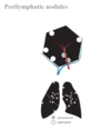
Perilymphatic Nodules - ddx
- Sarcoidosis is by far the most common cause of perilymphatic nodules, typically with an upper-lobe distribution. The nodules may become confluent creating the galaxy sign.
- Pneumoconioses (silicosis and coal workers pneumoconiosis) are reactions to inorganic dust inhalation. The imaging may look identical to sarcoidosis with perilymphatic nodules, but there is usually a history of exposure (e.g. a sandblaster who develops silicosis).
- Lymphangitic carcinomatosis.
Mandell, Jacob. Core Radiology (p. 15). Cambridge University Press. Kindle Edition.

How do random nodules spread? What is the DDx?
- Spread hematogenously
- DDx
- Hematogenous metastases.
- Septic emboli. Embolic infection has a propensity to cavitate but early emboli may be irregular or solid.
- Pulmonary Langerhans’s cell histiocytosis (PLCH), a smoking-related lung disease that progresses from airway-associated and random nodules to irregular cysts. PLCH is usually distinguishable from other causes of random nodules due to the presence of cysts and non-angiocentric distribution.
Mandell, Jacob. Core Radiology (p. 16). Cambridge University Press. Kindle Edition.
*

What causes tree-in-bud nodules?
- The linear branching structures represent the impacted bronchioles, which are normally invisible on CT, and the nodules represent impacted terminal bronchioles. Tree-in-bud nodules are due to mucus, pus, or fluid impacting bronchioles and terminal bronchioles.
- Mandell, Jacob. Core Radiology (p. 17). Cambridge University Press. Kindle Edition.
What is the ddx of tree-in-bud nodules?
- Mycobacteria tuberculosis and atypical mycobacteria.
- Bacterial pneumonia.
- Aspiration pneumonia.
- Airway-invasive aspergillus. Aspergillus is an opportunistic fungus with several patterns of disease. The airway-invasive pattern is seen in immunocompromised patients and may present either as bronchopneumonia or small airways infection.
Mandell, Jacob. Core Radiology (p. 17). Cambridge University Press. Kindle Edition.
DDx for diffuse GGO in a central predominant distribution?
- Pulmonary edema.
- Alveolar hemorrhage.
- Pneumocystis jiroveci pneumonia.
- Alveolar proteinosis.
DDx for GGO in a peripheral distribution
- Organizing pneumonia.
- Chronic eosinophilic pneumonia, typically with an upper lobe predominance.
- Atypical or viral pneumonia.
- Pulmonary edema.
- Peripheral pulmonary edema tends to be noncardiogenic in etiology, such as edema triggered by drug reaction. Peripheral consolidation/ground glass is unusual for cardiogenic pulmonary edema.


