Gross Brain Topography 2: blood supply, meninges, ventricular system, and CSF week 2 Flashcards
The brain receives a dual blood supply and anastamoses are a critical component of this arrangement.
What areterie(s) does anterior circulation come from? What arterie(s) supply the posterior circulation? What is the anastamosis of these arteries called?
The anterior circulation (from the Internal carotid Artery) joins the posterior circulation (from the Vertebrobasilar system) at the circle of Willis.
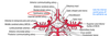
Identify the indicated arteries.

Notes: The posterior communicating artery is btwn the internal carotid artery and the posterior cerebral artery.
There are “feeder” arteries coming off of the basilar aa to supply the lateral pons called pontine arteries.
The anterior inferior cerebellar arteries (AICA) are brancehs of the basilar artery while the posterior inferior cerebellar arteries (PICA) are branches of the vertebral arteries.
The vertebral arteries form one anterior spinal artery. Each vertebral artery gives off one posterior spinal artery (so that there are 2 posterior spinal arteries and one anterior spinal artery).

Identify the indicated arteries.


Anerusyms in what vessels may compress the oculumotor nerve (CN III)? What is the name for the condition this would cause?
The oculmotor nerve exits the midbrain btwn the posterior cerebral aa and the superior cerebellar aa. Aneurysms (dilations in wall of arteries) in these vessels may compress this nerve causing 3rd Nerve palsy.

What are the symptoms of 3rd nerve palsy? Other than aneursym(s) in the PCA and/or SCA arteries, what else may cause 3rd nerve palsy?
3rd Nerve palsy: Severe Ptosis, Eye is down and out, pupil dilated and loss of efferent limb of pupillary light reflex.
Note close proximity of the Uncus to CN III: also may occur with Uncal herniation (due to increased Intracranial Pressure- ICP).


Identify the parts of the internal carotid aa.


What does the anterior cerebral artery travel in?
What parts of the brain does the anterior cerebral artery supply? If applicable, name specific branches.
The anterior cerebral artery courses through the longitudinal fissure supplying superficial branches to the medial and ventral portions of the frontal and parietal lobes (including lower trunk and leg area of primary motor and sensory cortex-paracentral gyrus), the cingulate gyrus and the anterior 4/5ths of corpus callosum (pericallosal branch). Note that it goes back to the parietoccipital sulcus.
The anterior cerebral artery gives off deep branches called the medial striate arteries (MSA) which supply anterior deep brain structures: head of caudate and anterior limb of internal capsule

____ ____ ____ forms part of the circle of Willis connecting the internal carotid artery (ICA) to the posterior cerebral artery (PCA).
Posterior Communicating artery (PCom) forms part of the circle of Willis connecting the internal carotid artery (ICA) to the posterior cerebral artery (PCA).

What artery does the opthalmic artery branch from? What is the branch of this artery called that supplies the retina?
Ophthalmic artery: to the eye and retina; retina supplied by branch called Central artery of retina.
The opthalmic artery is a branch of the internal carotid artery. This artery travels in the optic canal with the optic nerve.
What does the middle cerebral artery (MCA) travel in?
What parts of the brain does the MCA supply? If applicable, name specific branches.
Why is the MCA also known as the “Artery of Stroke”?
What part of the circle of Willis does the MCA contribute to?
Middle Cerebral artery (MCA): is a large continuation of the internal carotid (NOT part of the circle of Willis). It runs in the lateral fissure and supplies superficial branches to the lateral cortical surface (sensorimotor areas of hand, face, upper trunk as well as auditory and speech areas). (Lateral portions of Frontal, Parietal and Temporal lobes) Note that this includes both Wernicke’s and Broca’s areas.This vessel also gives rise to deep branches, the lenticulostriate arteries (LsA), which supply deep brain structures (basal ganglia nuclei: putamen and globus pallidus and internal capsule: genu and posterior limb). see slides 16-17 of notes for pictures
Also known as “The Artery of Stroke” because it is in direct line for entry of dislodged thrombi.
What arteries do the vertebral arteries branch from? Explain their pathway to the brain.
Before the vertebral arteries unite, what structures do they supply? Via what branches?
State what structures the basilar artery as well as its branches supply.
What are the 2 terminal branches of the basilar arteries?
A stroke in what artery causes Wallenberg’s syndrome?
The vertebral arteries branch from the subclavian arteries in the neck, course through foramina in the transverse processes of the cervical vertebrae, pass through the foramen magnum, and ultimately unite to form the basilar artery.
Before uniting the vertebral arteries will supply the spinal cord (via the anterior and posterior spinal arteries), cerebellum, and the medulla (medial medulla via the anterior spinal artery and lateral medulla via the posterior inferior cerebellar artery (PICA). The PICA supplies the cerebellum, choroid plexus of the fourth ventricle, and the dorsolateral medulla (Wallenberg’s Syndrome caused by stroke in this vessel).
The basilar artery courses along the midline of the pons and gives rise to anterior inferior cerebellar arteries (AICA), superior cerebellar arteries (to the cerebellum and pons), and pontine branches which are small feeder arteries at the midline of the pons. The basilar artery ends by dividing into two terminal branches, the posterior cerebral arteries.

What parts of the brain do the posterior cerebral arteries supply?
Supplies ventral and medial surfaces of temporal and occipital (visual) cortex to the parieto-occipital sulcus.
Also branches to the midbrain and thalamus.

Why is there normally little blood flow in the circle of Willis? How can the circle of Willis prevent neurological damage in the case of stroke?
Normally, little blood flows around this circle because arterial pressure in the Internal Carotid Artery and the PCA is the same (with minimal blood flow in Posterior Communicating arteries). If a major vessel within or just proximal to the Circle becomes occluded, however, there is the potential for flow through the communicating arteries, sometimes preventing neurological damage. Rarely can this compensate for an abrupt blockage. Variability exists in the circle of Willis and arterial origins. Note the MCA is NOT part of it.

summary of cortical blood supply
see reverse


Generally, where are watershed zones located?
In what type of situation are these areas subject to ischemic damage?
What are symptoms of “man in a barrel”? These symptoms arise as a result of the watershed zone in between what arteries?

Watershed zones are located at the margins of territories of each major vessel. These are domains of the distal vascular bed and therefore less robust perfusion. They are therefore subject to ischemic damage in systemic hypotensive episodes (e.g. cardiac arrest).
One clinical symptom sometimes referred to as “man in a barrel” results from bilateral cortical damage because of systemic hypotension. The hands and feet are spared, but the shoulder and trunk musculature is paralyzed bilaterally, along with corresponding sensory loss. The damaged “shoulder and trunk” representation in the pre- and post-central gyri corresponds to the watershed zone between the middle and anterior cerebral artery circulations. Note that there will be other functions affected because these arteries also supply deep brain structures.

What are the ways in which blood flow to the brain is controlled?
The brain is highly metabolically active but does not possess the capacity to store glucose. Therefore a constant supply of blood flow is needed, but also this flow needs to respond to increased demands of particular regions of the brain at a given time. How is blood flow maintained?
1) Autoregulation: The cerebral vessels act to maintain constant blood flow. The smooth muscle cells in these vessels respond to stretch and thus respond to changes in blood pressure (i.e. constrict when blood pressure increases and dilate when blood pressure decreases).
2) Response to Increased Neural Activity: This is a collaborative effort of neurons, astrocytes (glia), and the cerebral vessels. The most well-studied component of this is the role of astrocytes. Astrocytes have “end-feet” that are directly applied to neurons and CNS arterioles and capillaries. A large majority of CNS synapses utilize glutamate so an increase in neural activity causes an increase in glutamate release. Some of this glutamate spills out of the synaptic cleft and reaches the astrocyte end-feet which also possess glutamate receptors. The binding of glutamate to these receptors causes release of vasodilating factors which cause increases in blood flow.
3) Autonomic Innervation of Cerebral Vessels: This factor is perhaps the least well understood and may have the most minor role in cerebral blood flow. It may play a role in stress responses, but likely is not a “major player” in maintaining cerebral flow under normal circumstances.
What supplies the dura mater with blood? What is the largest and most significant of these arteries?
What artery does it branch from? Through what foramina does it enter the skull?
What can cause rupture of this vessel?

The Dura mater receives blood supply from the meningeal arteries. The largest and most significant of these is the middle meningeal artery, a branch of the maxillary artery. It enters the cranial cavity through the foramen spinosum and cuts a groove in the bone on the floor and lateral wall of the middle cranial fossa. This vessel can rupture in trauma to the head (e.g. fractures of temporal bone).

What nerves innervate the dura mater? What kind of innervation do they provide? What parts of the dura do they innervate?
What are headaches associated with increased intracranial pressure due to?
What is the embryological origin of the Leptomeninges (arachnoid and pia mater)?
The dura mater receives sensory innervation (GSA fibers) from the Trigeminal nerve (CN V) for the anterior and middle cranial fossa and the Vagus (CN X) for the posterior cranial fossa.
Headache associated with increased intracranial pressure (ICP) results from stretching of the dura, n_ot because of compression of brain tissue itself_.
The Leptomeninges (Arachnoid and Pia mater are derived from neural crest cells): The arachnoid mater creates the subarachnoid space containing cerebrospinal fluid (CSF). The pia mater follows the contours of the brain and spinal cord.
What is found within the subarachnoid space?
cerebral arteries and veins as well as CSF
Where does blood collect in an epidural hemorrhage?
What typically causes epidural hemorrhages?
Epidural hemorrhage
- Due to skull fracture and tearing of meningeal arteries (which run in the periosteal dura layer)
- Bleeding occurs between the two layers of dura and/or between the periosteal layer of dura and the skull.
- life threatening condition: hemorrhage and associated swelling compresses brain tissue and vasculature to the brain

Where does bleeding occur in a subdural hemorrhage? What is a subdural hemorrhage due to?
In what population are subdural hemorrhages more common in than others?
Subdural: a bleed btwn the dura and arachnoid mater. Results form rupture of bridging cerebral veins as they pierce the dura to enter the dural venous sinuses.
Note the Bridging Veins which traverse the subarachnoid space to enter the Superior sagittal sinus in attached figure.
In the elderly there is natural shrinkage of brain tissue, stretching the veins and increased risk of Subdural bleeds/hematomas.

Where does bleeding occur in a subarachnoid hemorrhage?
What causes subarachnoid hemorrhages?
What symptom typically accompanies this type of hemorrhage?

- Bleed in the subarachnoid space.
- typically result from head trauma or rupture of arterial aneurysm (dilated arterial wall)
- Accompanied by a terrible, intense “worst headache of my life” from stretching of the meninges

What is a cerebral aneursym? Where do they commonly occur?
What can cerebral aneurysms cause?
What are Berry or “saccular” aneurysms?
What predisposes individuals to development of aneurysms?
Cerebral Aneurysm is a dilated arterial wall of varying sizes that frequently arise at bifurcation point of vessel; they can grow, compressing brain tissue or can rupture causing a subarachnoid hemorrhage.
If detected can be surgically corrected. Berry or “saccular” aneurysms are those that occur at bifurcations in the circle of Willis.
CV disease and HTN predispose people to development of aneurysms.


