GI Anatomy of Abdominal Wall Flashcards

1
Q

What is the abdominal wall subdivided into?

A
  • The anterior wall
  • Right and left lateral walls
  • Posterior wall
2
Q

Why is the term anterolateral abdominal wall used?

A

Because the boundary between the anterior and lateral walls is indefinite

3
Q

What is the anterolateral abdominal wall bounded by superiorly?

A
  • The cartilages of the 7th-10th ribs
  • The xiphoid process of the sternum
4
Q

What is the anterolateral abdominal wall bounded by inferiorly?

A
  • The inguinal ligament
  • The superior margins of the anterolateral aspects of the pelvic girdle
5
Q

What are the anterolateral aspects of the pelvic girdle?

A
  • Iliac crests
  • Pubic crests
  • Pubic symphysis
6
Q

What does the anterolateral abdominal wall consist of?

A
  • Skin
  • Subcutaneous tissue
  • Muscles and their aponeuroses
  • Deep fascia
  • Extraperitoneal fat
  • Parietal peritoneum
7
Q

What does the subcutaneous tissue of the anterolateral abdominal wall consist of?

A

Superficial fasica and fat

8
Q

Label this diagram

A
  • A - Skin (cut edge)
  • B - Superficial fatty layer of subcutaneous tissue (Camper fascia)
  • C - Deep membranous layer of subcutaneous tissue (Scarpa fascia)
  • D - Investing (deep) fascia- superficial, intermediate, and deep
  • E - Parietal peritoneum
  • F - Endoabdominal (transversalis) fascia
  • G - Extraperiteoneal fat
  • H - Transversus abdominis
  • I - Internal oblique
  • J - Internal oblique
9
Q

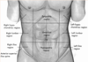
What is the umbilicus?

A

An obvious feature of the anterolateral abdominal wall at spinal level L3

10
Q

What is the epigastric fossa also known as?

A

Pit of the stomach

11
Q

What is the epigastric fossa?

A

Slight depression in the epigastric region

12
Q

Where is the epigastric fossa found?

A

Just inferior to the xiphoid process

13
Q

What is the epigastric fossa particularly noticeable?

A

When the person is in the supine position

14
Q

Why is the epigastric fossa particularly noticeable when the person is in the supine position?

A

Because the abdominal organs spread out

15
Q

What is commonly felt on the side of the epigastric fossa?

A

Heartburn

16
Q

What forms the linea alba?

A

Aponeurosis of the abdominal muscles

17
Q

What does the linea alba separate?

A

The left and right rectus abdominis

18
Q

Who is the linea alba visible in?

A

Lean individuals

19
Q

Why is the linea alba visible in lean individuals?

A

Because of the vertical skin groove superficial to it

20
Q

What is the name of the condition where the linea alba is lax?

A

Divarication of recti

21
Q

What happens in divarication of recti?

A

When the rectus abdominis contract, the muscles spread apart

22
Q

What is the pubic crest?

A

The upper margins of the pubic bones

23
Q

What is the pubic symphysis?

A

The cartilaginous joints that unite the pubic bones

24
Q

Where can the pubic crest be felt?

A

At the inferior end of the linea alba

25
What is the inguinal groove?
A skin crease that is parallel
26
Where is the inguinal groove found?
Parallel and just inferior to the inguinal ligament
27
Where does the inguinal groove run?
Between the ASIS and pubic tubercle
28
What does the inguinal groove mark?
The division between the abdominal wall and the thigh
29
What are semilunar lines?
Tedinous line on either side of rectus abdominis
30
What shape are the semilunar lines?
Slightly curved
31
In whom are the tendinous intersections of rectus abdominis clearly visible?
In persons with well developed rectus muscles
32
As well as the tendinous intersections of the rectus abdominis, what else is visible in persons with well-developed rectus muscles?
The interdigitating bellies of the serratus anterior and external oblique muscles
33
What is the arcuate line also known as?
Douglas' line
34
What is the arcuate line?
Where the fibrous sheath stops at the inferior limit of the posterior layer of the rectus sheath
35
Where is the arcuate line?
1/3 of the way from the umbilicus to the pubic crest
36
Label this diagram
* A - Site of xiphoid process * B - Right costal margin * C - Umbilicus * D- Iliac crest * E - Anterior superior iliac spine * F - Inguinal ligament * G - Site of pubic tubercle * H - Epigastric fossa * I - Site of linea alba * J - Semilunar valves * K - Site of pubic symphysis * L - Serratus anterior * M - External oblique * N - Left rectus abdominis * O - Location of linea alba * P - Umbilicus *(level of L3 vertebrae)* * Q - Location of inguinal ligament and groove
37
How many muscles are there in the abdominal wall?
5
38
How are the muscles in the anterolateral abdominal wall related to each other?
They are bilaterally paired
39
What shape are the muscles in the anterolateral abdominal wall?
* Three flat * Two vertical
40
What are the flat muscles in the anterolateral abdominal wall?
* External oblique * Internal oblique * Transversus Abdominis
41
What are the vertical muscles in the anterolateral abdominal wall?
* Rectus Abdominis * Pyramidalis
42
What orientation are the fibres in the flat muscles in the anterolateral abdominal wall?
Varying orientation; * Fibres of obliques run diagonally and perpendicular to each other * Fibres of transversus running transversely
43
Label these diagrams
* A - Free posterior border of external oblique * B - Internal oblique * C - Anterior superior iliac spine * D - External oblique * E - Rectus sheath *(anterior layer)* * F - Inguinal ligament * G - External oblique *(cut)* * H - Attachment of external oblique to iliac crest * I - Internal oblique * J - Inguinal ligament * K - External oblique *(cut)* * L - Thoracolumbar fascia * M - Cut attachments of internal oblique * N - Transversalis abdominis
44
What is the origin of the external oblique muscle?
External surfaces of the 5th to 12th ribs
45
What is the insertion of the external oblique muscle?
* Linea alba * Pubic tubercle * Anterior half of iliac crest
46
What is the origin of the internal oblique?
* Thoracolumbar fasica * Anterior two thirds of iliac crest * Connective tissue deep to lateral third of inguinal ligament
47
What is the insertion of the internal oblique?
* Inferior borders of the 10th to 12th ribs * Linea alba * Pectin pubis via conjoint tendon
48
What is the origin of the transversus abdominis?
* Internal surfaces of the 7th to 12th costal cartilages * Thoracolumbar fascia * Iliac crest * Connective tissue deep to lateral third of inguinal ligament
49
What is the insertion of the transversus abdominis?
* Linea alba with aponeuroses of internal oblique * Pubic crest * Pectin pubis via conjoint tendon
50
What happens to all three of the flat muscles of the anterolateral abdominal wall?
They are continued anteriorly and medially as strong, sheet-like aponeuroses
51
What happens to the aponeuroses of the flat muscles of the anterolateral abdominal wall?
They form the tough, aponeurotic, tendinous rectus sheath
52
Where is the rectus sheath formed?
Between the mid-clavicular line and the midline
53
What does the rectus sheath enclose?
The rectus abdominis
54
Label these diagrams
* A - Fibres of left external oblique aponeurosis, *which run deep on the right side and running superficially on the left side* * B - Umbilical ring * C - Deep fibres of right external oblique aponeurosis * D - Deep fibres of left external oblique aponeurosis * E - Right external oblique muscles * F - Fibres of right external oblique aponeurosis * G - Fibres passing from superficial to deep at linea alba * H - Fibres of left internal oblique aponeurosis * I - Left internal oblique muscle
55
What happens once the aponeuroses' of the flat muscles of the anterolateral abdominal wall have formed the rectus sheath?
They interweave with their fellows of the opposite side
56
What is formed when aponeuroses interweave?
The linea alba
57
Where does the linea alba extend?
From the xiphoid process to the pubic symphysis
58
What is the interweaving of aponeuroses between?
* Between right and left sides * Between intermediate and deep layers
59
Label this diagram
* A - Linea alba * B - Rectus abdominis * C - Rectus sheath * D - Linea alba, *with fibres passing from superficial to deep, and vice versa* * E - Linea alba * F - Rectus sheath * G - Aponeurosis of transversus abdominis muscle * H - Parietal peritoneum * I - Transversalis fascia * J - Transversus abdominis muscle * K - Internal oblique * L - External oblique * M - Subcutaneous tissue * N - Aponeurosis of external oblique * O - Laminae of external oblique * P - Skin * Q - Parietal peritoneum * R - Extraperitoneal fat * S - Transversalis fascia * T - Membraneous layer * U - Fatty layer
60
What is contained within the rectus sheath?
The two vertical muscles of the anterolateral abdominal wall; * Rectus abdominis * Pyramidalis
61
Label this diagram
* A - 12th rib * B - Quadratus lumborum *(posterior abdominal wall)* * C - Rectus abdominis * D - Costal cartiages * E - Tendinous intersections * F - Pubic crest
62
What do we want when designing an incision?
One that will closer and provide long-lasting strength
63
What is the importance of designing an incision that will provide long lasting strength?
Minimising the incidence of incisional herniae
64
What happens if we try to sew muscle together?
The sutures will 'cut out'
65
When is a midline incision used abdominally?
Surgeons suture the linea alba together
66
When is a transverse incision used abdominally?
Surgeons suture the external oblique aponeuroses together
67
Where are incisions made for an appendicectomy?
* McBurney's Point * 2/3rds of the distance between the umbilicus and ASIS
68
How is the incision through 2/3 of the distance between the umbilicus and the ASIS made?
Throguh a 'gridiron' muscle-splitting incision
69
How is a girdiron incision conducted?
Put scissors in, and open and close them to separate out the muscle fibres, followed by the next two layers
70
What muscle fibres have to be seperated out when performing an appendectomy?
Those of the external and internal oblique's, and the transversalis
71
Give 6 disorders that are consequences of developmental defects in the abdominal wall
* Patent Urachus * Gastroschisis * Omphalocoele * Problems of the Vitelline duct resulting in: * Meckel's Diverticulum * Vitelline cyst * Vitelline fistula
72
What is the most common GI abnormality?
Meckel's Diverticulum
73
When does a patent urachus present?
Can present at birth, or later in life in men
74
What can cause a patent urachus to present later in life?
A bladder outflow obstruction
75
What causes a bladder outflow obstruction that can cause a patent urachas to present later in life?
Benign prostatic hypertrophy
76
What does the persistance of a vitelline duct result in?
A number of different abnormalities
77
What is Meckel's Diverticulum also known as?
Ilieal Diverticulum
78
What is Meckel's Diverticulum?
A 'cul-de-sac' in the ileum
79
What rule does Meckel's Diverticulum follow?
The rule of 2's
80
Why is it said that Meckel's Diverticulum follows the rule of 2's?
* 2% of population affected * 2 feet from ileocecal valve * 2 inches long * Usually detected in under 2's * 2:1 male:female
81
What can Meckel's Diverticulum contain?
Ectopic gastric or pancreatic tissue
82
What will the ectopic tissue in Meckel's Diverticulum do?
Secrete enzymes and acids into tissue not protected from them
83
What is the result of the secretion of enzymes and acid from Meckel's Diverticulum?
Causes ulceration
84
Draw a diagram illustrating Meckel's Diverticulum
85
What happens in a vitelline cyst?
The vitelline duct forms fibrous stands at either end
86
Draw a diagram illustrating a vitelline cyst
87
What happens in a vitelline fistula?
There is direct communication between the umbilicus and the intestinal tract
88
What is the result of a vitelline fistula?
Faecal matter comes out of the umbilicus
89
Draw a diagram illustrating a vitelline fistula
90
What is omphalocoele?
The persistance of physiological herniation
91
What happens in omphalocoele?
A part of the gut tube fails to return to the abdominal cavity following its normal herniation into the umbilical cord
92
What covers the defect in omphalocoele?
An epithelial layer
93
Why does an epithelial layer cover the defect in omphalocoele?
Since the umbilical cord is covered by a reflection of the amnion
94
Draw a diagram illustrating omphalocoele
95
What is gastroschisis?
Failure of closure of the abdominal wall during folding of the embryo, leaving the gut tube and its derivatives outside of the body cavity
96
What covers the gut tube and its derivatives in gastroschisis?
Nothing
97
Where do the gut tube and its derivatives herniate in gastroschisis?
Directly into the abdominal cavity
98
What is somatic referred pain?
Pain caused by a noxious stimulus to the proximal part of a somatic nerve that is perceived in the distal dermatome of the nerve
99
Give an example of a condition that gives somatic reffered pain?
Shingles
100
Why does shingles cause somatic referred pain?
Shingles affects nerves, and pain is felt distally along the nerves from the problem
101
How does the CNS perceive visceral pain?
As coming from the somatic portion of the body supplied by the relevant spinal cord segments
102
Why does the CNS perceive viseceral pain as coming from a somatic portion of the body?
Because in the thorax, visceral afferent pain fibres follow sympathetic fibres back to the same spinal cord segments that give rise to the preganglionic sympathetic fibres
103
What causes visceral pain?
* Ischaemia * Abnormally strong muscle contractions * Inflammation * Stretching
104
What does **not** cause visceral pain?
* Touch * Burning * Cutting * Crushing
105
Draw a diagram illustrating how visceral reffered pain may arise?
106
Where is pain originating from the foregut referred to?
Epigastric region
107
Where is pain originating from the midgut referred to?
Periumbilical region
108
Where is pain originating from the hindgut referred to?
Suprapubic region
109
Show where gall bladder pain presents
110
Show where hepatic pain presents
111
Show where splenic pain presents
112
Show where gastric and duodenal pain presents
113
Show where oesophageal pain presents
114
What structures can cause central back pain?
Retroperitoneal structures, *e.g. pancreas and abdominal aorta*
115
Show where pancreatic and abdominal aorta pain presents
116
Where is the main in early appendicitis?
Begins at the umbilicus
117
Why does appendicitis pain begin at the umbilicus?
Since the innervation of the appendix enters the spine at level (T10)
118
What happens as the appendix becomes more inflamed in appendicitis?
It irritates the surrounding bowel wall, localising the pain to the right lower quadrant
119
Why does the appendicitis later cause pain in the right lower quadrant?
Irritation to the somatic nerve
120
Show where acute appendicitis pain presents
121
How bad is the pain in small or large bowel colic?
The patient will be doubled over in pain
122
Where does small bowel colic present?
Periumbilical region
123
Why does small bowel colic present in the periumbilical region?
Because it is derived from the midgut
124
Show where pain from small bowel colic presents
125
Where does large bowel colic present?
Suprapubic region
126
Why does large bowel colic present in the suprapubic region?
It is derived from the hindgut
127
Show where pain from large bowel colic presents
128
How bad is the pain in renal or uteric colic?
* Patient rolls around on the floor * Pain worse than child birth
129
Show where pain from renal or uteric colic presents
130
Show where uterine or ovarian pain presents
131
Show where bladder pain presents
132
What can cause referred diaphragmatic irritation?
* Ruptured spleen * Ectopic pregnancy * Perforated ulcer
133
How does referred diaphragmatic irritation occur?
Blood pools in pelvis, giving pain. The patient feels faint due to the loss of blood, so lies down, causing the blood to rush up to the diaphragm (C3/4/5). The presence of blood here results in referred pain the left shoulder.
134
Why does referred diaphragmatic irritation not lead to pain in the right shoulder?
Because the liver is in the way of the blood
135
Show where referred diaphragmatic irritation causes pain
136
Label this diagram
* A - Mouth * B - Tongue * C - Larynx * D - Trachea * E - Oesophagus * F - Gallbladder * G - Pylorus * H - Duodenum * I - Pancreas * J - Transverse colon * K - Ascending colon * L - Jejunum * M - Ileum * N - Cecum * O - Appendix * P - Anal canal * Q - Rectum * R - Sigmoid colon * S - Descending colon * T - Stomach * U - Liver * V - Pharynx
137
Label this picture of the undisturbed abdominal contents
* A - Thoracic *(descending)* aorta * B - Oesophagus * C - Inferior vena cava * D - Diaphragm * E - Falciform ligament * F - Stomach * G - Liver * H - Costodiaphragmatic recess *(pulmonary cavity)* * I - Round ligament of liver * J - Fundus of gallbladder * K - Gastrocolic ligament *(greater omenum)*
138
Label this diagram of the anterior view of the abdominal viscera
* A - Right dome of diaphragm * B - Liver * C - Fundus of gallbladder * D - Pylorus * E - Outline of duodenum * F - Ascending colon * G - Cecum * H - Anterior superior iliac spine * I - Apex of heart * J - Spleen * K - Outline of pancreas * L - Stomach * M - Transverse colon * N - Jejenum * O - Ileum * P - Descending colon * Q - Urinary bladder
139
Label this diagram of the posterior view of the abdominal viscera
* A - Scapula * B - Left dome of diaphragm * C - Spleen * D - Left suprarenal gland * E - Left kidney * F - Outline of pancreas * G - Descending colon * H - Small intestine * I - Small intestine in pelvis * J - Sigmoid colon * K - Oesophagus * L - Right dome of diaphragm * M - Liver * N - Right suprarenal gland * O - Right kidney * P - Ascending colon * Q - Ureter * R - Cecum * S - Appendix * T - Rectum * U - Urinary bladder
140
What is the peritoneal cavity?
A potential space of capillary thickness between the parietal and visceral layers of the peritoneum
141
Does the peritoneal cavity contain organs?
No
142
What does the peritoneal cavity contain?
A thin film of peritoneal fluid
143
What absorbs peritoneal fluid?
Lymphatic vessels, particularly on the interior surface of the diaphragm
144
How does the peritoneal cavity differ in males and females?
In males it is completely closed, but in females there is a communication pathway
145
Where is the peritoneal cavity communication pathway in females?
Through the uterine tubes, cavity, and vagina
146
What does the peritoneal cavity communication pathway constitute?
A potential pathway of infection from the exterior
147
Label this diagram
* A - Lesser omnentum * A1 - Portal triad in hepatoduodenal ligament * A1i - Hepatic artery * A1ii - Bile duct * A1iii - Hepatic portal vein * A2 - Hepatogastric ligament * B - Stomach * C - Visceral peritoneum *(covering stomach)* * D - Gastrosplenic ligament * E - Visceral peritoneum *(covering spleen)* * F - Parietal peritoneum * G - Spleen * H - Splenorenal ligament * I - Left kidney * J - Abdominal aorta * K - Inferior vena cava * L - Right kidney * M - Parietal peritoneum * N - Omental foramen * Light - Greater sac * Dark - Omental bursa *(lesser sac)*
148
What is the peritoneum?
A continous, two layered member
149
What are the layers of the peritoneum called?
* The parietal peritoneum * The visceral peritoneum
150
What does the parietal peritoneum line?
The internal surface of the abdominal wall
151
What does the visceral peritoneum do?
Invest viscera, *such as the stomach and intestines*
152
What do both layers of peritoneum consist of?
Mesothelium
153
What is mesothelium?
A layer of simple squamous epithelial cells
154
What separates the parietal peritoneum from the muscular layers of the abdominal wall?
Extraperitoneal connective tissue
155
How is the parietal peritoneum covering the anterior abdominal wall and pelvis walls attached?
Generally, loosely by the extraperitoneal connective tissue
156
What does the loose connection of parietal peritoneum and abdominal and pelvis walls allow for?
The considerable change in size of the bladder and the rectum
157
What does extraperitoneal tissue frequently contain?
A large amount of fat
158
Who in particular has a large amount of fat in their extraperitoneal tissue?
Obese men
159
How does the extraperitoneal tissue behind the linea alba and on the inferior surface of the diaphragm differ from that around the abdominal and pelvic walls?
It is denser and more firmly adherent
160
What is the blood, lymphatic, and somatic nerve supply of the parietal peritoneum?
The same as the region of wall it lines
161
What is the result of the parital peritoneum having the same nerve supply as the region of the wall it lines?
It is sensitive to pressure, pain, heat, cold, and laceration
162
Is parietal pain well localised?
Generally, yes, apart from on the inferior surface of the central part of the diaphragm
163
What innervates the inferior surface of the central part of the diaphragm?
Phrenic nerve *(C3/4/5)*
164
Where does the inferior surface of the central part of the diaphragm refer pain?
Shoulder
165
Why does the pain from appendicitis shift to over the appendix?
As the parietal peritoneum becomes inflamed, localising the pain
166
What is the blood, lymphatic, and somatic nerve supply of the visceral peritoneum?
The same as the organ it covers
167
Is the visceral peritoneum sensitive to touch, heat, cold, and laceration?
No
168
How is the visceral peritoneum stimulated?
Primarily by stretching and chemical irritation
169
Is visceral pain well localised?
No
170
Where is visceral pain referred to?
The dermatomes of the spinal ganglia providing the sensory fibres
171
Is the peritoneal cavity a simple shape?
No, it is complex
172
What terms are used to describe the parts of the peritoneum that connects organs with other organs or to the abdominal wall?
* Mesentery * Omentum * Greater omentum * Lesser omentum * Peritoneal ligament
173
What is a mesentery?
A double layer of peritoneum that occurs as a result of the invagination of the peritoneum by an organ
174
What does mesentery constitute?
A continuity of the visceral and parietal peritoneum
175
What does a mesentery connect?
An intraperitoneal organ to the body wall *(usually the posterior abdominal wall)*
176
What is an omentum?
A double-layered extension or fold of peritoneum that passes from the stomach and proximal parts of the duodenum to adjacent organs in the abdominal cavity
177
What is the greater omentum?
A prominent, four-layered peritoneal fold that hangs down like an apron from the greater curve of the stomach
178
What happens to the greater omentum after descending?
It folds back and attaches to the anterior surface of the transverse colon and its mesentery
179
What is the lesser omentum?
A much smaller, double-layered peritoneal fold that connects the lesser curvature of the stomach and the proximal part of the duodenum to the liver. It also connects the stomach to the portal triad
180
What is the peritoneal ligament?
A double layer of peritoneum that connects an organ with another organ, or to the abdominal wall
181
What to peritoneal ligaments connect the liver to?
* Anterior abdominal wall * Stomach * Duodenum
182
What ligament connects the liver to the anterior abdominal wall?
The falciform ligament
183
What ligament connects the liver to the stomach?
Hepatogatric ligament
184
What is the hepatogastric ligament?
The membranous portion of the lesser omentum
185
What ligament connects the liver to the duodenum?
The hepatoduodenal ligament
186
What is the hepatoduodenal ligament?
The thickened free edge of the lesser omentum
187
What does the thickened free edge of the lesser omentum conduct?
The portal triad
188
What is the portal triad?
* Portal vein * Hepatic artery * Bile duct
189
What do peritoneal ligaments connect the stomach to?
* Inferior surface of the diaphragm * Spleen * Transverse colon
190
What ligament connects the stomach to the inferior surface of the diaphragm?
The gastrophrenic ligament
191
What ligament connects the stomach to the spleen?
Gastrosplenic ligament
192
What ligament connects the stomach to the transverse colon?
Gastrocolic ligament
193
Where does the gastrocolic ligament originate from?
Greater omentum
194
Why do organs have bare areas?
To allow for entrance and exit of neurovascular structures
195
Label this diagram
* A - Bristle in epiploic foramen * B - Stomach * C - Transverse colon * D - Greater omentum * E - Small intestine * F - Uterovesical excavation * G - Bladder * H - Vagina * I - Superior layer of coronary ligament * J - Bare area of liver * K - Inferior layer of coronary ligament * L - Pancreas * M - Duodenum * N - Aorta * O - Mesentary * P - Uterus * Q - Rectovaginal excavation * R - Rectum
196
Label this diagram
* A - Liver * B - Lesser omentum * C - Pancreas * D - Falciform ligament * E - Stomach * F - Tranverse colon * G - Duodenum * H - Mesentery * I - Tranverse mesocolon * J - Transverse colon * K - Greater omentum * L - Jejenum * M - Parietal peritoneum * N - Ileum * O - Visceral peritoneum * P - Rectum * Q - Rectovesical pouch * R - Urinary bladder
197
Label this diagram
* A - Liver * B - Falciform ligament * C - Stomach * D - Gallbladder * E - Greater omentum * F - Ascending colon * G - Gallbladder * H - Greater omentum * I - Transverse colon * J - Mesentery * K - Lesser omentum * L - Transverse colon * M - Descending colon * N - Small intestine
198
How can the structures of the peritoneum be classified?
* Intraperitoneal * Retroperitoneal/extraperitoneal
199
When is an organ considered to be intraperitoneal?
When it is completely covered by peritoneum
200
Is an intraperitoneal organ completely enclosed?
No
201
Why is an intraperitoneal organ not completely enclosed?
Due to mesentery
202
When is an organ considered to be retroperitoneal/extraperitoneal?
When it is outside the peritoneal cavity, and thus only partially covered by the parietal peritoneum
203
Give an example of a retroperitoneal organ
The kidneys
204
What are the retroperitoneal organs?
* Kidneys, ureters, and bladder * The aorta and IVC * The oesophagus * The duodenum *(except from proximal part)* * Most of the pancreas * The ascending and descending colon, and rectum
205
Where are the kidneys found?
On the anterior surface, between parietal peritoneum and the posterior abdominal wall
206
What can the abdominal wall be divided into?
9 regions
207
How is the abdomen divided?
By the midclavicular lines vertically, and the subcostal and transtubercular lines horizontally
208
Draw a diagram illustrating the division of the abdominal wall
209
What is the transverse mesocolon?
The mesentery of the transverse colon
210
What does the transverse mesocolon do?
Divide the abdominal cavity into a supracolic compartment and an infracolic compartment
211
What does the supracolic compartment contain?
* Stomach * Liver * Spleen
212
What does the infracolic compartment contain?
* Small intestine * Ascending and descending colon
213
Where does the infracolic compartment lie?
Posterior to the greater omentum
214
What is the infracolic compartment divided into?
The right and left infracolic spaces
215
What divides the infracolic compartment?
The mesentery of the small intestine
216
How does free communication between the supracolic and infracolic compartments occur?
Through the paracolic gutters
217
Label this diagram
* A - Superior recess of omental bursa * B - Lung * C - Liver * D - Lesser omentum * E - Falciform ligament * F - Subhepatic space * G - Pancreas * H - Stomach * I - Duodenum * J - Transverse mesolon * K - Transverse colon * L - Inferior recess of omental bursa * M - Mesentery of small intestine * N - Greater omentum * O - Jejenum * P - Ileum * Q - Viseral peritoneum * R - Parietal peritoneum * S - Rectovesical pouch * T - Urinary bladder * U - Rectum
218
Label this diagram
* A - Transverse colon * B - Supracolic compartment * C - Transverse mesocolon * D - Phrenicocolic ligament * E - Left colic fixure * F - Tenia coli * G - Root of mesentery of small intestine * H - Descending colon * I - Infracolic compartment * Ii - Right paracolic gutter * Iii - Right infracolic space * Iiii - Left infracolic space * Iiiii - Left paracolic gutter * Ascending colon * Right colic fixture
219
What is the greater sac made up of?
The supracolic and infracolic compartments
220
What is the lesser sac also known as?
The omental bursa
221
What is the lesser sac?
An extensive sac-like cavity
222
Where does the lesser sac lie?
Posterior to the stomach, lesser omentum, and adjacent structures
223
What does the lesser sac have?
* Superior recess * Inferior recess
224
What is the superior recess of the lesser sac limited by superiorly?
The diaphragm and posterior layers of the coronary ligament of the liver
225
Where is the inferior recess of the lesser sac?
Between the superior parts of the layers of the greater omentum
226
What does most of the inferior recess of the lesser sac become?
Sealed off from the main part *(posterior to the stomach*)
227
Why does most of the inferior recess of the lesser sac become sealed off?
Because of adhesion of the anterior and posterior layers of the greater omentum
228
What does the lesser sac permit?
Free movement of the stomach on the structures posterior and inferior to it
229
Why does the lesser sac permit free movement of the stomach on the structures posterior and inferior to it?
Because it's anterior and posterior walls slide smoothly over one another
230
How do the greater and lesser sacs communicate?
Through the omental foramen *(epiploic foramen)*
231
What is the omental foramen?
An opening situated posterior to the free edge of the lesser omentum *(hepatoduodenal ligament)*
232
How can the omental foramen be located?
By running a finger along the gallbladder to free to edge of the lesser omentum
233
How many fingers does the omental foramen usually admit?
Two
234
Label this diagram
* A - Diaphragm * B - Falciform ligament * C - Liver * D - 7th rib * E - Porta hepatis * F - Lesser omentum * G - Finger is inserted through omental foramen into omental bursa * H - Gallbladder * I - Costodiaphragmatic *(pleural)* recess * J - 10th rib * K - 11th costal cartilage * L - Trasversus abdominis muscle * M - Transverse colon appearing in an unusual gap in the greater omentum * N - Anastomosis between right and left gastro-omental arteries * O - Greater omentum, gastrocolic portion * P - Stomach * Q - Diaphragm * R - Oesophagus
235
Where does the right subphrenic space lie?
Between the diaphragm and the anterior, superior, and right lateral surfaces of the right lobe of the liver
236
What is the right subphrenic space bounded by on the left side?
The falciform ligament
237
What is the right subphrenic space bounded by behind?
The upper layer of the coronary ligament
238
What is the right subphrenic space a relatively common site for?
Colletions of fluid after right-sided abdominal inflammation
239
Where does the left subphrenic space lie?
Between the diaphragm, the anterior and superior surfaces of the left lobe of the liver, the anterosuperior surface of the stomach, and the diaphragmatic surface of the spleen
240
What is the left subphrenic space bounded by on the right side?
The falciform ligament
241
What is the left subphrenic space bounded by behind?
The anterior layer of the left triangular ligament
242
When is the left subphrenic space much enlarged?
In the absence of a spleen
243
What is the left subphrenic space a common site of?
Fluid collection, particularly after a splenectomy
244
Is the right or left subphrenic space bigger?
Left
245
Why is the left subphrenic space bigger?
Liver is on the right
246
Where does the peritoneum pass in females?
From the rectum to the posterior vaginal fornix and then back to the uterine cervix and body as the recto-uterine fold
247
What does the recto-uterine fold do once it has reached the uterine cervix and body?
Descends to form the recto-uterine pouch *(of Douglas)*
248
What does the peritoneum spread over in females?
The uterine fundus
249
How far does the peritoneum spread over the uterine fundus?
To its anterior surface, as far as the junction of the body and cervix
250
What happens to the peritoneum at the junction of the body and cervix?
It is reflected forwards to the upper surface of the bladder
251
What is formed when the peritoneum is reflected forwards to the upper surface of the bladder?
A shallow vesico-uterine pouch
252
Where does the peritoneum leave the rectum in males?
At the junction of the middle and lower thirds
253
What happens once the peritoneum leaves the rectum in males?
It passes forwards to the upper poles on the seminal vesicles and superior aspect of the bladder
254
Where does the peritoneum form the rectovescial pouch in males?
Between the rectum and the bladder
255
What is the the mesentery of the small intestine?
A broad, fan shaped fold
256
What does the mesentery of the small intestine connect?
The coils of the jejenum and ileum to the posterior abdominal wall
257
What is found between the two sheets of the mesentery of the small intestine?
* Blood vessels * Lymph vessels * Nerves
258
What does the mesentery of the small intestine allow?
The parts of intestine it connects to move relatively freely within the abdominal cavity
259
What is the root of the mesentery?
The attached, parietal border of the mesentery of the small intestine
260
Where is the root of the mesentery found?
About 15cm from the duodenojejenal flexture at the level of left side L2, obliquely *(towards inferior right)* to the ileocaecal junction
261
What does the root of the mesentery cross?
The second and third parts of the duodenum, abdominal aorta, inferior vena cava, right ureter, right psoas major muscle, and right gondal artery
262
What is the sigmoid mesocolon?
A peritoneal fold attaching the sigmoid colon to the pelvic wall
263
What shape of the attachment of the sigmoid mesocolon?
An inverted V
264
Where is the apex of the sigmoid mesocolon?
Near the division of the left common iliac artery
265
Where does the left limb of the sigmoid mesocolon descend?
Medial to the left psoas major
266
Where does the right limb of the sigmoid mesocolon pass?
Into the pevlis
267
Where does the rihgt limb of the sigmoid mesocolon end?
In the midline at the level of the third sacral vertebra
268
What runs between the layers of the sigmoid mesocolon?
Sigmoid and superior rectal vessels
269
Where does the left ureter descend?
Into the pelvis behind the apex of the sigmoid mesocolon