Gastrointestinal Physiology Flashcards
(39 cards)
Describe the 4 processes of the digestive system
SECRETION
- transfer of water and ions from ECF through GI epithelial cells to the digestive tract lumen
DIGESTION
- Chemical and mechanical breakdown of foods into smaller units that can be taken across the intestinal epithelium into the body
MOTILITY
- Movement of material in the GI tract as a result of muscle contraction
ABSORPTION
- Active/passive transfer of substances from the lumen of the GI tract to the extracellular fluid

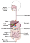
Draw the anatomy of the GI tract with the following labels:
- Mouth
- Salivary glands
- Esophagus
- Stomach
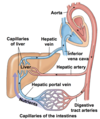
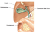
- Liver
- Gall bladder
- Pancreas
- Small intestine
- Duodenum
- Jejenum
- Illeum
- Large intestine
- Appendix
- Rectum
Draw the anatomy of the upper GI tract with the following labels:
- Parotid gland
- Pharynx
- Oral cavity
- Tongue
- Sublingual gland
- Submandibular gland

What is mastication?
“Chewing” - mechanical manipulation of food into a lump of food (“bolus”)
What is peristalsis?
- Movement of bolus down the esophagus
- Coordinated contractions & relaxations of both circular & longitudinal muscles
- Under the control of medulla oblongata ⇒ Involuntary
- Secondary peristalsis initiated if food is lodged in the esophagus
What is saliva composed of?
Saliva is mostly made up of water containing proteins (enzymes), ions, and mucus (produced by mucous cells found in the mouth)
Name the three exocrine glands that secrete saliva and describe the types of saliva secreted by these glands.
Saliva is secreted by 3 exocrine glands:
-
Parotid gland:
- secrets watery liquid that contains salivary amylase
- breaks down carbs
- secrets watery liquid that contains salivary amylase
-
Submandibular gland:
- secrets a thicker liquid that contains mucus and salivary amylase
- breaks down carbs
- secrets a thicker liquid that contains mucus and salivary amylase
-
Sublingual gland:
- secrets more mucus and less amylase
- ligual lipase
- breaks down fat
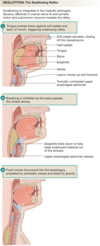
List and describe the 3 stages of swallowing
Voluntary Stage
- food is moistened with saliva and chewed
- bolus is formed
- tongue pushes bolus to the back of the throat (the pharynx)
- This process is under neural control of several areas of cerebral cortex (+motor cortex)
Pharyngeal Stage
- swallow reflex is initiated under involuntary control
Esophageal Stage
- bolus is propelled down the esophagus by peristalsis - a wave of muscular contraction that pushes the bolus ahead of it.
Draw the anatomy of the stomach with the following labels:
- Diaphragm
- Esophagus
- Fundus
- Antrum
- Pylorus
- Rugae

What is the function of the stomach?
- Reservoir for food before it enters the intestines to be absorbed
- Bolus is liquified to enhance enzymatic digestion
- mixed thorougly through coordinated muscular contractions to mechanically breakdown contents of the stomach
- ~2-3L of gastric juices are secreted into the stomach each day
- from different exocrine gland cells
What are the 3 types of exocrine cells found in the stomach?
Mucus neck cells
- secretes mucus & bicarbonate
Chief cells
- secretes pepsinogen and gastric lipase
Parietal cells (aka oxyntic cells)
-
intrinsic factor, H+ and Cl<span>-</span> (HCl)
- intrinsinc factor helps digest vitamin B12
These glands also contain a type of **enteroendocrine cell **(aka G cell).
- secrete gastrin (hormone) - involved in gastric motility and function
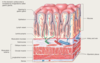
What are the 4 types of layers the stomach wall is composed of?
Note: These layers are similar to that of the rest of the GI tract
Mucosa
- inner lining (apical)
- single layer of epithelial cells
- entire wall is crumpled into folds called rugae
- Rugae increases SA to enhance absorption
Submucosa
- layer adjacent to the mucosa
- composed of connective tissue with larger blood and lymph vessels
- contains submucosal plexus:
- one of the two major nerve network of enteric NS (helps coordinate digestive function)
Smooth muscle (muscularis externa)
- outer wall consists of primarily 2 layers of smooth muscle:
- inner circular layer
- outer longitudinal layer
- 3rd incomplete layer of oblique muscle between circular & submucosa
- contains myenteric plexus (second nerve network of enteric NS)
- between longitudinal & circular muscle layers
- controls & coordinates motor activity of muscularis externa
Serosa
- outer convering of the entire stomach
Explain both the mechanical and chemical digestion that occurs in the stomach.
Digestion in the stomach = converting bolus into chyme
Mechanical Digestion:
- gentle mixing waves w/ secretion of gastric glands
- more vigorous mixing
- from body of the stomach and intensifying towards the pylorus
- small amount of chyme empties into duodenum through slightly opened pyloric sphincter
- most of chyme is pushed back into the body of stomach for more mixing
Chemical Digestion:
- acidic gastric juices (HCl):
- inactivate salivary amylase (inhibit carb digestion)
- activates lingual lipase (fat digestion)
- activates pepsin (protein digestion):
- pepsinogen (inactive protein) comes in contact with HCl to produce active enzyme pepsin
Note: Production of HCl is essential as it is the pH of the stomach
⇒ either activates/inactivates enzymes
What are the functions of acid in the stomach?
- Activates lingual lipase
- fat digestion
- Activates pepsin
- protein digestion
- Inactivates salivary amylase
- stops carb digestion
- Kills microbes
- kills bacteria and other organisms
- Denatures proteins
- It activates pepsin and it denatures the proteins by breaking disulfide and hydrogen bonds that hold the protein in its tertiary structure. Unfolding protein chains make the peptide bonds between amino acids accessible to pepsin.
- Stimulates secretion of hormones
- G cells, found deep in the gastric glands, secrete the hormone gastrin into the blood.
- stimulated by the presence of amino acids and peptides in the stomach, by distension of the stomach, and by neural reflexes mediated by gastrin-releasing peptide.
- Coffee (even if decaffeinated) also stimulates gastrin release
- G cells, found deep in the gastric glands, secrete the hormone gastrin into the blood.
List and describe the 3 regions of the small intestine.
-
Duodenum
- where most digestion occurs
- functions to regulate how fast/slow digestion & absorption occurs
-
Jejunum
- where most nutrient absorption occurs (due to many villi)
-
Ileum
- less nutrient absorption (less villi) - variable
There are many enzymes that are secreted from the pancreas that are necessary for proper chemical digestion of macromolecules. What would happen if the pancreas was suddenly unable to secrete digestive enzymes?
If the mouth & stomach were unable to secrete their digestive enzymes, digestion would still occur fairly normally.
However, if the pancreas was unable to secrete digestive enzymes, digestion would be severely disrupted, quickly leading to starvation due to improper nutrient absorption.
The liver is also important, but the loss of pancreatic secretions into the duodenum has the most severe consequences to digestion.
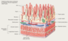
List and describe the layers of the small intestine.
Mucosa
- inner lining (apical)
- single layer of epithelial cells
- entire wall is crumpled into folds called plicae
- projects into the lumen in small finger-like structures called villi
Submucosa
- layer adjacent to the mucosa
- composed of connective tissue with larger blood and lymph vessels
- contains submucosal plexus
- one of the two major nerve network of enteric NS (helps coordinate digestive function)
Muscularis
- outer wall consists of primarily 2 layers of smooth muscle:
- inner circular layer
- contraction of this layer decreases the diameter of the lumen
- outer longitudinal layer
- contraction of this layer shortens the tube
- inner circular layer
- contains myenteric plexus
- second nerve network of enteric NS
- between longitudinal & circular muscle layers
- controls & coordinates motor activity of muscularis externa
Serosa
- connective tissue membrane that is a continuation of the peritoneal membrane (peritoneum) lining the abdominal cavity
- peritoneum also forms sheets of mesentery that hold the intestines in place so they do not become tangled as they move
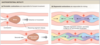
Explain the segmentation and peristalsis of the small intestine
Segmentation
- Segments of intestine alternately contract and relax
- Churns chime ⇒ mixes chyme
- Increase interactions of food particles in chyme with absorptive cells in the intestinal mucosa
Peristalsis
- progressive waves of contraction that move chyme forward from one section of the small intestine to the next after it has been thorougly mixed (segmentation)
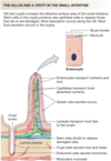
List the cell types of the intestinal wall and their function.
Absorptive cells
- epithelial cells with microvili
Goblet cells
- secrete mucus
Intestinal gland cells
- secrete intestinal juice (a watery mucus that is slightly alkaline)
Paneth cells
- secrete lysozyme
S cells
- secrete secretin
CCK cells
- secrete cholecystokinin (CCK)
K cells
- secrete glucose-dependent insulinotrophic peptide (GIP)
What are microvilli (“brush borders”) and crypts?
- finger-like structures that increase SA for digestion & absorption
- contains several digestive enzymes within it
- crypts also add to SA, but the crypt cells are specialized for fluid and hormone secretion
List the brush border enzymes and what they digest.
Disaccharidases (maltase, sucrase, lactase) digests end products of carbs:
- maltose (glucose + glucose)
- sucrose (glucose + fructose)
- lactose (glucose + galactose)
Aminopeptidase
- digests proteins
Dipeptidase
- digests dipeptides
(single/two peptide bonded molecule)
What is the main function of the large intestine?
- finish process of absorption
- responsible for production of certain vitamins and formation of feces that will be expelled from the body
- feces is NOT composed of any of the macronutrients absorbed:
- mostly made up of fibre, old epithelial cells (lasts ~2 days), old bacteria in large intestine
- macronutrients are generally all absorbed
- feces is NOT composed of any of the macronutrients absorbed:
Draw the main anatomy of the large intestine with the following labels:
- Ascending colon
- Transverse colon
- Descending colon
- Ileum
- Ileocecal valve
- Haustra
- Rectum

Describe the motility in the large intestine
Gastroileal reflex
- involuntary neuronal connection that opens the ileocecal valve for more food to enter the stomach
Haustral churning
- mixes contents of the intestine around allowing for final nutrient absorption
Peristalsis & mass peristalsis
- moving contents out of the intestine