Foot & Ankle Flashcards

1
Q

Where are the Lisfranc Joints Located?

A

Between midfoot and forefoot (between metatarsals and cuneiforms/cuboid)

2
Q

Where are the chopart joints located?

A

Between the hindfoot and midfoot (between talus &calcaneus, and the navicular & cuboid

3
Q

List the Ligaments of the Syndesmosis

A
  1. Anteroinferior Talofibular Ligament (AITFL)
  2. Posteroinferior Talofibular LIgament (PITFL)
  3. Inferior Transverse Ligament (ITL)
  4. Interosseus Ligament (IOL)
  5. Interossesus Memebrane
4
Q

Which tendons attach to the talus?

A

None

5
Q

What are the facets/parts of the Talus?

A
  • Parts: Head, Body & Neck
  • Facets: Anterior, Medial, Posterior
6
Q

Describe the Arteries Providing Blood Supply to the Talus (6)

A
  1. Artery of the Tarsal Canal
    • From Posterior Tibial A.
    • Supplies Body
  2. Deltoid Artery
    • From Artery of the Tarsal Canal
    • Supplies Medial Body
  3. Direct Posterior Artery
    • From Peroneal Artery
    • Supplies Posteiro Process/Body
  4. Direct Superomedial Artery
    • From Dorsalis Pedis
    • Supplies Head & Neck
  5. Artery of the Tarsal Sinus
    • From the Dorsalis Pedis & Peroneal A
    • Supplies Neck & Lateral Body
  6. Perforating Peroneal A.
    • From Peroneal A.
    • Supplies Head & Neck

In general:

  • Dorsalis Pedis A. - Supplies Head & Neck
  • Peroneal A. - Supplies Head & Lateral Body
  • Posterior Tibial A. - Supplies Tarsal Canal A. & Medial Body
    • The main blood supply to the talus
7
Q

Describe the location, boarders and contents of the Sinus Tarsi

A
  • Location: Just distal to the tip of the fibula between the posterior and middle facets
  • Boarders
    • Superior: Talus
    • Inferior: Calcaneus
    • Anterior: Talocalcaneonavicular Joint
    • Posterior: Posterior facet of subtalar joint
  • Contents:
    • Sinus Tarsi A.
    • Tarsal Canal A.
    • Cervical Ligament
    • Interosseus Talocalcaneal Ligament
    • Medial, Intermediate Lateral origins of plantar retinaculum.
    • Fat
8
Q

Parts of the Calcaneus

A
  1. Anterior Facet
  2. Middle Facet (on sustentaculum)
  3. Posterior Facet
  4. Anterior Process (articulates with cuboid)
  5. Calcaneal Tuberosity
9
Q

What is the main motion at the Chopart Joints?

A

Abduction/Adduction

10
Q

List the Ligaments of the Tarsometatarsal (TMT) Joints

A
  1. Transverse Ligaments: attaches base of metatarsals 2-5 (none between 1&2)
  2. Longitudinal Ligaments (connects base of MT to midfoot cuneiforms or cuboid)
  3. Oblique Ligament (From medial cuneiform to base of 2nd metatarsal)
    • aka LISFRANC LIGAMENT
11
Q

What are the components of the Lisfranc Ligament? Which is strongest? Weakest?

A
  1. Dorsal - weakest
  2. Interosseus - true “lisfranc ligament”. Strongest.
  3. Plantar
12
Q

Describe the mechanism for Hallux Valgus.

A
  1. Failure of medial ligments (MCL and medial sesamoid)
  2. Valgus at joint, sesamoid subluxes laterally
  3. Medial sesamoid sits on cristae and further erodes it causing further lateral subluxation and valgus deformity at MTP joint
  4. EHL & FHL move lateral to the axis of the ray, worsening the deformity
  5. EHL, FHL, Add Hallicus overpower the foce of Ab Hallicus further causing valgus deformity
13
Q

Describe the Angles Measured in Hallux Valgus

A
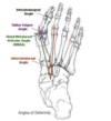
  1. Intermetatarsal Angle (IMA) - angle between 1st & 2nd metatarsals.
    • Normal <= 9 degrees
  2. Hallux Valgus Angle (HVA) - angle between 1st metatarsal and proximal phalanx D1
    • Normal <= 15 degrees
  3. Distal Metatarsal Articular Angle (DMAA)- Between 1st MT long axis and line along base of distal articular cap
    • Identifies MTP joint incongruity
    • Normal <= 10 degrees
  4. Hallux Valgus Interphalangeus Angle (HVI): between long axis of distal phalanx and proximal phalanx
    • Normal <=10 degrees
14
Q

What is the order of tendons crossing the ankle joint anteirorly, from medial to lateral?

A
  1. Tibialis Anterior
  2. EHL
  3. EDL
  4. Peroneus Tertius
15
Q

What is the order of tendons crossing the ankle posteriorly from medial to lateral?

A
  1. Plantaris
  2. Achilles
16
Q

What is the order of tendons behind the medial mallolus from ant to post, medial to lateral?

A
  1. Tibialis Posterior
  2. Flexor Digitorum Longus
  3. Flexor Hallicus Longus

*Tom Dick & Harry

17
Q

What is the Knot of Henry?

A
  • Distal crossing of FHL and FDL in the foot
  • Important site for graft harvest
  • FHL comes medial to FDL, FDL corssess superifical to FHL.
  • Tendon sheaths communicate at the knot
18
Q

Where is Kager’s Fat Pad?

A

Fat pad anterior to the achilles tendon.

19
Q

What is the order of the tendons crossing the ankle laterally both above and below the ankle.

A
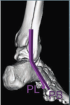
  • Above ankle: peroneus longus superficial to peroneus brevis
  • Distal to fibula: brevis is anterior to longus
    • They share a common sheath proximally but split into individual sheaths distally
20
Q

What are the primary restraint to lateral instabiliy of the peroneal tendons? Where are they located?

A
  1. Superior Peroneal Retinaculum
    • Primary restraint
    • 3.5mm proximal to the tip of the fibula
  2. Inferior Peroneal Retinaculum
    • Distal over the lateral wall of the calcaneus
21
Q

What are the components of the fibroosseus tunnel of the posterior fibula.

A
  1. Posterior Fibular Groove
  2. Fibrocartilage Ridge
  3. Superior Peroneal Retinaculum
  4. Ligaments
    1. CLF
    2. PITFL
    3. PTFL

Contents:

  • Peroneus Longus & Brevis
22
Q

What are the boarders of the contents of the tarsal tunnel?

A
  • Tarsal Tunnel:
    • Fibroosseus tunnel formed by:
      • Posterior medial malleolus
      • Medial wall of calcananeus & talus
      • Flexor retinaculum
  • Contents
    • Tibialis Posterior
    • FDL
    • Posterior Tibial A. & V.
    • Tibial N.
    • FHL

*Tom Dick & Very Nervous Harry

23
Q

List the Lateral Ligaments of the Ankle. What stresses do they combat?

A
  • Anterior Talofibular Ligament (ATFL)
    • Lateral malleolus to neck of talus
    • Resists anterior translation
    • Most commonly injurued
  • Calcanofibular Ligament (CFL)
    • Peroneal tubercle of calcaneus to tip of fibula
    • Deep to peroneal tendons
    • Resists inversion forces
  • Posterior Talofibular Ligament (PTFL)
    • Lateral mallelleolus to lateral tubercle of posterior process of talus
    • Resists dorsiflexion and inversion forces
24
Q

In which postion are ATFL, CFL and PTFL the tightest?

A
  • ATFL: plantarflexion
  • CFL: Inversion
  • PTFL: Dorsiflexion
25
Q

What are the compontents of the Deltoid?

A
  • Superficial
    1. Anterior Tibiotalar Ligament - runs from anterior colliculus of medial malleolus to anteriomedial talus
    2. Tibionavicular Ligament- tibia to navicular tuberosity
    3. Tibiocalcaneal Ligament - tibia ot sustentaculum talus
  • Deep
    1. Posteriortibiotalar Ligament - posterior colliculus of medial malleolus to medial tubercle of talus
26
Q

List the important intertarsal ligaments (6)

A
  1. Interosseus Talocalcaneal - aka Cervical ligament
    • Strongest of the subtalar joint
  2. Calcaneocuboid/Calcanonnavicular - aka Bifurcate
  3. Calcanometararsal - aka Long Plantar
  4. Plantar Calcanocuboid - aka Short Plantar
  5. Palntar Calcanonavicular - Spring Ligament
  6. Oblique Tarsometatarsal - Lisfranc
27
Q

List 3 common accessory ossicles of the foot.

A
  1. Os Trigonum
    • Off posterior process of talus
    • in 5-15% of population
  2. Accessory Navicular
    • aka Os Tibale Externum
    • in 10% of population
  3. Os Peroneum
    • Plantar to calcanocuboid joint
28
Q

Contents of Layers of the Foot

A
  • 1st Plantar Layer - two abductors, one flexor
    • Abductor Hallicus
    • Flexor Digitorum Brevis
    • Abductor Digiti Minimi
    • Plantar Fascia
  • 2nd Plantar Layer - two muscles two tendons
    • Lumbricles
    • Quadratus Plante
    • Flexor Hallicus Longus
    • Flexor Digitorum Longus
  • 3rd Plantar Layer - Two Flexors, one ADductor
    • Flexor Hallicus Brevis
    • Flexor Digiti Minimi
    • Adductor Hallicus
  • 4th Plantar Layer- Interossei & 2 tendons
    • Dorsal Interossei (4)
    • Plantar Interossei (3)
    • Peroneus Longus Tendon
    • Tibialis Posterior Tendon
29
Q

What are the components of the Plantar Fascia?

A
  • Central Band
    • Medial calcaneal tuberosity to flexor tendon sheath & Transverse MT ligaments
  • Medial Band - supports abductor hallicus brevis
  • Lateral Band - supports abductor digiti minmi. Inserts on base of 5th metatarsal
30
Q

Between which plantar layers of the foot does the tibial nerve run?

A

1st & 2nd

31
Q

What are the plantar branches of the Tibial Nerve?

A
  1. Medial Calcanenal Nerve
  2. Medial Plantar Nerve - deep to Adductor Hallicus
  3. Lateral Plantar Nerve
32
Q

Where does the tibial nerve bifurcate in 90% of cases?

A

In the tarsal tunnel.
In the remaining cases, will bifurcate proximal.

33
Q

What is Baxter’s Nerve and what is it’s clinical relevance?

A
  • 1st branch of the lateral plantar n.
    • Innervates Abductor Digiti Minimi
  • Impingment of nerve causeas medial heel pain
34
Q

What is the course of the Deep Peroneal Nerve in the Foot?

A
  1. Emergeces deep to inferior extensor retinaculum with dorsalis pedis
  2. Travels lateral to EHL (lateral to)
  3. Drops branches to EHB, EDB
  4. Continues onto 1st webspace
35
Q

What are the branches of the Dorsalis Pedis Artery?

A
  • Medial & Lateral Tarsal Branches
  • Arcurate A. - runs oover dorsal aspect of TMT joints
  • Deep Plantar A. - Diverts deep between base of 1st and 2nd metatarsal. Forms deep plantar arch.
  • Ends as digital artery.
36
Q

Which Plantar Layer of the Foot Do You Find the Following?

  1. Knot of Henry:
  2. Tibial N.:
  3. Tib Post Tendon:
  4. Plantar Fascia:
A

Which Layer of the Foot Do You Find the Following?

  1. Knot of Henry: 2
  2. Tibial N.: Between 1 & 2
  3. Tib Post Tendon: 4
  4. Plantar Fascia: 1
37
Q

Which Nerve injury causes deficits in the following:

  1. Hindfoot Eversion
  2. Hindfoot Inversion
  3. Great Toe Extension
  4. Great Toe Flexion
  5. Ankle Plantarflexion
A

Which Nerve injury causes deficits in the following:

  1. Hindfoot Eversion: SPN
  2. Hindfoot Inversion: Tibial
  3. Great Toe Extension: Deep Peroneal
  4. Great Toe Flexion: Tibial
  5. Ankle Plantarflexion: Tibial
38
Q

Which two joints make up the Chopart Joints?

A
  1. Calcanocuboid
  2. Talonavicular
39
Q

Which facet of the subtalar joint is most commonly affected in tarsal coalition?

A

Middle Facet

40
Q

Which two intrinsic foot muscles are innervated by the Deep Peroneal Nerve?

A
  1. Extensor Hallicus Brevis
  2. Extensor Digitorum Brevis
41
Q

Describe the 4 most common arthroscopic portals of the ankle. What dangers are associated with each portal.

A
  1. Anteriolateral
    • Medial to lateral mallolus
    • Dangers: Dorsal internal cutaneous branch of SPN
  2. Anteromedial
    • Medial to Tib Ant Tendon, lateral to MM
    • Dangers: Saphenous N. & V.
  3. Posterolateral
    • Medial to peroneus tertius, lateral to achilles
    • Dangers: Sural N. Short Saphenous V.
  4. Posteromedial
    • Through any interval
    • Watch NVB
42
Q

What attaches to the posterior process of the talus?

A

PTFL- attaches to the lateral tubercle of the posteior process

43
Q

What are the contents of the sinus tarsi?

A
  1. Interosseus Talocalcaneal Ligament
  2. Cervical Ligament
  3. Medial, Intermediate and Lateral roots of inferior extensor retinaculum
  4. A. of tarsal sinus
44
Q

What is an Os Naviculae? 3 types?

A
  • Accessory Navicular
  1. Sesamoid in distal part of the tib post tendon
  2. Attachement to the post tib tendon, separated from actual navicular by synchrondrosis
  3. Enlargement of the navicular
45
Q

What benign structure can be confused with a lisfranc injury?

A
  • Os intermetatarseum
  • Ossicle between base of 1st and 2nd metatarsals which can be mistaken for a fleck sign. Clinical correlation of symptoms required.
46
Q

Which structure does the sural nerve run with? On what side?

A
  • Runs lateral to short saphenous vein.
47
Q

What is the innervation of peroneus tertius?

A

Deep peroneal nerve.

48
Q

What are the normal ankle syndesmosis measurements on xray. On which view?

A
  • Tibiofibular Overlap
    • Normal >6mm on AP, >1mm on mortise
  • Tibiofibular Clearspace
    • Located 1cm proximal to the plafond. Measure from anterior incisiura to medial fibula
    • Normal <5mm on both AP and mortise
  • Medial Clearspace
    • <= 4mm on mortise
49
Q

What are the parts of the posterior process of the talus? Attachements?

What is the epmonymous name for fractures here?

A
  • Parts: Medial & Lateral Tubercle
    • PTFL attaches to lateral tubercle
    • posterior deep 1/3 of deltoid attaches to medial tubercle
  • Sheppard’s Fracture
    • Injury to the lateral tubercle
    • Caused by inversion or extreme equinus
  • Cedell’s Fracture
    • Injury to medial tubercle
    • Caused by forced dorsifleion and pronation
50
Q

What are the talar and calcaneal bony contributions to the the sinus tarsi?

A
  • Talar- sulcus tali ( talar sulcus)
  • Calcaneus - calcaneal sulci

Two halves above form sinus tarsi.

51
Q

Describe a compartment release of the foot.

A
  1. Midfoot Dorsal Incisions
    1. Medial to 2nd MT
      • Release medial, deep central, 1st and 2nd interosseus
    2. Lateral to 4th MT
      • Release 3rd & 4th interosseus, lateral compartment, superficial middle compartment and middle central compartment
  2. Medial Calcaneus Incision
    • May add to decompress calcaneal compartment

Can also do a single medial incision to release all 9 compartments.

52
Q

What are the Compartments of the Foot? (9)

A
  1. Medial - Abductor Hallicus, Flexor Hallicus Brevis
  2. Lateral - Abductor Digiti Minimi, Digiti Minimi Brevis
  3. Interosseus x4
  4. Superficial Central - Flexor Digitorum Brevis
  5. Middle Central - Quadratus Plantae
  6. Deep Central - Adductor Hallicus, Posterior Tibial NVB
53
Q

What is a Chaput Fragment?

Volkman’s Fragment?

Wagstaff Fragment?

A
  • Chaput: Avulsion fracture of the anteriolateral tibial plafond secondary to AITFL avulsion
  • Volkman’s: Avulsion fracture of the posterolateral tibial plafond secondary to PITFL avulsion
  • Wagstaff: Avulsion fracture of the anterior distal fibula secondary to AITFL avulsion
54
Q

What is the Constant Fragement of the Tibia? Calcaneus?

A
  • Tibia - Volkmann’s Fragment
  • Calc - Sustentaculum Tali

*Called that because it stays in it’s anatomic location. Can be used to build to.

55
Q

What is Morton’s Neuroma?

A

Neuroma of the digital nerve commonly between 3rd and 4th MT.

56
Q

What are the primary stabilizers of the MTP joints?

A
  1. Plantar Plate (primary)
  2. Collaterals (MCL, LCL)
57
Q

Which muscle inserts onto the tibial and fibular sesamoids of the 1st metatarsal head?

A

Flexor Hallicus Brevis

58
Q

What are the terminal branches of the Posterior Tibial Artery.

A

Medial and Lateral Plantar Artery.

*They follow the same path as the corresponding nerves.

59
Q

What runs in the plantar groove of the cuboid?

A

Peroneus Longus

60
Q

What tendons run above the peroneal tubercle? Below?

A

Preoneal Tubercle: bony protuberance on the lateral aspect of the calcaneus.

  • Above: Peroneus Brevis
  • Below: Peroneus Longus