Foot and Ankle Flashcards
What joints comprise the Chopart joint?
[JAAOS 2016;24:379-389]
Calcaneocuboid and talonavicular
- aka. Transverse tarsal joint

What is the most common accessory bone in the foot?
[JAAOS 2016;24:379-389]
Accessory navicular
What is the differential for lateral ankle pain?
[AAOS comprehensive review 2, 2014]
- Acute or chronic lateral ankle instability
- Lateral talar process fracture
- Osteochondral lesion of the talus
- Anterior process of the calcaneus fracture
- Fifth metatarsal base fracture
- Peroneal tendon pathology
- Subtalar instability
- Soft tissue impingement
- Bone impingement lesion
- Tarsal coalition
What is the differential for heel pain?
[Mann’s Surgery of the Foot and Ankle 2014]
- Plantar fasciitis
- Calcaneal stress fracture
- Entrapment of Baxter’s nerve
- Subcalcaneal bursitis
What are the three components of the spring ligament?
[Mann’s Surgery of the Foot and Ankle 2014]
AKA Plantar calcaneonavicular ligament
- Superomedial (largest)
- Medial plantar oblique
- Plantar inferior

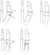
Describe the coleman block test?
- The heel, lateral border of the foot and the 4th and 5th rays are placed on a 2.5-4cm block with the 1st-3rd rays free
- If the hindfoot varus corrects = hindfoot is flexible, forefoot driven deformity
- If the hindfoot varus does not correct = hindfoot is rigid
***Used for varus/cavovarus foot
What is the most common compressive neuropathy of the lower limb?
[JAAOS 2016;24:1-10]
Common peroneal nerve palsy
What is the most common site of compression of the CPN?
[JAAOS 2016;24:1-10]
Fibular neck
What are potential causes of CPN palsy?
[JAAOS 2016;24:1-10]
- Compressive (most common)
- Others:
- Knee dislocation
- Severe ankle inversion injury
- Laceration
- Blunt trauma
- Iatrogenic
What is the classic gait associated with a CPN palsy?
[JAAOS 2016;24:1-10]
Steppage gait
- Ipsilateral knee is lifted higher than normal during the swing phase to avoid dragging the toes on the ground, followed by slapping the forefoot on the ground after heel strike
What resulting foot deformity occurs in untreated CPN palsy?
[JAAOS 2016;24:1-10]
Equinovarus deformity
Following a postoperative or traumatic CPN palsy when should an EMG/NCV study be performed?
[JAAOS 2016;24:1-10]
2-6 weeks
- Repeated every 3 months to monitor for improvement or deterioration
Why is the EMG/NCV study recommended to be delayed 2-6 weeks?
[HSS J. 2006 Feb; 2(1): 19–21.]
- The degree of muscle denervation can only be accurately determined once Wallerian degeneration is complete
- This is a length dependent process where longer distal segments take longer to degenerate
- Evident as fibrillations
- If the study is done too early it may underestimate the extent of injury
What is the management of a postoperative compression CPN palsy?
[JAAOS 2016;24:1-10]
- Initial nonsurgical management
- Activity modification (eg. cessation of leg crossing, padding of the fibular head, avoid squatting, night splints)
- Physiotherapy
- Stretch plantarflexors and invertors
- Strengthening dorsiflexors and evertors
- AFO (allows clearance of foot during ambulation)
- Surgical decompression
- Considered if no improvement after trial of nonoperative treatment (minimum 3 months) or if motor loss is rapidly progressive
- May also be considered over initial nonoperative treatment if EMG/NCV studies show severe conduction loss or disruption of motor innervation
- Tendon transfer
- Considered if no improvement with nonoperative and surgical decompression
- Tendon transfer = posterior tibial tendon (PTT) transfer to lateral cuneiform or cuboid
- Medial cuneiform can be used if only the anterior muscle compartment is affected
- 4-IncisionTechnique
- Incision distal to medial malleolus (extends 5cm distal)
- PTT is harvested subperiosteally from distal to proximal at the naviculocuneiform joint
- Incision ~15cm proximal to the medial malleolus
- The soleus and FDL are retracted posteriorly to expose the PTT, the PTT is then pulled through the proximal incision and tagged with suture
- Incision along the anterior border of the fibula
- EDL is retracted medially and a ~4cm of interosseous membrane is dissected off the fibula and excised
- The PTT is then passed through the window created
- Incision over the lateral cuneiform
- PTT is then tunneled subcutaneously to this incision and anchored to the lateral cuneiform with an interference screw
- Incision distal to medial malleolus (extends 5cm distal)
What conditions cause charcot arthropathy?
[JAAOS 2009;17: 562-571]
- Diabetic neuropathy (most common)
- Others
- Alcohol
- Leprosy
- Tabes dorsalis (tertiary syphilis)
- Myelomeningocele
- Congenital insensitivity to pain
What is the pathogenesis of charcot arthropathy?
[JAAOS 2009;17: 562-571]
- Neurotraumatic theory
* Abnormal sensation prevents normal protective mechanisms after single or repetitive trauma leading to delay in presentation and typical Charcot changes - Neurovascular theory
* Autonomic dysfunction leads to increased blood flow resulting in increased bone turnover - Inflammatory cytokines also implicated in bone resorption
What is the clinical presentation of Acute Charcot Arthropathy of the foot and ankle?
[JAAOS 2009;17: 562-571]
- Hot and swollen foot and ankle
- Bounding distal pulses
- Pain is present ~50% of the time
- May have a history of traumatic episode
What is the radiographic and clinical classification of Charcot Arthropathy?
[JAAOS 2009;17: 562-571]
Eichenholtz classification
- Stage 0
- Xrays – normal
- Clinical
- Swelling
- Erythema
- Warmth
- Dependent rubor decreases with leg elevation (cellulitis does not)
- Stage 1 (fragmentation phase)
- Xrays
- Osteopenia
- Periarticular fragmentation
- Subluxation
- Dislocation
- Clinical
- Swelling
- Warmth,
- Erythema
- Increased ligamentous laxity
- Xrays
- Stage 2 (coalescence phase)
- Xrays
- Absorption of debris
- Early fusion
- Sclerosis
- Clinical
- Decreased swelling, warmth, erythema
- Xrays
- Stage 3 (reconstruction phase)
- Xrays
- Joint arthrosis
- Osteophytes
- Subchondral sclerosis
- Clinical
- Absence of inflammation
- Xrays
What is the anatomic classification based on pattern of collapse in charcot arthropathy of the foot?
[JAAOS 2009;17: 562-571]
Brodsky classification (Trepman modification – added 4 and 5)
- Type 1
- Collapse of tarsometatarsal joints (most common)
- Leads to fixed rocker bottom foot with valgus angulation
- Develop exostosis increasing risk of ulceration
- Collapse of tarsometatarsal joints (most common)
- Type 2
- Collapse of subtalar and Chopart joints
- Unstable, requires prolonged immobilization
- Type 3a
- Collapse of the ankle joint
- Late deformity leads to severe varus or valgus collapse
- Can lead to ulceration and osteomyelitis
- Collapse of the ankle joint
- Type 3b
- Involves fracture of the posterior calcaneal tuberosity
- Type 4
- Combination of above
- Type 5
- Collapse of the forefoot only

How can charcot arthropathy be distinguished from osteomyelitis?
[JAAOS 2009;17: 562-571]
- There is no definitive imaging test to differentiate
- Bone scan followed by WBC scan has sensitivity 93-100% and specificity 80% in localizing osteomyelitis
What is the management based on the Eichenholtz classification?
[JAAOS 2009;17: 562-571]
Stage 0
- Protected WB and foot care
- Serial radiographs to monitor for Stage 1 changes
Stage 1
- Total contact casting with nonWB or partial WB
- Serial radiographs and exam until swelling, warmth and erythema resolve
Stage 2
- Protected WB with total contact cast or CROW (charcot restraint orthotic walker) or clamshell AFO
Stage 3
- If plantigrade foot – custom inlay shoes
- Recurrent ulceration – exostectomy, achilles tendon lengthening if plantar ulcerations
- Severe deformity – arthrodesis
- Recurrent ulceration, infection or failed previous surgeries - amputation
What is the definition of hallux valgus?
[Miller’s, 6th ed.]
Lateral deviation of the great toe with medial deviation of the first metatarsal
Describe the pathoanatomy of hallux valgus?
[Miller’s, 6th ed.]
- Lateral deviation of the proximal phalanx
- Medial deviation of the metatarsal head
- Medial capsular attenuation
- Lateral capsule contraction
- Abductor hallucis migrates plantar and lateral (causes phalanx plantar flexion and pronation)
- Adductor hallucis contracture (deforming force)
- Lateral deviation of the EHL and FHL (deforming force)
- Lateral displacement of the sesamoids relative to the metatarsal head (the crista gradually erodes)
- The medial eminence develops with lateral migration of the proximal phalanx, but it is not characterized by new bone formation or hypertrophy of the medial first metatarsal head [Mann’s Surgery of the Foot and Ankle 2014]

What is the insertion of the adductor hallucis?
Oblique and transverse head insert onto the fibular sesamoid and lateral base of the proximal phalanx