Final Exam Flashcards
first 3 first for elbow
Observation, Fracture Screen, AROM
in acute elbow presentation, what are you observing for (in the first 3 first)
Antalgia . . .
o Atrophy
o Bruises/Cuts
o Scars
o Swelling
o Observations unique to elbow
▪ Cubitus varum (Gunstock Elbow) / cubitus valgum
▪ Pronated/Supinated forearm . . .
in acute elbow, what are you doing in the fracture screen
Light palpation, Percussion*, Tuning fork*, U/S (moving towards elbow)
Humerus:
- Shaft of humerus
- Medial and lateral supracondylar ridges
- Medial and Lateral epicondyles*
Ulna:
- Ulnar styloid*
- Ulnar ridge*
- Olecranon Process*
Radius
- Radial styloid*
- Radial shaft
- Radial head*
and Torsion Test
in acute elbow, what do you do for torsion test?
Step 1-
- Pt seated/standing with elbow @ 90° (if possible)
- Pt keeps his/her elbow at their side
- Doc instructs patient to “meet” doc’s hand with their hand
- IR stress
- ER stress
Step 2-
- Doc instructs patient to “meet” doc’s handin handshake position
- Pronation stress
- Supination stress
AROM for elbow flexion / extension / pronation / supination
Flexion (150°)
Extension (0° to -5°)
Pronation (80°)
Supination (80°)
perform Elbow Valgus Stress Test
Elbow Valgus Stress Test
- At 0˚ elbow flexion
- At 20˚-30˚ elbow flexion

perform Moving Valgus Stress Test
- Patient is supine or standing with the shoulder abducted to 90° and the elbow fully flexed.
- Doctor maintains a valgus stress to the elbow while quickly extending the elbow.
+ Reproduction of patient’s pain between 70°-120°, likely partial tear to medial collateral ligament
- Specificity 75%
- Sensitivity 100%

perform Elbow Varus Stress Test
- At 0˚ Elbow Flexion withforearm pronation
- At 20˚-30˚ elbow flexion, with forearm pronated

PE findings that if (+)ve suggest lateral epicondylitis / tennis elbow (4)
Palpation
- Thorough palpation of the common extensor bundle
- Also look at MFTPs that may refer to the lateral elbow
Cozen (Method 1)
Mill’s Test (Method 2)
Long Finger Test (Method 3)
perform Cozen’s Test (Method 1)
what muscle does this load primarily?
what is (+) sign?
- Examiner pulls towards wrist flexion, contacting radial side of hand
- Patient resists
This test primarily loads the extensor carpi radialis brevis (ECRB) whose contribution to the common extensor tendon is often inflamed in lateral tennis elbow (lateral epicondylitis)
Alternate options include having the patient make a fist while loading as above and palpating over the common extensor tendon.

perform Mill’s test (Method 2)
what is (+) sign?
what does it mean?
This is a passive stretch test.
The patient is seated, the examiner fully extends and pronates the elbow while simultaneously flexing the wrist to stretch the common extensor tendon.
(+)ve Pain at lateral condyle = lateral epicondylitis.
↓ROM… arthritis, capsularadhesion or tendon contracture
This test also compresses the radial nerve, causing similar symptoms to tennis elbow. Electrodiagnostic studies can differentiate the two conditions.

perform Long (middle) Finger Test (Method 3)
what is (+) sign?
Support patient’s forearm
Press down on middle finger, while patient resists flexion
(+)ve Pain at lateral epicondyle

peform bible/heavy book test
DDX if (+)ve sign is at lateral vs medial epicondyle?
Lifting a heavy book with the affected arm may reproduce lateral or medial epicondyle pain
+ Pain at lateral epicondyle = Lateral Epicondylitis
+ Pain at medial epicondyle = Medial Epicondylitis

PE exams that suggest medial epicondylitis / golfers elbow
Palpation
Reverse Cozen’sTest
Reverse Mill’s Test
Book/Bible Test
perform Reverse Cozen’s
what is (+)ve sign?
Muscle testing wrist flexors (with ulnar deviation)
+ Pain at medial epicondyle

perform Reverse Mill’s
what is (+)ve sign?
what does it suggest?
Support elbow, extend supinated wrist passively
(+)ve Pain at medial epicondyle
suggests medial epicondylitis

perform Pronator teres provocation
What is (+) ve sign?
Patient sits with elbow flexed to 90˚
The examiner strongly resists pronation as the elbow is extended.
Tingling or paresthesia in the forearm and hand = +

perform tinel’s on ulnar nerve
where is this location?
Location: cubital tunnel
Tapping the nerve at exposed sites may indicate nerve irritation/entrapment
- Use finger pads (in this area)
- Look for reproduction of symptoms (ulnar distribution)

perform Elbow Flexion / Hyperflexion Test
what is suggested if there are Ulnar Sx? Median Sx?
Elbow flexion, wrist extension
Overpressure
Stretches Ulnar, compression
Median at elbow
Ulnar Symptoms = Cubital tunnel
Median Symptoms = Pronator teres entrapment

perform tinel’s at the wrist
A) median nerve
B) ulnar nerve
Median: fingertips on carpal tunnel
Ulnar: fingertips on tunnel of guyon

olecronon bursitis can be cause by? (3)
Trauma, Infection, Pressure
first 3 first of the wrist and hand: what might you observe?
OBSERVATION:
o Swelling, Atrophy, Bruises, Cuts, Scars…
o Boutonniere Deformity (flexion at PIP and extension at DIP- extensor tendon rupture at PIP joint)
o Swan Neck Deformity (extension at PIP, flexion at DIP- associated with RA or volar PIP ligament lesion)
o Mallet Finger (hyperflexion at DIP – associated with ED rupture at DIP)
o Heberden’s Nodes (associated with OA)
o Buchard’s Nodes (associated with OA, RA)
o Bishop’s Hand (associated with median neuropathy)
o Madelung’s Deformity (anterior translation of wrist/hand –associated with volar growth plate issue)
o Dinner Fork Deformity (associated with Colle’s Fx)
Fx screen of wrist and hand - where will you palpate / percuss / fork?
Ulna (full length)
- Olecranon/ Ulnar ridge/Ulnar Styloid*
Radius (full length)
- Radial head/Radial shaft/Radial styloid*
Carpals
- Scaphoid* (M/C carpal Fx)
- Carpal rows*
Metacarpals and Phalanges
- 1st through 5th *
how do you perform torsion test on wrist and hand?
- Doctor instructs patient to “meet” doctor’s hand in handshake position
- Pronation stress
- Supination stress
AROM for
- wrist: flexion / extension / radial deviation / ulnar deviation
- MP joint: flexion / extension / abd / add
- finger DIP: flexion / ext
- Thumb M/P joint: flexion / ext
- thumb I/P joint: flexion / ext
AROM: Wrist
- Flexion (85˚)
- Extension (65˚)
- Radial Deviation (20˚)
- Ulnar Deviation (30˚)
M/P Joint
- Flexion (90˚)
- Extension (45˚)
- Abduction (20˚)
- Adduction (20˚)
Finger PIP Joint
- Flexion(1000),Extension(00)
Finger DIP
- Flexion (70˚)
- Extension (0˚)
Thumb M/P joint
- Flexion (60˚)
- Extension (20˚)
Thumb I/P joint
- Flexion (70˚)
- Extension (0˚)
perform Phalen’s Test
tests for?
hold for 1 minute, look for reproduction of hand symptoms
tests for: Carpal Tunnel

perform Phalen’s Compression Test
Compress the carpal tunnel with thumb pressure, hold for 20 seconds and observe for symptoms
suggests: carpal tunnel

perform Reverse Phalen’s
The patient maintainsa position of full wrist and finger extension for two minutes.
- The pressure on the carpal tunnel increases after 10 seconds (compared to 20-30 seconds for the standard Phalen test).
- The longer the position is held, the greater the pressure on the wrist and carpal tunnel.

perform Thumb to Index Pinch Test
(+)ve sign?
what is this a test for?
Pt firmly pinches thumb to index while maintaining a circle. Doc does the same. Doc pulls.
(+)ve Inability of Pt to hold fingers together indicates anterior interosseous neuropathy (innervated by median n.)
Test for median nerve pathology

perform Thumb Opposition Test
(+)ve sign?
(+)ve test suggests?
Pt hold thumb against head of 5th metacarpal. Examiner attemps to pull thumb away while grasping base of thumb.
Tests for median nerve pathology

perform Thumb Opposition – Paper Test
(+)ve suggests what pathology?
alternative method of Thumb Opposition Test where Pt holds paper and examiner pulls on paper as indicated
(+)ve = inability to hold paper
Test suggests Median Nerve pathology

perform tinel’s for median nerve
firmly tap carpal tunnel

perform Finger Abduction
(+)ve?
patient abducts fingers
(+)ve sign is inability / weakness
(+)ve test suggests ulnar nerve pathology

perform Froment’s Cone
(+) tests for?
(+)ve suggests ulnar nerve pathology

perform Froment’s Pinch
(+) is?
(+) test suggests?
Pt pinches thumb against side of index; both thumb and index must remain straight as indicated. Examiner attempts to pull thumb and index apart.
(+)ve sign is weakness
(+)ve test suggests ulnar nerve pathology

perform Froment’s Pinch (Paper test)
(+)ve test suggests?
thumb is held straight
(+)ve suggest ulnar nerve pathology

perform Thumb Grinding Test
what is (+)
(+) test suggests?
examiner applies firm long axis compression at the base of the thumb and circumducts attempting to provoke CMC joint
(+)ve painful crepitus
(+)ve suggests arthritis of CMC joint

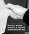
perform Scaphoid Fracture Test
squeeze the scaphoid in the anatomical snuff box
perform Abduction Stress Test part 1 and 2
(+)ve acute test suggets?
(+)ve chronic test suggets?
- examiner applies pressure toward abd w/ thumb straight
- examiner applies pressure w/ thumb flexed
Acute = skier’s thumb
Chronic = gamekeeper’s thumb

perform Finger Collateral Ligaments Abduction/ Adduction Stress
examiner contacts either side of the injured IP joint; distal contact carefully bends distal contact from side to side to stress collateral ligaments

perform Finkelstein’s Test
(+)ve sign?
(+) suggests what problem?
Pt makes fist with thumb inside. Pt ulnar deviates wrist and, if necessary, Examiner add gentle over-pressure as indicated to stretch EPB and APL
(+)ve sign = significant lateral wrist pain
(+) suggests DeQuervains Tenosynovitis

what is the Sweater Sign (Jersey Finger)
what muscle is indicated?
patient makes a fist - also, check knuckle alignment on posterior side
sweater sign = FDP tendon rupture
FDP = flexor digitorum profundus

perform FDP tendon test (flexor digitorum profundus)
(+) sign?
(+) test suggests?
examiner extends finger and stabilizes the intermediate phalanx; Pt attemps to flex distal phalanx.
(+)ve sign = inability to flex distal phalanx of 1 finger
(+) suggests rupture of FDP tendon

perform FDS Test (Flexor Digitorum Superficialis)
Examiner holds all fingers except the 1 in extension. Wrist is extended also. Pt attempts to flex injured finger.
(+)ve sign is inability to flex injured finger
(+)ve test suggests FDS tear/avulsion

check for Lunate Instability
(+) sign?
(+)ve sign is excessive A-P motion

perform Tabletop test
(+) sign?
(+) test suggests?
(+)ve sign is inability to lay palm flat
(+) test suggests Dupuytren’s contractures
As the disease progresses, patients will be unable to rest a flat palm on a table-top.

