Eye Exam Flashcards
What is this condition called?

Esophoria
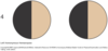
What is this condition called?

Exophoria
How do you evaluate exophoria and esophoria?
The cover test.
Identify these structures


Identify these structures.


Identify these structures


Identify these structures


What are the element of the examination of the eye
(8)
1•Visual Acuity
2•Color blindness
3•Visual field testing
4•External examination
5•Conjunctiva and sclera
6•Cornea, lens and pupil
7•Extra-ocular movement
8•The ophthalmoscopic examination
20/100
What do these two numbers mean?
What are the acronyms for the eyes?
20: distance of patient from chart
100: The distance a normal eye could read the section of the chart visible to the patient.
O.S.: Left
O.D.: Right
O.U.: Both
What is this condition called?
What test is used to identify this condition?

Homonymous Hemianopsia
Visual Field test by confrontation
What is this condition called?

Bitemporal Hemianopsia
What is this condition referred to as?

Quadrantic defects
If a patient can read a newspaper or magazine at 14 inches, what is their vision?
20/40
What is this condition called? What causes it?

Exopthalmos
Hyperthyroidism - Grave’s Disease
What is this condition?

Entropion - inwardly turned eyelids
What is this condition?

Ectropion - Outwardly turned eyelid
What are the following/where are they located?
Conjunctiva –
Bulbar conjunctiva –
Palpebral conjunctiva –
Conjunctiva – clear mucus membrane which covers the eye
   Bulbar conjunctiva – covers the anterior eye

   Palpebral conjunctiva – lines the eyelids
What does this person have?

Osteogenesis Imperfecta
What is anisocoria?
unequal pupils greater than 0.5 mm
Define the following:
Direct Reaction –
Consensual Reaction –
Accommodation –
Convergence –
Direct Reaction – Constriction of the
same pupil
Consensual Reaction – Constriction of the
opposite pupil
Accommodation – Change in pupil and lens
for near and far objects
Convergence – Eyes look inward to focus on
a near object
What is the mnemonic for normal pupillary reaction to light? What does it stand for?
PERRLA
Pupils equal, round, react to light and accomodation
What is nystagmus?
fine rhythmic oscillations of the eyes at the extreme lateral gaze
What does EOMI stand for?
Extra ocular movement intact
How does one assess extra ocular movement?
The H test.
During opthalmoscopic examination what are the three aspects of the optic disc we are looking for?
a. Clarity of the disc margin
b. Color of the disc
c. Central physiologic cup
     (A small whitish depression

     within the optic disc)
Which line depicts the blind spot?


How would a lesion at position three present in a patients field of view?


How would a lesion at position 1 present.


What would a lesion at position 2 look like in a patients field of view?


How would a lesion at position 4 present in a patients field of view?

How would a lesion at position 5 present?


How would a lesion at position 6 present in a patients field of view?


What is this condition called? Describe it.

Pinguecula – small nodule on the
bulbar conjunctiva, does not cross
over to the cornea.
What is this condition called? Describe it.

Pterygium – thickening of the bulbar conjunctiva which grows across the cornea.
What is this condition?

Sty – infection at the margin
of the eyelid
Name and describe this condition

Chalazion – painless nodule involving
the Meibomian gland
What is this condition called? What is it associated with? What is the treatment?

Xanthelasma – flat yellow plaques
Found under the eye.
Associated with hyperlipidemias.
No treatment, investigate lipids and
Cholesterol.
What is this condition called? What can cause it?

Ptosis – drooping of the upper eyelid.
Horner’s Syndrome – ptosis, miosis and
anhydrosis – sympathetic innervation
Bell’s Palsy – CN VII
Name this condition. What causes it? What are the symptoms and how do you treat?

Conjunctivitis – infection or
inflammation of the conjunctiva.
Discomfort, discharge.
Topical antibiotics.
What is this condition? What causes it?

Subconjunctival hemorrhage – leakage of
Blood under the conjunctiva.
Painless, sharply demarcated, resolves on
its own.
From left to right, name the conditions.
What is the overall type of condition? Describe it.

corneal injury or infection, Acute Iritis, Glaucoma
Ciliary injection – inflammation of the radiating vessels around the limbus.
Very painful, vision affected.
Ocular emergency.
What is this condition called? What causes it?

Hyphema – Blood in the anterior chamber.
Due to trauma.
This is an example of papilledema, describe this condition.

Papilledema – disc is swollen with blurred
margins. Physiologic cup is not visible.
Increased intracranial pressure.
This is an example of glaucomatous cupping, describe the condition.

Increased intraocular pressure
Causes increased disc cupping.
The physiologic cup is enlarged
occupying more than half of the
Disc’s diameter.
What are the Hypertensive changes of the artery to the light reflex?
Focal or generalized narrowing – the arterial
walls thicken and light reflex is narrowed.
Describe AV Nicking. What is this related to?
Related to hypertension,
Arterial walls become thickened
and lose transparency. Atherosclerotic
changes.
The veins appear to taper as the
artery crosses.

What condition is this related to? Identify the indicated phenomenon from top to bottom.

Once again, hypertensive retinopathy. (Three slides on this one… for what thats worth)
Top: Cotton wool patches - infarcted nerve fibers
Middle: Copper or silver wiring
Bottom: AV Nicking
What does condition does this horror show depict?

Ocular Melanoma
