Exam 1 (X-Ray Images A) Flashcards

1
Q

What do you do when someone comes in with a skull trauma?

A

REFER out IMMEDIATELY They need a CT

2
Q

Skull Fractures

A

Skull is anatomically complex interpretation is very different less than 10% of skull fracture detected on X-rays

3
Q

Types of Skull Fractures

A

Depression fractures compound fractures Hairline fractures Subdural hematoma

4
Q

What’s the diagnosis?

A

Linear skull fractures are the most commin skull fractures

5
Q

Pathology

A

Linear Skull Fracture

-Most common skull fracture

6
Q

Pathology

-What advanced imaging would help confirm out findings?

A

Linear skull fracture

-CT is the best imaging utilized for skull fractures

7
Q

Pathology

A

Depression Skull Fracture

8
Q

Pathology

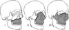
-What name is given to this fractured portion of the face?

A

Zygomaticomaxillary Complex Fracture (a.k.a. Tripod Fracture)

-Called an “Elephant’s Head” where the ear is the lateral margin of the orbit, the trunk the temporal process of the zygomatic bone and the eye is the infraorbital foramen

9
Q

Pathology

-What findings help support our Dx?

A

Blowout Fracture

  • Fracture of the floor of the orbit
  • Fluid inside the right maxillary sinus (should normally be black from air)

-

10
Q

What type of classification of facial trauma are these known as?

A

Le Fort Fractures

11
Q

What type of Le Fort fracture is this?

A

Type I

-Maxilla is separate from the face

12
Q

Pathology

A

Subdural hematoma

-***Convex shaped***

13
Q

Pathology

A

Epidural Hematoma

-***Concave***

14
Q

What are all the mechanisms of trauma that can cause Spinal trauma?

A
  • Hyperflexion
  • Hyperflexion and rotation
  • Hyperextension
  • Hyperextension with rotation
  • Vertical compression
  • Lateral flexion
  • Other
15
Q

Trauma associated with hyperflexion injuries

-Which types of trauma are most common and most significant?

A
  • Simple wedge (compression) fracture (MC by far)
  • Bilateral interfacetal dislocation
  • Flexion teardrop fracture (most significant by far)
  • Clay shoveler’s fracture
  • Anterior subluxation
  • Dens fracture
16
Q

Traumas associated with hyperflexion and rotation injuries

A

Occurs along the same side as rotation

  • Unilateral interfacetal dislocation
  • Unilateral interfacetal fracture–dislocation
17
Q

Traumas associated with hyperextension injuries

A
  • Avulsion of the anterior tubercle of C1 (rare)
  • Hyperextension fracture-dislocation
  • Hyperextension dislocation
  • Posterior arch fracture of C1
  • Extension tear drop fracture
  • Hangman’s Fracture
  • Lamina fracture
  • Dens fracture
18
Q

Traumas associated with hyperextension-rotation injuries

A
  • Pillar fracture
  • Pedicolaminar fracture
19
Q

Traumas associated with Vertical compression forces

-Can neurologic problems arise from this type of trauma?

A
  • Jefferson’s Fracture of C1
  • Burst fracture of the lower cervical spine
  • YES, these types of injuries may cause paraplegia or quadriplegia
20
Q

Traumas associated with Lateral flexion injuries

A
  • Unilateral fracture, lateral mass of C1
  • Transverse process fracture
  • Uncinate process fracture
21
Q

Most common locations for spinal trauma

  • How common is spinal cord injuries?
  • Where is the spine is neurologic injury most common?
A

C1-C2, C5-C7, T12-L1

  • Spinal cord injuries = 10-14% overall
  • Neurologic injury = 40% cervicals
22
Q

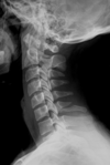
Correct order for the Davis Series for the Cervical Spine

-Which film is used to rule out 90% of spinal trauma to the cervicals?

A

7 views (least to most invasive)

1) Lateral (90% of spinal trauma will be seen here)
2) A-P Open Mouth
3) A-P Cervical

4-5) Left and Right Obliques

6-7) Flexion and Extension

8) Swimmers (OPTIONAL)

23
Q

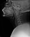
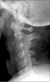
Why is this film not an acceptable lateral cervical film for the Davis series?

A

Does NOT show base of occiput to the top of T1

-If unable to get C7-T1 on the lateral film, should do a Swimmers view

24
Q

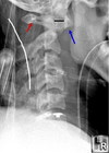
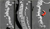
Most significant finding

A

Increased Retropharyngeal Space

( >7 mm is abnormal)

-Many causes including intubation, fracture, infection, SOL, etc.

25
Q

What is the primary function for obtaining these views?

A

Check for instability of the cervical spine and increased ADI

-Very invasive, which is why it’s done last

26
Q

What is this?

A

Denis 3 Column Theory of Stability

  • >1 column disrupted = Unstable
  • 1 column disrupted only = Stable
27
Q

What abnormal soft tissues can let us know if a fracture of the cervical spine has occured?

A
  • Retropharyngeal space ( >7 mm)
  • Retrotracheal space ( >22 mm)
  • Pre vertebral fat stripe (VERY subtle finding)
  • Tracheal air shadow (should always be present)
28
Q

Pathology

A

Increased Retrotracheal Space ( >22 mm)

29
Q

T/F The retrotracheal space should never exceed 14 mm in children younger than 15

A

True

30
Q

What abnormal vertebral aligment are signs of spinal trauma

A
  • Loss of lordosis
  • Acute kyphotic angulation
  • Widened interspinous spaces
  • Vertebral rotation
  • Torticollis
31
Q

Findings

A
  • Widening of the interspinous distance (Fanning)
  • Loss of parallelism between facet joints
  • Horizontal displacement > 3.5 mm
  • Angular displacement (sagittal plane rotation) > 11 degrees compared with adjacent interspaces

Dx: Sprain/Strain of the cervical spine consistent with a flexion/extension mechanism of injury (Whiplash)

32
Q

Pathology

A

Whiplash

33
Q

Pathology

-How long would it take this injury to heal

A

Internal Decapitation

  • normal spinal fracture healing is 3-6 months
  • this injury is almost always fatal

+notice the increased ADI, increased Occipital-Atlas articulation, and the intubation tube causing an increase RPI

34
Q

Pathology

-What type of injury mechanism can cause this?

A

Posterior Arch Fracture of C1

-Hyperextension mechanism

35
Q

Pathology

A

Agenesis of the posterior arch of C1

  • NORMAL VARIANT
  • Note megaspinous sign of C2
  • DDX Posteiror arch fracture
36
Q

Pathology

-How extensive is the myelopathy associated with this condition?

A

Posterior Arch fracture of C1

-Does NOT cause myelopathy because the fracture will increase the size of the sagittal canal which if anything takes less pressure of the cord

37
Q

Is this a Posterior Arch Fracture of C1?

-Why or why not?

A

NO! This is a Jefferson Fracture (of C1)

  • a.k.a. Burst fracture of C1
  • Jefferson Fracture and Posterior arch fracture of C1 look the same on a Lateral film. Need the CT or APOM to help DDX
38
Q

Pathology

-What type of mechanism of injury is associated with this?

A

Jefferson Fracture of C1

  • a.k.a Burst fracture of C1
  • Axial compression mechanism

+ “Over Hang Sign” = Jefferson Fracture

39
Q

In order for a Jefferson Fracture to be considered unstable, the lateral masses of C1 must not extend larger than ____ mm. What ligament helps to stabilize the lateral masses of C1?

A

Under 7 mm = Stable

  • Transverse Atlantal Ligament
  • > 7 mm indicates a tear in the TAL making it unstable
40
Q

Tiny fractures often obscured on plain X-ray but may become more apparent on advanced imaging like CT

A

Occult Fractures

41
Q

T/F The TAL is more likely to shear off the odontoid than rupture secondary to trauma

A

True

-TAL is one of the strongest ligaments in the body

42
Q

If an increased ADI is noticed on a lateral plain film, what 3 conditions are the most likely culprit in order?

A

1) RA
2) RA variant (AS, Reiters, etc.)
3) Trauma
- Increased ADI = >3 mm in adults and >5 mm in children

43
Q

Pathology

A

None

  • Congenital variant (Accessory ossicle of C1)
  • DDX Avulsion fracture of the anterior tubercle of C1 (RARE)

+Notice how the cortex around the ossicle is well corticated = old injury/congenital variant

44
Q

Etiology factors of an Increased ADI

A
  • Normal variant (RARE)
  • Trauma (RARE)
  • Down’s Syndrome (ligamentous instability)
  • Major upper cervical anomalies (Klippel-Feil)
  • Inflammatory arthopathies (RA = #1)
45
Q

If an enlarged ADI is suspected, how do we determine if the area if stable or not?

A

Flexion/Extension X-rays

  • Flexion is best for evaluating the ADI
  • Neutral may show a subtle anteriority for the spinolaminar line of C1
46
Q

Pathology

A

Increased ADI

47
Q

Pathology

A

Type II Odontoid Fracture

  • MC injury at C2
  • MC type of odontoid fracture
48
Q

Pathology

A

Type II Odontoid Fracture

  • Watch out for the Mach Effect
  • Advanced imaging like CT would confirm DX
49
Q

Pathology

A

None

  • Normal congenital variant Os Odontoideum
  • Well corticated bone with no pain or history of trauma
  • DDX Type II Odontoid fracture
50
Q

Which types of odontoid fractures are stable and/or unstable?

A

Type I = Stable (rare)

Type II = Unstable (MC)

Type III = Can be either stable or unstable

51
Q

Pathology

A

Type 1 Odontoid Fracture

  • Rare, but stable
  • DDX Os Terminale
  • Fragment is irregular and top of dens is not uniformly corticated
52
Q

What causes a type 1 odontoid fracture?

A

The avulsion of the tip results from a distraction caused by the apical and alar ligaments

53
Q

30 year old male who came in complaining of upper neck pain that started after his car accident yesterday (being rear ended).

-Pathology

A

None seen on this view

  • Patient has a normal Os terminale that would produce no signs or symptoms
  • Well corticated structures rules out odontoid fractures
54
Q

T/F Most os odontoideum are old ununited dens fractures

A

True

55
Q

Pathology

A

Type III Odontoid Fracture

  • Unstable
  • Best seen on lateral plain film or with advanced imaging
56
Q

Pathology

-What radiographic image is disrupted?

A

Type III Odontoid Fracture

-Harris’ ring is disrupted

Disrupted Harris’ Ring = Type III Odontoid Fracture

57
Q

Pathology

A

Type III Odontoid Fracture

58
Q

Pathology

-What mechanism of injury is associated with this?

A

Hangmans Fracture

  • Bilateral pedicle fracture of C2 with anterior displacement of C2
  • Associated with hyperextension injury
59
Q

Pathology

-What other fracture can occur from the same mechanism of trauma that causes this fracture?

A

Hangmans Fracture

-May also see posterior arch fracture of atlas since that is also caused by a hyperextension injury

60
Q

Where in the spine is the only place where a one time trauma can produce a spondylolisthesis?

-What neurologic problems associated with this type of fracture?

A

C2

  • Hangmans Fracture
  • Almost no neurologic problems since the fracture will cause an increase in the sagittal canal
61
Q

Acute trauma present?

A

No!

  • Nonunion Hangmans Fracture
  • Well corticated bone indicated a nonacute injury
62
Q

Pathology

A

Compression Fracture of C5

  • Decreased height in the Anterior 1-2/3 of the vertebral body
  • Superior endplate is most commonly affected

-

63
Q

Pathology

-What mechanism of injury is most likely to cause this?

A

Compression Fracture of C5

-Flexion injury

STABLE!!!

64
Q

Why are compression fractures most common in the anterior 1-2/3 of the vertebral body?

A

Cortical trabeculation of the vertebral body is less evident in the anterior 1-2/3 of the vertebral body

-Figure B

65
Q

Helpful “clues” to determine old vs. new fractures

A
  • Degenerative changes = old or new
  • Intact cortex = old
  • Cortical disruption = new
  • Blurry/Hazy endplates = new
  • Fracture line = new
  • Line of impaction = new
  • Step defect = new
66
Q

Old or new fracture?

A

New

  • Decreased intensity on T1 (less marrow)
  • Increased intensity on T2 (lots of fluid/edema)
67
Q

Old or new injury?

A

New

-Increased signal on bone scan indicates increased vascular flow consistent with edema

68
Q

What radiographic signs are evident?

-What is the Dx?

A

Step defect

Zone of impaction (superior endplate)

-Acute Compression Fracture of L4

69
Q

Pathology

A

None

Limbus Vertebrae

-Normal variant, especially in pediatrics

70
Q

A faint black line is found traveling through the body of C2 to separate the dens from the rest of the body on the lateral cervical film of a 1 month old baby. What is the most likely pathology?

A

None

  • Faint line is NOT a Type III odontoid fracture.
  • Faint line is present because C2 has not fully ossified yet and the cartilage between the ossified bones does not absorb as much X-ray as the developed bones do.
71
Q

Pathology

-What type of mechanism of injury is most indicated?

A

Burst Fracture of C5

-Axial compressive forces

-

72
Q

T/F Burst fractures are generally stable and seldom present with neurologic compromise

A

FALSE.

  • Unstable because the vertebrae is blown apart
  • Neurologic compromise is likely because fragments can affect the cord (myelopathy) or nerve roots (neuropathy)
73
Q

What activity is the most likely cause of an axial compression mechanism type of fracture?

A

Diving into a shallow pool/water

74
Q

Pathology

A

Burst Fracture

  • Flatened central body
  • Vertical splitting (invagination) of the vertebral body
  • Increased sagittal diameter

-

75
Q

Pathology

A

Burst Fracture

76
Q

Pathology

A

Cervical Burst Fracture

77
Q

Patients presents with severe neurologic compromise in all his extremities

  • X-ray was negative
  • What should we do next?
A

Given neurologic problems, further imaging is needed (CT or MRI) to rule out other complications

-Problem is clear as day with CT

78
Q

Pathology

A

Spinal Cord Transection

79
Q

Pathology

-What mechanism of injury is associated with this?

A

Unilateral Facet Dislocation

  • Flexion and rotation injury
  • Anterior body displacement 25%
  • Decreased laminar length
  • Spinous rotation to the side of dislocation
80
Q

Radiographic Sign

-What pathology is indicated?

A

Bow Tie Sign

-Non-superimposed articular pillars of the involved segment

Dx: Unilateral facet dislocation

81
Q

T/F People with unilateral facet dislocations can not turn their head to the side of injury. The affected side is however mechanically stable

A

True

82
Q

What film view is best utilized to confirm a diagnosis of unilateral facet dislocation?

A

Obliques

-No IVF seen at the location of the dislocation

83
Q

Pathology

A

Unilateral Facet Dislocation at C6

  • Short lamina
  • Bow Tie Sign
  • Anterior displacement of C6 to C7 by 25%
84
Q

Radiographic Sign

-What pathology is indicated?

A

Inverted Hamburger sign (seen only on Axial CT)

-Dx: Unilateral facet dislocation

85
Q

Pathology

-What mechanism of injury is most indicated?

A

Bilateral Facet Dislocation

-Hyperflexion mechanism

*C4-C7 most commonly*

-Anterior displacement by > 50%

86
Q

Is this a unilateral facet dislocation?

A

No!

  • No Bow Tie Sign
  • Anterior displacement is NOT 25%
  • Regular length lamina

Rules out Unilateral Facet dislocation

+++Dx: Bilateral Fecet Dislocation

87
Q

Radiographic signs of Bilateral facet dislocation

A
  • Anterior displacement of vertebral body > 50%
  • Perched facets (incomplete bilateral facet dislocation)
  • Interlocking facets (complete facet dislocation)
  • Widened interspinous space
88
Q

Pathology

A

Complete Bilateral Facet Dislocation

89
Q

Pathology

A

Bilateral Facet Dislocation

90
Q

T/F Bilateral Facet Dislocations are likely to present with neurologic problems

A

True

-Decrease in the sagittal canal

91
Q

Radiographic sign

-What pathology is indicated?

A

Bilateral Inverted Hamburger Sign

-Dx: Bilateral Facet Dislocation

92
Q

Most severe injury that can occur to the cervical spine

A

Flexion Tear Drop Fracture

93
Q

Pathology

A

Flexion Tear Drop Fracture

  • Triangular fragment at the anterior-inferior part of the body
  • Facets are fully subluxated
  • Leads to full paralysis or death
94
Q

Pathology

A

Flexion Tear Drop Fracture

95
Q

T/F Tear drop fractures and burst fractures appear so similar on X-ray. The major difference will be that tear drop fractures will produce tremendous neurologic problems while burst fractures will sometimes produce problems

A

True

-Tear drop injury = fully paralysis or death

96
Q

Pathology

-What mechanism of injury is most likely to cause this?

A

Extension Teardrop Fracture

  • Extension mechanism
  • Triangular fragment at the anterior inferior aspect of the vertebrae
  • Unstable
97
Q

Patient walks into your office complaing of severe neck pain after slipping in his bathroom and hitting his chin on the sink. Your colleague thinks the patient has a flexion tear drop injury and orders a CT. What is the Diagnosis?

A

Extension Teardrop Fracture

-Wrong location and lack of neurologic problems to be Flexion tear drop fracture (would be at C5/C6 with either full paralysis or death)

98
Q

What ligament becomes buckled following a extension teardrop fracture?

A

Ligamentum Flavum

99
Q

Pathology

A

Extension Teardrop Fracture

  • Triangular bone fragment anterior inferior aspect of C2
  • Located at C2
  • Increased RPI
  • C2 posterior body line is posterior to C2

*** Classic Extension Teardrop Fracture***

100
Q

Pathology

-What mechanism of injury can cause this?

A

Clay Shoveler’s Fracture

  • Avulsion fracture of the spinous process
  • Flexion injury mechanism
  • MC at C7, then C6, then T1
101
Q

Pathology

-Is this stable?

A

Clay Shoveler’s Fracture

-Stable

102
Q

Radiographic Sign

-What is the diagnosis?

A

Double Spinous Sign

-Clay Shoveler’s Fracture

103
Q

Pathology

A

None!

  • Congenital Nonunion of the Secondary Growth Center of the Spinous Process
  • Normal variant (well corticated margins rules out fracture)
104
Q

Pathology

A

Clay Shoveler’s Fracture

105
Q

T/F All spinous fractures occur from a flexion mechanism type of trauma

A

FALSE

  • Clay shoveler’s fracture is the only one to occur due to a flexion mechanism
  • Other spinous fractures can occur either due to a extension mechanism or from direct trauma
106
Q

Spinous process fractures are most common in the mid _________, thoracic (except __), and _______

A

Mid cervicals

T1

Lumbars

107
Q

Pathology

-What mechanism of injury most likely caused this?

A

Laminar Fractures

  • Hyperextension
  • MC in lower cervicals
  • STABLE
108
Q

Pathology

A

Lamina Fracture

109
Q

Is this a Clay Shoveler’s Fracture of Lamina Fracture?

A

Clay Shoveler’s Fracture

-Lamina fracture would cause posterior displacement of the spinolaminar line

110
Q

T/F Lateral flexion injuries (uncinate process, transverse process, and unilateral lateral mass of C1 fractures) are rare

A

True

111
Q

T/F X-ray is the best tool to see articular pillar fractures

A

FALSE

  • Seeing a pillar fracture on X-ray is rare (A-P only)
  • CT is the best choice
112
Q

Pathology

-What mechanism of injury is associated with this pathology?

A

Whiplash

-Hyperflexion and Hyperextension

113
Q

What Radiographic findings are seen on X-ray?

-What is the Dx?

A
  • Widened interspinous spaces
  • Widened facets
  • Flex/Extension views to stress ligaments

Dx: Whiplash

114
Q

Pathology

A

Whiplash

  • Increased signal intensity on T2 indicates edema
  • Edema is present in the posterior soft tissue structures and along the ligaments surrounding the vertebral bodies indicating soft tissue damage
115
Q

Pathology

-What is the prognosis?

A

Occipital Atlantal Dislocation

  • a.k.a. Internal Decapitation
  • Almost always fatal
116
Q

Is this a Flexion Teardrop Fracture?

A

NO! Extension Tear Drop Fracture

-Wrong segment

Flexion = C5-C6

Extension = C2-C3