Exam 1 Flashcards
Overview of the Nervous System
- Sensory receptors (the afferent system) and motor neurons/effectors are part of the PNS
- Sensory info feeds into the CNS, where info is processed and sent to the effectors
Cells of the Nervous system
-Neurons- main cell responsible for integration and relay of messages -Glia- support neurons
Neurons
-Cell body and dendrites receive input -Axon hillock= summation of APs, integrative -Axon= conductive -Terminal branches= output -Metabolically compartmentalized- proteins made in cell body and dendrites only *Specialized cells that conduct APs over long distances (quickly)
How do neurons vary?
-Number of dendrites -Branching pattern of dendrites -One axon, but vary in # of collaterals
What are the two types of neurons?
-Projection neurons (sensory, motor, tract) -10% pop -Take info from one place to another (large distances) -Largest and best studied -Local Interneurons -90% -Unmyelinated -Modify info within local/small area -Small, difficult to study
Pseudounipolar cells
-One cell body, axon that starts as one and then branches into 2 collaterals -Ex: sensory neurons
Multipolar cells
-multiple dendrites and an axon -Ex: motor neuron– synapses to muscle
Local Interneuron
-no myelin -Multipolar
Nissl Stain
-Shows ribosomes bound to ER and nucleolus (site of RNA synthesis 4 ribosomes -Doesn’t stain axon -Stains proximal dendrites and cell body
Neuronal Cytoskeleton
-Microtubule- Largest, hollow tube, support axons, laid end to end -Neurofilament- stable, middle size -Microfilament- actin, found in parts of neurons that rapidly change (dendrites)
Axoplasmic Transportation
-How materials are moved within the cell -Use molecular motors and microtubules -Various rates of transportation -Anterograde- cell body to synapse -Retrograde- synapse to cell body
Features of the synapse
-Synaptic vesicles/secretory granules present at presynapse–> Hold NTMs -Postsynaptic density- proteins connecting the two synapses -Glia surround the synapse -Mitochondria at synaptic button produce ATP
Dendritic Spines
-Dendrites have numerous spines to maximize surface area to receive synapses -Neurons receive thousands of synapses which summate to create an AP
Glial Cells
CNS- -Ependymal cells -Microglia -Astrocytes -Oligodendrocytes PNS -Schwann cells -Satellite cells
Ependymal cells
-Line the ventricular system -Fluid-filled part of CNS -Cilia face inward to keep CSF moving
Astrocytes
-Most numerous -Surround neurons -Protoplasmic in gray matter -Fibrillar in white matter -Control extracellular environment (K+) -Take up NTMs -Break down glucose and pass it down to neurons
Oligodendrocytes
-Myelinate in CNS only, more than one axon -Each axon requires more than one oligodendrocyte
Microglia
-Derived from immune system, not nervous system -Phagocytes–> clean up cells -Respond to Injury -Found in resting state
PNS Glia
-Schwann cells myelinate only 1 axon in PNS —-Need many schwann cells to myelinated the same axon -Satellite cells- Support cells around neurons in ganglia
Grey vs White Matter
-G: Little myelin, mostly cell bodies and dendrites -W: a lot of myelin and oligodendrocytes
Early Nervous System Development
-Develops from ectoderm (outermost germ layer) -Begins to form at day 19- ectoderm worlds due to thickening of the neural plate -Neural groove–> neural tube–> CNS cells -Neural folds–> neural crest cells–> PNS
Neural Crest Cells
-Migrate to diff places and make ganglia -Become: —Sensory ganglia (dorsal root ganglia) —Autonomic ganglia (sympathetic, para-) —Enteric ganglia of the digestive system —Arachnoid and Pia covering (CNS) —Schwann cells —Adrenal medulla
Ganglia
-A collection of nerve cell bodies in the PNS -Formed from neural crest cells
Neural Tube
-Cells become CNS structures —Cerebrum- cerebral cortex and deep nuclei —Diencephalon, Midbrain, Pons, Cerebellum, Medulla —Spinal cord —Glial Cells of CNS
Neural Tube at week 4
-3 primary brain vesicles form –Forebrain/Prosencephalon (rostral most) –Midbrain/Mesencephalon –Hindbrain/Rhombencephalon (caudal most)
Neural Tube at week 5
- Forebrain splits to Telencephalon and Diencephalon
- Midbrain/Mesencephalon
-Hindbrain splits to Metencephalon and myelencephalon
Telencephalon
-Outgrows all other parts of the brain–> becomes cerebrum —Expands and fold to increase surface area
Sulci and Fissures
-Small grooves/valleys= sulci -Larger, deeper grooves= fissures
Major Lobes in the Adult Brain
-Frontal lobe -Parietal lobe -Temporal lobe -Occipital lobe —-Central sulcus separates frontal and parietal —-Lateral fissure separates temporal from frontal and parietal
Major Divisions of the Adult Brain
-Cerebrum -Brainstem -Cerebellum -Spinal cord
Ventricular System of the Adult Brain
-Fluid filled space within vesicles -Choroid plexus-> produces CSF -Inside= lumen
Neural Tube Defects
-Occur when neural tube fails to close in development -Spina Bifida–> failure to fuse at caudal end —Meningocele–CNS coverings exposed to outside of body —Meningomyelocele– Menigies and neural components exposed to outside of body -Anencephaly-Failure of rostral neural tube to develop–> no brain formation **Caused by folic acid deficiency, high glucose levels, retinoid acid
Neural tube breakdown
-Prosencephalon–> Telencephalon + Diencephalon
—Tel–> Cerebrum, cerebral hemispheres (cortex, white matter, basal nuclei), lateral ventricles
—Diencephalon–> Thalamus, hypothalamus, epithalamus, 3rd Ventricle
- Mesencephalon- Brain stem, midbrain, cerebral aqueduct
- Rhombencephalon–> Metencephalon + Myelenchepalon
—Metencephalon- brainstem, pons, cerebellum, part of 4th V
—Myelencephalon- Brainstem, medulla oblongata, 4th V -Spinal cord + central canal
Cerebrospinal Fluid
-Fluid w/in neural tube -Produced by choroid plexus -Clear fluid; similar to interstitial fluid, but diff from plasma -Potassium, calcium, bicarbonate, and glucose levels tightly regulated–Brain not subject to variations seen in blood -Volume- 150ml; Daily Production= 500ml -Circulates in spaces within brain (ventricular system) and through subarachnoid space around brain
Choroid Plexus
-Specialized ependymal cells that extract fluid and solutes from plasma and filter it -Located in the Lateral Ventricles
Ventricular System Flow
Lateral Ventricles–> Foramen of Monro–> 3rd V–> cerebral aqueduct–> 4th ventricle–> foramen of Magendie and Luschka (L and R)–> Superior Sagittal Sinus–> Arachnoid granulations
Location of Ventricles
-Lateral Vs-> under cerebral cortex -3rd-> midline btwn L and R thalamus -Cerebral aqueduct-> midline of midbrain -4th-> btwn pons/upper medulla, cerebellum
Problems in CSF circulation
-Blockage of circulation -Damage of arachnoid granulations -Overproduction– rare *Hydrocephalus–Not as dangerous in children cuz bones haven’t fused *Chiari malformation–> CSF directed down central canal to spinal cord, compressing spinal cord tissue
Spinal Taps
-How CSF samples are obtained -Needle inserted through skin btwn L4 and L5 -Test 4 abnormal: pressure, protein, glucose, blood cells, immunoglobulin
Divisions of the cerebrum
-Cortex: Outermost grey matter; perception, thought, conciousness -Limbic system- white matter structures encircling ventricular system; learning, memory, emotion -Basal ganglia- deep grey matter; motor control
Association Fibers
-Connect cortical areas on the same side of the brain -White fiber bundle
Commissural Fibers
-Connect cortical areas on opposite sides of brain -White fiber bundle -Ex: corpus callous
Projection Fibers
-Connect cortex w subcortical structures and other brain areas -Ex: Internal capsule and corona radiata
Internal Capsule and Corona Radiata
-Long projection fibers btwn the cortex and inferior structures that make up an array of vertically directed axons– corona radiata -Internal capsule- fibers from the corona radiata funnel into this compact fiber bundle lateral to the thalamus and caudate nucleus
Hippocampus
-Temporal Lobe Structure -Related to inferior horn of lateral v’s -Involved in learning, memory, spatial localization
Amygdala
-Temporal lobe structure -Related to inferior horn of lateral v’s -Receives info from all sensory systems -Mediates intense emotions such as anger and fear
Basal Ganglia
-A collection of nuclei -Regulates starting, stopping, and amplitude of activities -Parts- Caudate nucleus, putamen, globes pallidus -Caudate associated w lateral wall of lateral ventricle
Basal Forebrain area
-Lies anterior and lateral to the hypothalamus on each side -Near front end of corpus callous
Thalamus
-Gateway to the cortex -Occupies a central position in the brain (btwn brainstem and cerebrum; R and L) *R and L sometimes (NOT ALWAYS) connected by interthalamic adhesion -On either side of 3rd V
Thalamic divisions
-Based off of fxn 1. Anterior nuclear group 2. Pulvinar 3. Medial geniculate nucleus 4. Lateral geniculate nucleus *Each group projects to a diff region of cortex for diff sensations (except smell)
Hypothalamus
-Central to the limbic system -Homeostasis (body temp, endocrine, behavioral reg) -Group of nuclei -Boundaries: optic chasm, anterior commissure, maxillary body, lamina terminals
Epithalamus
-Contains pineal gland (melatonin production, regulates circadian rhythms) -Habenula- pain and reward pathways -Posterior commissure (connect L and R sides of the midbrain)
Subthalamus
-Nucleus -Part of basal ganglia circuitry; internal structure
Laminar organization of the cortex
-Sheet-like, horizontal organization of the cortex -Difference in thickness of the layers= basis for brodmann areas
Development of the Cerebral Cortex
-Cells are born in the ventricular zone -cells differentiate into neurons and “climb” radial glia to cerebral surface -Newer cells= top layer, older on bottom -Adult cerebral cortex= 6 layers —-Layer 1= Pia matter
Vertical Organization of the Cerebral cortex
-The cortical column- built around pyramidal cells —Columns of cells oriented perpendicular to the surface of the cortex that are individual functional units *Columns encompassing all 6 layers will fire when receptive field is reached
Layer 5 of the cortex
-Location of pyramidal cells -Output to subcortical areas -Contains Betz cells -Projection fibers
Pyramidal cells
-Dictates whether that part of the cortex will send an output -Define boundaries of one cortical column -Long dendrites that extend to layer 1–> apical dendrites -Basal dendrites extend to bottom of layer 5 -Recurrent collaterals- project into layer 6 -Axon projects into subcortical areas
Layer 1 of the cortex
-Plexiform/molecular layer -Fibers parallel to the surface
Layers 2/3 of the cortex
-Small and medium pyramidal layers -Intracortical connections -Short and long association fibers -Commissural fibers —Stellate and pyramidal neurons with intracortical connections
Layer 4 of the cortex
-Inner granular layer -Stellate neurons receive INPUT from thalamic nuclei
Layer 6 of the cortex
-Fusiform/multiform layer -Cortical output to specific thalamic nuclei —-inputs and outputs to thalamus
Motor areas and sensory areas in regard to cortex layers
- Motor areas have large layer 5, small layer 4 -Sensory areas have large layer 4, small layer 5
- Association ares= in between
Brodmann’s areas
-Defined specific cortical areas based on their histology and functional correlates
Functional areas of the cerebral cortex
-Primary areas–> devoted to only 1 function —-Primary visual, auditory, motor, and somatosensory cortices —-Described w maps -Associated areas–> make sense of info —Interpretive, integrative, complex processing
Lateralization of the cerebral cortex
-Processes info related to opposite side of body -L: analytical, precise, organized, literal -R: creative, imaginative, conceptual, intuitive -Dominance: often, one side more strongly controls an activity -Ex: language predominates left side
Occipital Lobe
-The visual cortex–Brod area 17 —-Map of contralateral retina–points in visual space -Visual association cortex- Brod area 18/19 —Interpretation
Dorsal and Ventral Pathways of Visual cortex
-Where= dorsal, what=ventral -Feeds info away from occipital lobe to other brain areas (memory areas) -Damage leads to visual deficits/ “cortical blindness”
The primary auditory cortex and association area
-Temporal lobe; temporal lobe gyrus inside lateral fissure -Brodmans area 41/42 -Map of tones; sounds mix in both hemispheres -Association area- interprets pattern of sound —-BA 22
Wernicke’s Area
-reading mentally, interpreting speech -Dominant side= left *Part auditory cortex, part association cortex -Damage–> sensory aphasia-> inability to understand speech; fluid nonsensical speech
Visual Association Cortex
-Temporal Lobe -ID of stimuli we recognize -Agnosias= difficulty recognizing stimuli, naming categories of things *Ex: prosopagnosia= inability to recognize faces *Dominant= left
Primary Somatosensory Cortex and Association area
-Parietal lobe -PSC- Lies on the post central gyrus, BA 3,1,2 —-Map of contralateral body —-Somatic sensation (touch, pain, temp) -AA- Superior parietal lobule (BA 5+7) —-Interprets sensations in space
Sensory Homunculus
-Neurons of the primary somatosensory cortex are topographically arranged and receive sensory signals from diff parts of the body -Greater area= greater sensory acuity
Subdivisions of the Frontal Lobe
-Motor region -Prefrontal region
Motor Region of the Frontal Lobe
-2 final areas: –1. Primary motor cortex= BA 4 ———precentral gyrus; map of skeletal muscles (primary motor strip) –2. Premotor cortex (rostral to primary motor) ——–Designs motor plan; activaion and synchronization of groups of muscles
Motor Homonculus
-Neurons of the primary motor cortex are topographically arranged and execute commands to skeletal muscles in diff parts of the body
Broca’s area
-Adjacent to speech muscle area of primary motor cortex on DOMINANT SIDE (left) -Damage gives “expressive aphasia” or “non-fluent aphasia”–> defective grammar/difficulty producing speech
Parts of the Prefrontal Cortex
-Complex behavioral areas 1. Dorsolateral PFC 2. Orbitofrontal cortex 3. Anterior cingulate
Dorsolateral PFC
-Working memory –Ex: remembering a phone # -Associative memory–> learn to associate an action w/ reward -Executive fxn
Orbitofrontal Cortex
-Involved in social and emotional decision making –Risk vs reward -Damage– loss of inhibition/ change in behavior *Last part of the brain to reach maturity
Anterior cingulate
-Involved in motivation -If damaged–> Akinetic mutism (not motivated to speak)
Tasks for executive system impairment
- Alternating sequences task —-Patient asked to draw a circle, then a triangle —Inability to disengage from stimulus —Preservation
Frontal vs. Parietal Association Cortex
-Posterior parietal cortex orients attention toward extra personal world (things around u/ ur body) -Prefrontal association cortex orients attention toward internal mental processes
Insular Cortex
-Feelings and self-awareness -How feelings are colored onto the body
4 types of organization w/in the brain
- Laminar/Horizontal 2.Cortical column/ Vertical 3. Functional (BAs) 4. Topographical (homunculi)
Peripheral Nerves
-Connect CNS to periphery
Spinal Nerves
-31 pairs of spinal nerves that connect peripheral structures to the spinal cord -Share similar organization; contain motor and sensory fibers -Cell bodies located in dorsal root ganglia -Mixed nerve–> sensory and motor mixed together
Cranial Nerves
-12 pairs that connect brain to periphery -Every cranial nerve is diff from the others -CN II is CNS tissue, formed from neural tube
Neuron Types in the PNS
-Sensory -Motor
Sensory Neurons
-carry info from receptor into CNS -Axon= sensory axon or sensory fiber; pseudounipolar -Cell body located in PNS ; joins w others to form ganglion *No dendrites
Types of Sensory Neurons
-Somatic sensory- Receive info from skin or skeletal muscle (axons= somatic sensory axons) -Visceral sensory-receive info from receptors in organs (axon visceral sensory axons)
Motor Neurons
-carry info out of CNS, into PNS -Axon= motor axon or motor fiber; unipolar -Cell body located within gray matter of CNS -Many dendrites; synapse on target cells in periphery
Types of Motor Neurons
-Somatic Motor- Carry info to skeletal muscle; release NTM onto it so muscle contracts -Autonomic motor/Visceral Motor- go to organs; chain of 2 neurons (1st= preganglionic, 2nd is postganglionic)
Nerve types in the PNS
-Further classified according to which types axons are mixed within the nerve 1.Motor nerve 2. Cutaneous nerve 3. Splanchnic nerve
Motor Nerve
-Nerve that enters muscle cells -Contains 2 axon types: —Somatic motor fibers (axon)= synapses on skeletal muscle cells —Somatic sensory fibers (axons)= collect info from sensory receptors in muscle and carry it back to the CNS
Cutaneous Nerve
-Nerve that enters a patch of skin -2 axon types: —Somatic sensory fibers (axons)- collect info from sensory receptors in skin; carry back to CNS (touch info) —Autonomic motor fibers (axons)- synapse to smooth muscle or gland cells in skin
Splanchnic Nerve
-Nerve that enters an organ -2 axon types: –Visceral sensory fibers (axons)- collect info from sensory receptors in organ and carry to CNS –> info helps CNS adjust conditions of organ –Autonomic motor fibers (axons)- Synapse on cardiac muscle cells, smooth muscle cells, or gland cells in organs to alter organ fan
Innervate
-When an axon contacts a cell or structure in the body -4 motor axons–> synapses on skeletal muscles -4 sensory axons–> synapses
Axons in the PNS
-Myelinated; produced by Schwann cells -Schwann cells also ensheath axon–> Hug axons w/o myelinating them
Thickness of Myelin Sheath and Axon Diameter
-Proportional to each other -Increased myelin or axon diameter increases conduction speed -Diff fxnal classes of peripheral axons conduct APs at diff speeds -Ex: A and B fibers= myelinated, C is unmyelinated (pain, temp, mechanoreception)
Structure of Peripheral Nerves
-Axons organized by specific connective tissue coverings —–Axons w or w/o myelin arranged into fascicles -Schwann cells -Blood vessels nourish cells *Axons could be sensory or motor, but look identical -Connective tissue layers –Around each axon= Endoneurium (deepest layer) –Around each fascicle– Perineurium –Around the entire nerve= Epineurium (tough outer coating)
Fascicles
-Group of axons bundled together with collagen-rich connective tissue -Have a common fxn *Only in PNS
Can one neuron be myelinated by oligodendrocytes and Schwann cells?
-Yes! -Part of axon may be in CNS–>myelinated by oligo -Part may be in PNS–> myelinated by Schwann
Differentiation within the neural tube (Dorsal-Ventral)
-Cells divide into the ventricular zone and migrate from the ventricle within the wall -In SC–> dorsal alar plate (eventually dorsal root) and ventral basal plate develop (ventral root) -Sulcus limitans= the grove in the lateral wall of the neural tube delineating alar from basal plates
Differentiation of the neural tube (Medial-Lateral)
-Hollow opening in center= ventricle -Ventricular layer= dividing cells -Mantle layer= neuroblasts —Cells migrate, differentiate -Marginal layer= axon growth
Development of the Spinal Cord
-Central grey portion–> Dorsal and ventral horns -Central canal does not enlarge -White matter surrounds the grey
Dorsal Root Entry Zone
-Medial DREZ= myelinated axons entering from dorsal root -Lateral DREZ= unmyelinated axons entering from the dorsal root
Spinal Cord at Diff Levels
-The overall cross-sectional size of the SC decreases from cervical to sacral levels -Volume of white matter decreases from cervical to sacral levels **Shape of grey matter changes at each level —Greatest volumes of dorsal and ventral horn at limb attachment -Some areas have lateral horns
Spinal Cord- White Matter
-Carry info up spinal cord (ascending tracts) OR -Down from brainstem or cerebrum–> descending tracts *No connective tissue separating tracts -Tracts are only sensory or motor, never both
Spinal Cord-Grey Matter
-Organized into nuclear cell columns OR Rexed’s Laminae
Rexed’s Laminae
- Marginal Zone 2-3- Substantia Gelatinosa 4- Nucleus Proprius 6-7-Column of Clark 9- Motor neurons *Different lamina are related to diff functional systems
Dorsal and Ventral Ramus
-Mixed- sensory and motor -Names refer to location of body they go to —Dorsal branches to dorsal skin and muscle —Ventral branches to ventral skin and muscle
Dorsal Roots and Rootlets
-Only sensory axons
Ventral roots and rootlets
-Only motor axons
Spinal Nerves are the first point where…
-First point where sensory and motor axons are bundled together
Comparison- CNS vs PNS: Terms describing clusters of neuron cell bodies
-CNS: gray matter, nuclei, centers, horns -PNS: Ganglia *All supported by glia
Comparison- CNS vs PNS: terms describing groups of axons
-CNS- white matter, tract, funiculi, fasiculi, fiber bundles (ex: corpus callosum) -PNS: Nerves, fascicle
Meissner’s Corpuscles
-Just under skin surface -Sensitive to fine touch -Lead to stimulation of 1st order sensory neurons *Rapid adapting- fire quickly and stop firing quickly
Pacinian Corpuscles
-Deep under surface of skin -Sensitive to pressure and vibration -Lead to stimulation of 1st order sensory neurons *Rapidly adapting- fire quickly and stop firing quickly
Merkel Cells
-Located just under the surface -Sense of fine discriminative touch -Lead to stimulation of 1st order sensory neurons *Slow adapting
Ruffini Endings
-Deep under skin -Provide touch directionality *Slow adapting
Free Nerve Endings
-Stimulate 1st order sensory neurons -Sensations of pain and temp *not a receptor* -Channels in axon at peripheral terminus change conductance in response to heat or cold/ local chemicals *Localized in skin, connective tissues, and organs
Sensory receptors of skeletal muscle
-1a fiber=muscle spindle —Detect stretch of mucle -1b fiber= tendon ending/ golgi tendon
Hierarchy in somatosensory pathway
Stimulus–> receptor–> 1st order neuron in PNS (dorsal root gang)–> 2nd order neuron in CNS (SC or brainstem)–> 3rd order neuron in thalamus–> neuron in primary sensory cortex–> conscious perception of stimulus *reflexes occur at 2nd order neuron
Divergence
-Ex; Somatosensory system; Muscle afferents -One input in CNS can be sent to many areas simultaneously -Info can be processed in multiple ways in multiple sensory pathways
3 Major Spinal Somatosensory Pathways
- Dorsal Spinocerebellar Pathway 2. Dorsal Column/Medial Lemniscal Pathway 3. Anterolateral pathway (spinothalamic)
Dorsal Spinocerebellar Pathway
-1a, 1b, Abeta afferents -Muscle length -Muscle contraction -Joint contraction *1st order neuron= Dorsal root gang cell *2nd= neuron in column of clark – dorsal spinal cerebellar tract in lateral funiculus (ipsilateral SC)–> *3rd: ipsilateral cerebellum
Column of Clark
-Only present from C8 to L2
Dorsal Column/ Medial Lemniscal Pathway
-1a, 1b, Abeta afferents -Fine touch, vibration, conscious proprioception -1st order- DRG sensory neuron *Dorsal column tract in SC* -2nd order- Gracile or cuneate nucleus (lower medulla) **CROSS THE MIDLINE TO OPPOSITE SIDE OF BRAINSTEM, medial lemniscus pathway in brainstem -3rd order- VPL nuc of thalamus *Sensory axons in this pathway travel in dorsal funicular of ipsilateral SC (don’t relay through dorsal horn nuc)
Gracile Fasciculus and Cuneate Fasciculus
*Parts of the dorsal funiculus -Gracile= feet/lower body/ below T6 -Cuneate= arms/ upper body/above T6 T6= bottom of sternum
VPL
-Relay nucleus of the Medial Lemniscus Pathway -Axons travel trough internal capsule and synapse in layer 4 of the primary somatosensory cortex
Anterolateral System
-Adelta (sharp pain), C afferents (dull itching or burning) –Pain, temp, itch, crude touch 1. DRG sensory neuron (free nerve endings stimulated) 2. Marginal nucleus or nucleus proprius (dorsal horn of ipsilateral sc) **Cross midline, ascends to brain via anterolateral tract** 3. VPL nucleus of thalamus –> relays to somatosensory cortex
Marginal Nucleus
-Alpha Delta fibers carrying sharp pain, some temp -Where dorsal root ganglion neurons synapase in the anterolateral pathway
Reticular Formation
-Responsible for autonomic and limbic connections of pain —Arousal —Emotional component of pain
Spinal Cord Sensory Pathways
-1st order neurons are DRG cells–> bring in info from mechanoreceptors and free nerve endings 2nd order neuron= CNS cells which relay to brain -If info is destined for cortex, 3rd order neuron= part of thalamus
Spinal MOTOR pathways
-DONT USE TERMS 1ST ORDER, 2ND etc -Upper motor neurons and lower motor neurons -Axons form descending tracts traveling in white matter
Lower Motor Neurons
-Cell bodies in ventral horn -Project to skeletal muscles
Upper Motor Neurons
-Cell bodies and axons in CNS -Regulate LMNs
Somatic motor neurons in the ventral horns
1.Alpha Motor Neurons 2.Gamma Motor Neurons -Project through motor nerves -Terminate on skeletal muscle at neuromuscular Jxn *Only source of skeletal muscle activity Axon damage= cells in motor unit cant contract Nerve damage= whole muscle cant contract
Alpha Motor Neurons
-Large in diameter -Rapidly conducting cells -Cause skeletal muscle cells to contract
Gamma Motor Neurons
-Smaller, slower conducting cells -Control tiny muscle cells within muscle spindle receptors -Modulate the sensitivity of MS receptors
Motor Unit
-One alpha motor neuron and all the muscle cells it innervates –Small: innervates about 10 cells –Large: innervates 1000 cells= less fine control
How Gamma and Alpha motor neurons work at a muscle spindle
-if only alpha motor neurons were activated, only extramural muscle would contract–> muscle spindle becomes slack and no APs fire–> cannot signal further length changes -When alpha and gamma coactivated–> extrafusal and intrafusal contract–> tension is maintained in muscle spindle and can signal changes in length


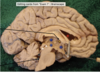
Choroid plexus


- Red circle= subthalamus
- Blue circle= epithalamus

Blue- Dorsolateral PFC
Red-Orbitofrontal PFC
White- Anterior cingulate


- Spinocerebellar Pathway
- Dorsal Column/Medial Lemniscal Pathway
- Anterolateral Pathway


The Meninges

- Protective covering of the CNS
1. Dura mater-tough outer membrane
2. Arachnoid mater- Thin, transparent membrane covering superficial blood vessels in the cortex
3. Pia Mater- layer adjacent to brain - Cannot be removed w/o tearing brain tissue

Spaces within the Skull
- Epidural space: between skull and dura
- Occurs upon injury- not normally there
- Subdural space: Btwn dura and arachnoid
–Not normally there
-Subarachnoid space- contains CSF
—Location of superior saggital sinus
Where are blood vessels located within the brain?
- W/in the dura (supply dura only)
- Btwn arachnoid and pia
—Supply blood to brain

- Yellow–> Cerebrum
- Green–> brainstem
- White–> Cerebellum
What fissure separates the left and right hemispheres?
-Interhemispheric fissure

- Red: Frontal lobe
- Blue dots= central sulcus
- Yellow: Parietal lobe
- White dots= Lateral fissure
- Black= temporal lobe
- Green= occipital lobe
Where can the insular cortex be found?
-At lateral fissure, pull temporal lobe apart from frontal and parietal lobes
- Back?
- Front/Stomach?
- Coccyx?
- Brain/head?

- Back= dorsal/posterior
- Front= ventral/anterior
- Coccyx= caudal
- Brain/head= rostral

- Separating hemispheres?
- Separating dorsal and ventral planes
- Vertical separation
- Separating superior and inferior portions
- Sagittal (midsaggital if perfect split, parasaggital if unequal)
- Coronal/Frontal
- Transverse
- Horizontal

- Flax cerebri= btwn cerebral hemispheres
- Tentorium cerebelli= btwn occipital lobe and cerebellum


What described the 90 degree angle that the brain and brainstem form?
-Mesencephalic Flexure
- Red= lateral ventricles
- Clip= Septum pellucidum–>membrane separating left and right LVs
- Yellow- Foramen of monro
- Blue-> 3rd V
- White= cerebral aqueduct
- Black= 4th Ventricle
What are the 3 drainage holes from 4th ventricle into subarachnoid space?
- Foramen of Magendie= midline
- Two foramen of Lushka- lateral

- Superficial Gray matter
- White matter
- Deep greymatter

Caudate, Putamen, and Globus Pallidus= Basal Ganglia *Grey matter


Red cross= optic chiasm
Blue= Anterior perforated substance
—Ventral surface where BVs penetrated

Blue circle- amygdala
Arrows= hippocampus

- Triangle= hypothalamus
- Blue= Thalamus
- Yellow= epithalamus
- Green= subthalamus

-interthalamic adhesion
*only some people have it

White= pons
Yellow= floor of the hypothalamus
Red= optic chiasm
-Black hole below optic chiasm= pituitary stalk

1.Parieto-Occipital fissure


Primary Motor Cortex/ Brodmann’s area 4/precentral gyrus
Motor Homunculus
- Tongue near insula
- Toes/leg near interhemispheric fissure


-Premotor cortex/BA 6/Part of superior and middle frontal gyri

- Broca’s area: BA 44 and 45
- Motor speech

-Prefrontal Cortex


-Primary somatosensory cortex/ BA 3,1,2/post-central gyrus
Somatosensory Homunculus
- Tongue, pharynx, intrabdominal–>insula area
- Leg= intrahemispheric fissure


Red=Somatosensory association cortex (BA 5and7)/ superior parietal lobule
-Blue= intraparietal fissure

-Inferior parietal lobule


- Primary visual (blue)- BA 17
- Calcarine fissure (red)
- Visual association cortex- White- BA 18 and 19
-


- Primary Auditory Cortex (green)- BA 41 and 42
- Auditory association cortex (yellow)- BA 22

Red= Broca’s are- BA 45 and 44
Yellow=Wernicke’s area= BA 22

-Parahippocampal gyrus

Uncus
Spinal Cord
- Continuation of the brainstem
- Bones of spine create a protective canal


- Intervertebral disk-> cartilage btwn each pair of vertebral bodies
- Vertebral canal= formed by alignment of vertebral foramina
- Intervertebral foramen- formed on each side between every pair of vertebrae

Organization of the spine
- 7 cervical vertebrae
- 12 thoracic vertebrae
- 5 lumbar vertebrae
- 1 sacrum (fusion of 5 sacral vertebrae)
- 2-4 coccygeal vertebrae–> coccyx
- Numbered
*Spines point posteriorly
*Bodies point anteriorly
-Spinal canal lies within the vertebral canal–> stops around L1/L2
Meninges surrounding the spinal cord

- Pia= adheres to SC
- Arachnoid- external to pia, attached by fine fibers
- Dura- surrounds arachnoid, extends out of the intervertebral foramina as dural sleeves
- Epidural, subdural, and subarachnoid spaces similar to brain, but dura not attached to bone
*Fat gives padding btwn dural sac and bone


- Space= central canal
- Grey matter is further divided into Rexed Laminae


- Rootlets merge into roots
- Roots move laterally, run within dural sleeve
- Spinal nerve and rami= mixed
- Ventral ramus= supplies anterior muscles and skin
—Larger than dorsal
-Dorsal ramus= supplies posterior muscles and skin


- White matter
- Grey matter
- Dorsal root
- Ventral root
- DRG
- Spinal nerve
- Anterior
- Posterior
Top arrow= dorsal ramus
Bottom arrow= ventral ramus
Lumbar Cistern
-Pool of CSF that surrounds the rootlets of the cauda equina in the dural sac
Why is a spinal tap performed at the L3/L4 or L4/L5 level?
-To reach the lumber cistern, needle isn’t likely to damage the cauda equina
What structures are in the dural sac at the L3/L4/L5 level?
- Cauda equina
- Lumbar cistern
Why are spinal taps not performed at the T12/L1 level?
S3/S4 level?
-Cannot be performed at T12/L1 cuz could damage the spinal cord
-S3/S4 would not work because dural sac ends b4 then
What meningeal layers must the needle pass through to collect CSF?
- Dura
- Arachnoid
*Goes into subarachnoid space

-Filum terminal= strand of pia mater; attached to bottom of dural sac

Numbering of the spinal nerves
-Numbered according to the vertebrae where they exit
-Cervical spinal nerves exit above the same numbered vertebrae
—-Ex: C3 SN exits between C2 and C3
-Thoracic, Lumbar, and sacral SNs exit below same numbered vertebra
–Ex:T5 exits between T5 and T6
*C8= exiyd below C7 and above T1
**Spinal nerves= organized topographically
Which levels of the spinal cord have the largest volume of grey matter? Why?
-Thoracic has the largest volume of grey matter because a lot of cell bodies are located there. Additionally, not as much white matter because not a lot of motor function occurs here.
Which levels of the spinal cord have the largest amount of white mater? Why?
-Cervical and lumbar because they are important for sensory and motor functions within the arms and legs
*Overall, WM greatest at cervical and least at sacral
Spinal Segments
- Give rise to dorsal and ventral rootlets, and spinal nerves
- For every spinal nerve, we can refer to its spinal segment

Plexus
-A network of nerves undergoing the sorting process
–Nerves branching from the plexus redistribute to muscles and skin
- Brachial plexus= Rostral end, formed from ventral rami C5-T1– Innervate muscles and skins in upper limbs
- Lumbosacral plexus- Caudal end, formed from L1-S5 ventral rami- innervate muscles and skin of lower limbs and pelvis
Organization of cell bodies in the ventral horn
- Medial ventral horn= motor neurons for chest and back
- Lateral VH= Motor neurons for arm and hands
Is there a relationship btwn size of gray matter and the amount of info being processed?
-Increased grey= increased info processed
Which levels of the SC process the most info? Why?
-L1-S5 and C5-T1 (brachial plexus and lumbosacral plexus) because they are processing touch and moving limbs


Which two named areas in the dorsal horn have the largest pop. of neurons? What are their Rexed Laminae?
-marginal zone (I) and nucleus proprious (IV)
Which cell group in the ventral horn has the largest pop of neurons? Rexed Laminae of this group?
-Alpha motor neurons in the ventral horn (XI)
