Exam 1 Flashcards
Fissured Tongue
(Strong association with geographic tongue, no treatment except brushing tongue)
Fordyce Granules
(Ectopic sebaceous glands, without the hair follicle, common in 80% of population)
Necrotizing Ulcerative Gingivitis (NUG)
Associated with specific bacteria: Fusobacterium nucleatum, Prevotella intermedia, Porphyromonas gingivalis, Treponema spp., and Selenomonas spp.
Frequently occurs in the presence of psychological stress
WWI; “trench mouth”
Immune suppression (AIDS, infectious mononucleosis)
Foul odor
Blunted papillae,“punched out”
Gray pseudomembrane
Pericoronitis
(Inflammatory process that arises within the tissues, surrounding the crown of a partially erupted tooth. Most commonly mandibular 3rd molars)
Scrofula
- “Scrofula” is a form of myobacterial infection caused by drinking contaminated milk (infected cow)
- Enlarged cervical lymph node
What causes a lateral facial cleft?
The lack of fusion of the maxillary and the mandibular processes.
What do exostoses arise from?
Cortical Plate
Chronic Hyperplastic Pulpitis
(Instead of inflammation going out the apex, it goes out the crown)
Periapical Cyst
How does Hypoplastic Amelogenesis Imperfecta happen?
Inadequate deposition of enamel matrix, but any matrix present is mineralized appropriately and radiographically contrasts well with underlying dentin.
Dental Fluorosis
(The critical period for clinically significant dental fluorosis is during the second and third years of life, when these teeth are developing. Optimum fluoridation of drinking water is around 0.7ppm. Adult toothpaste has 100 parts per million, and fluoride varnish contains several thousand parts per million, but these shouldn’t be ingested, but fluorosis happens during tooth development anyway. Fluorosis is permanent; it is intrinsic staining and doesn’t go away)
Eagle’s Syndrome (Or Stylohoid Syndrome and Carotid Artery Syndrome)
- Styloid process is a slender bony projection that originates from the inferior aspect of the temporal bone.
- It is connected to the lesser cornu of the hyoid bone by the stylohyoid ligament.
- Classic Eagles Syndrome occurs after a tonsillectomy
- Calcification
- Symptoms: vague facial pain while swallowing, turning the head, or opening the mouth. Dysphagia, dysphonia, headache, dizziness.
Dilaceration
(Most common in mandibular 3rd molars)
Denture Stomatitis
- “Chronic atrophic candidiasis”
- Localized to denture-bearing areas of a maxillary removable denture
- Striking clinical appearance, but asymptomatic
- Pt wears denture continuously
- Denture harbors most of the organism
- Remember to treat both the soft tissues and the denture (recurrence)
How does Hypomaturation Amelognesis Imperfecta happen?
Enamel matrix is laid down appropriately and begins to mineralize, but the defect is in the maturation of the enamel’s crystal structure.
Treacher-Collins Syndrome
(Defects of structures derived from 1st and 2nd branchial arches.
- Hypoplastic zygoma
- Coloboma (notch on outer portion of lower eyelid)
- Underdeveloped manbible
Dentinogenesis Imperfecta
(Mutation of the dentin sialophosphoprotein gene (DSPP). If occurs with osteogenesis imperfecta, it is termed osteogenesis imperfecta with opalescent teeth. Bulbous crowns, cervical constriction, thin roots, early obliteration of root canals)
Condylar Hyperplasia
Multibacillary Leprosy (lepromatous pattern)
Leprosy is found in armadillos as a host, common in the south, caused by Mycobacterium leprae
Just know the two types of Leprosy, Tuberculoid (just small skin or oral lesions) and Lepromatous (collapse of bridge of nose, hair loss)
Median Rhomboid Glossitis
This is one of the 5 types of Erythematous candidiasis (which usually isn’t white, is more common than pseudomembranous, and often overlooked)
- AKA Central papillary atrophy
- Disease of adults
- Well-demarcated erythematous zone affecting the midline, posterior dorsal tongue (It looks like a bald patch in the middle of the posterior tongue)
- Anterior to the circumvallate papilla
Symmetrical
Surface may be smooth or lobulated
Erythema is due to loss of filiform papillae
Palatal Exostoses. They develop from the lingual aspect of the maxillary tuberosity.
What is the name of the disease with kinky hair, osteosclerosis, brittle nails, and brittle teeth?
Tricho-dento-osseus syndrome
Coronoid Hyperplasia
Pseudomembranous Candidiasis
- Best recognized form
- Thrush
- Cottage cheese white plaques
- Can remove with gauze
- If underlying mucosa bleeds, more likely of lichen planus occuring as well
- Can happen from antibiotics, immunocompromised, asthma inhalers
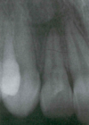
Dens Invaginatus (dens-in-dente)
(Frequency: Maxillary Permanent lateral incisors > central incisors > premolars > canines > molars)
Pilar Cyst
- Occur on the scalp and derived from hair follicle
- F>M
- Tx = conservative surgical excision
Impetigo
(Caused by GAS and Staph aureus, bullous and nonbullous forms, increased prevalence in HIV and immunocompromised)
The nonbullous form is more common, school aged children, contagious, amber colored crusts. The bullous form is more in infants, can lead to meningitis or pneumonia, the bullae can rupture and develop a thin brown crust “lacquer.”
Lupus Vulgaris
This is Tuberculosis of the skin. Tuberculosis is caused by Mycobacterium tuberculosis, is acid fast, hits immunocompromised, spreads airborne.
With TB, you can get painless, chronic ulcerations on tongue and palate, which is similar to syphilis, but oral cancer is usually painful. The histology shows granulomas with caseating granulomatous inflammation, which is a granuloma with a central area of necrosis, and multinucleated giant cells (macrophages).
Buccal Exostoses
What are some nonsyndromic environmentla factors for clefts?
Maternal alcohol
Folic Acid deficiency
Corticosteroid use
Anticonvulsant therapy
Hypoplastic Amelogenesis Imperfecta
Osteomyelitis
(This is chronic osteomyelitis. Usually due to bacterial infection. Result in: Expanding lytic destruction, Suppuration, Sequestra formation (The pooling of inflamed/broken bone). Uncommon in developed countries. Most cases arise after odontogenic infections or traumatic facture. 75% in Males, mostly mandible
Symptoms of acute infection:
Fever
Lymphadenopathy
Sensitivity
Soft tissue swelling
Acute infection doesn’t produce XRAY changes
During acute infection, fragments of necrotic bone may become surrounded by new, vital bone = involucrum
Coronoid Hyperplasia
(The mandible deviates toward the affected side on opening)
Syphilis
- Caused by Treponema pallidum
- Primarily through sexual contact and from mother to fetus
- 6x more common in men and African Americans
- Primary syphilis - characterized by the chancre that develops at the site of inoculation (3-90 days after inoculation) - oral lesions are only 2%, more common in anus and genitalia.
- Secondary Syphilis
- AKA disseminated syphilis
4-10 weeks after initial infection
Systemic symptoms:
Painless lymphadenopathy
ALWAYS: diffuse, painless, maculopapular, cutaneous, widespread rash - Tertiary Syphilis
- After 2nd stage, patients enter latent syphilis and are free of lesions/symptoms for 1-30 years
30% of pts progress to tertiary syphilis after latent syphilis
Possible cardiac complications
Aneurysm of the ascending aorta
Congestive heart failure
CNS involvement
Paralysis
Psychosis
Death
Ocular lesions
Tongue lesions (syphilitic glossitis)
Gumma – characteristic; scattered foci of granulomatous inflammation
Intraoral lesions usually affect the palate or tongue
- Congenital Syphilis - Hutchinson’s Triad + Mulberry Molars
Localized juvenile spongiotic gingival hyperplasia (The Nickname is Wright’s Lesion (his professor))
“Localized Juvenile Spongiotic Gingivitis”
Idiopathic, but it has been suggested that the alteration represents an isolated patch of exteriorized junctional or sulcular epithelium that may be altered secondarily by local factors (mouth breathing)
The most common presentation is a small bright red velvety or papillary alteration that often bleeds easily upon manipulation
Predilection for the maxillary anterior facial gingiva with a female predominance.
Vast majority occur under 20 with a median age of 12 years.
How does Hypocalcified Amelogenesis Imperfecta happen?
Matrix is laid down appropriately but no mineralization occurs.
Dentin Dysplasia Type I
(Rootless teeth, mobile clinically)
Dentinogenesis Imperfecta
Sinusitis
- Caused by blockage of the ostiomeatal complex.
- Acute can become chronic
- Calcification can occur in chronic = antrolith
- Maxillary sinusitis:
- Increased pain when head is upright
- Decreased pain when patient is supine
- *A sinus infection should be strongly considered when patients complain of pain form several teeth. (Due to seasonal allergies, because the root tips sit next to the sinuses and can cause referred pain)
- Radiographic imaging helps diagnose chronic disease
- Sinus is cloudy with increased density
- Treatment of acute sinusitis:
- Self-limiting
- Treatment of chronic sinusitis:
- Antibiotics
- If unresponsive, surgical correction to enlarge the ostial openings
- Surgery should try to be avoided in children
- Decreased distance between orbit and brain
Wha tis it called when a tooth fails to fully erupt?
Ankylosis
Paracoccidiodomycosis
This is really South American Blastomycosis
- Paracoccidioides brasiliensis
- Nine-banded armadillo harbors the organism (similar to the situation seen with leprosy)
- Thought to be attributable to a protective effect of female hormones (because β-estradiol inhibits the transformation of the hyphal form of the organism to the pathogenic yeast form.
- Most patients infected are employed in agriculture
- Generally self-limiting
- Histology: granulomatous inflammation (characterized by collections of macrophages and multinucleated giant cells.
- The organism often show multiple daughter buds on the parent cell, resulting in an appearance described as resembling “mickey mouse” or the spokes of a ship’s steering wheel (“mariner’s wheel”) (This is on boards a lot)
Periapical Cyst
(Inflammatory stimulation of epithelium in the area (Rests of Malassez), Can be found on side of the root – Lateral apical periodontal cyst. Cyst remains following extraction of the tooth – Residual apical periodontal cyst. The histology has white areas with epithelium around it)
What forms the medial and lateral nasal processes?
The proliferation of ectomesenchyme on both sides of each placode.
Drug-Related Gingival Overgrowth
Abnormal growth of gingival tissues secondary to use of systemic medication:
Cyclosporine (transplant therapy) ~25%
Phenytoin (anticonvulsant) ~50% (meaning half of patients with Phenytoin get this)
Nifedipine (calcium channel blocker) ~25
Cat-Scratch Disease
- Caused by Bartonella henselae
- Begins in the skin; classically spreads to adjacent lymph nodes
- Most common cause of chronic regional lymphadenopathy in children
- Arises after contact with a domestic cat or kitten
- Scratches
- Licks
- Bites
- 80% occur in patients younger than 21
- Primary lesions adjacent to eye can result in a conjunctival granuloma with preauricular lymphadenopathy = oculoglandular syndrome of Parinaud
- Diagnosed via serologic tests
- Self-limiting condition; palliative treatment only unless severely involved, then ABX are appropriate
- Resolves in 4 months
When do crowns of permanent dentition occur?
6 months to 15 years
Zygomycosis
- Aka Mucormycosis
- Found on decaying organic material
- Most important to oral health care provider = rhinocerebral form
- Growth is enhanced by iron
- Patients at risk:
- Diabetic patients who are uncontrolled (Ketoacidosis inhibits iron binding to transferrin, allowing serum iron levels to rise)
- Patients taking deferoxamine (iron-chelating agent)
- Immunocompromised patients
- Presenting symptoms of rhinocerebral zygomycosis are varied:
- Nasal obstruction
- Bloody nasal discharge
- Facial pain/HA
- Facial swelling/cellulitis
- Visual disturbances/proptosis
- If untreated, massive tissue destruction may result
- Appears black and necrotic
- Treatment
- Radical surgical debridement
- High doses of amphotericin B
- 60% of patients with rhinocerebral zygomycosis succumb to their disease
Acute Atrophic Candidiasis
- “Antibiotic sore mouth” – follows a course of broad-spectrum ABX
- Mouth feels as though a hot liquid scalded it
- Diffuse loss of filiform papillae of dorsal tongue (appears bald).
- Similar appearance symptomology is noted in xerostomia patients
Concrescence
(Union of two adjacent teeth by cementum alone without confluence of underlying dentin.)
Intraoral Sinus Tract
(Also known as “Parulis” or “gum boil”)
What causes the median cleft of the upper lip?
Failure of fusion of the medial nasal processes, which are both involved with formation of the primary palate.
Abrasion
(Wearing away of tooth structure, most commonly with tooth brushing. Demastication: exhibits features of both attrition and abrasion. (chewing tobacco between opposing teeth))
Lingual Thyroid
(Thyroid gland begins as an epithelial proliferation in the floor of the pharyngeal gut during week 3-4 of embryogenesis, If primitive gland doesn’t descend normally, ectopic thyroid can be found between the foramen cecum and the epiglottis, Dx is established via thyroid scan (using iodine isotopes or technetium-99m), CT, and/or MRI, biopsy is avoided due to hemorrhage)
Histoplasmosis
- Most common systemic fungal infection in US
- Caused by Histoplasma capsulatum
- Dimorphic
- Yeast at body temperature in human host
- Mold in natural environment
- Found in humid areas with soil enriched by bird or bat excrement
- Endemic in the Ohio and Mississippi River Valley
- Up to 90% of people in these areas are infected
- Airborne spores are inhaled, passed into the terminal passages of the lungs, and germinate
- 99% of those infected have either no symptoms or such mild symptoms that the patient doesn’t seek medical care
- Mild symptoms are flulike and can last for 1-2w
- Most who become exposed are healthy and inhale a small number of sporesP
- Patients:
- Older
- Debilitated
- Immunocompromised
- Up to 10% of AIDS patients develop disseminated disease in endemic areas
- Most oral lesions occur with disseminated form
- Appears as a solitary, variably painful ulceration of several weeks duration
- Ulcerated lesions have firm, rolled margins
- Clinically indistinguishable from malignancy (When it has rolled margins)
- May appear erythematous or white with an irregular surface
- Histopathology shows granulomatous inflammation
- Treatment - Acute
- None or palliative care
- Treatment - Chronic
- Require treatment despite the fact that 50% recover spontaneously
- Pulmonary damage is progressive if untreated
- 20% succumb to untreated disease
- Treatment of choice is IV amphotericin B, but can cause kidney damage
- Treatment – disseminated
- 90% will succumb to disease if untreated
- Amphotericin B is treatment of choice
Apert Syndrome
(Downward slant lateral palpebral fissures-so the outside of the eye slants down. A in Aspert for Angled.
Syndactyly of 2nd, 3rd, and 4th digits of hands and feet (webbing of the hands and feet)
Mental retardation)
Hyperdontia
(Mesiodens is an example. You have too many teeth.
Taurodontism
(An enlargement of the body and pulp chamber of a multi-rooted tooth, with apical displacement of the pulpal floor. Can be isolated or syndromic. The 3 syndromes associated with this are 1)Kleinfelter’s Syndrome, 2)Amelogenesis Imperfecta, and 3)Tricho-dento-osseus syndrome)
What are the three main drugs used to treat Candidiasis?
- Nystatin (antifungal)
- Oral use as suspension or lozenge
- Bitter taste, so sugar is added
- Don’t use in xerostomia-related candidiasis
- Effectiveness depends on direct contact so multiple daily doses are necessary
- Amphotericin B
- IV treatment for life-threatening systemic fungal infections
- Imidazole Agents – Clotrimazole
- One brand name is Mycelex
- Formulated as a troche (lozenge)
- Effectiveness depends on direct contact so multiple daily doses are necessary
- Very few side effects
- Candidiasis should resolve easily following antifungal therapy
- If infection recurs after treatment, investigate possible predisposing factors
Gemination
(This is an example of bilateral gemination. Single enlarged tooth or joined tooth in which the tooth count is normal when the anomalous tooth is counted as one)
Condensing Osteitis
Localized area of bone sclerosis associated with apices of teeth with pulpitis (The bone tries to get hard and form bone and become more radiopaque to get rid of the infection and protect itself). Association with inflammation is critical in diagnosis (radiographically identical to idiopathic osteosclerosis).
XRAY changes:
Increased RO adjacent to tooth apex that has a thickened PDL or apical inflammatory lesion
No RL border (distinguishes from focal cemento-osseous dysplasia)
Not separated from apex (distinguishes from idiopathic osteosclerosis)
No clinical expansion of bone
85% regress after odontogenic infection is eliminated
Residual areas are termed bone scars