Equine Appendicular Skeleton Flashcards
Name the views you would obtain on an equine radiograph.
- Lateromedial
- Dorsopalmar/plantar
- DMPLO
- DLPMO
When you take a DMPLO radiograph which side would you see?
- Dorsolateral
- Palmar/plantaromedial
When you take a DLPMO radiograph which sides would you see?
- Dorsomedial
- Palmar/plantarolateral
Where would you place a marker on the lateromedial radiograph?
A. Medial
B. Lateral
C. Dorsal
D. Palmar/Plantar
C. Dorsal
Where would you place a marker on the dorsoplantar, DMPL, DLPM radiograph?
A. Medial
B. Lateral
C. Dorsal
D. Palmar/Plantar
B. Lateral
Which joint is the most commonly affected in horses with hindlimb lameness?
Tarsus joint
Which view is depicted here?

DLPMO

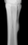
Which view is depicted here?

DMPLO (prominent indentation of the lateral trochlear ridge)

In the horse, where is the most common place to see OCD?
tarsocrural joint (intermediate ridge of the distal tibia DIRT)
What is the best view to see OCD lesions?
DMPLO
Which disease is indicated here and in what view?

OCD and DMPLO
This disease is the most common cause of lamenes and is a degenerative joint disease of the tarsus. Periarticular osteophyte formation is the most common finding.
Bone Spavin
What are the most common joints involved with bone spavin?
Distal intertarsal and tarsometatarsal joints
What disease is indicated here?

Bone Spavin
Advanced disease may have subchondral bone lysis and narrowing of the joint space (can progress to sclerosis and ankylosis)
Is this the equine tarsus or carpus?

Carpus
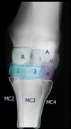
Which part of the carpus is indicated by the light blue star?
A. Radial carpal bone
B. Intermediate carpal bone
C. Ular carpal bone
D. Third carpal bone

B. Intermediate carpal bone

Which part of the carpus is indicated by the pink star?
A. Radial carpal bone
B. Intermediate carpal bone
C. Ular carpal bone
D. Third carpal bone

A. radial carpal bone

Which part of the carpus is indicated by the green star?
A. Radial carpal bone
B. Intermediate carpal bone
C. Ular carpal bone
D. Acessory carpal bone

D. Accesory carpal bone

Which part of the carpus is indicarted by the orange star?
A. Radial carpal bone
B. Intermediate carpal bone
C. Ulnar carpal bone
D. Accesory carpal bone

C. Ulnar carpal bone

Which part of the carpus is indicarted by the dark blue rectangle?
A. 2nd carpal bone
B. 3rd carpal bone
C. 4th carpal bone
D. Metacarpal bone 3

B. 3rd carpal bone

Which part of the carpus is indicated by the purple rectangle?
A. 2nd carpal bone
B. 3rd carpal bone
C. 4th carpal bone
D. Metacarpal bone 3

C. 4th carpal bone

Which part of the carpus is indicated by the light green rectangle?

A. 2nd carpal bone
B. 3rd carpal bone
C. 4th carpal bone
D. Metacarpal bone 3
A. 2nd carpal bone
Which part is indicated by the green circle?
A. MC2
B. MC3
C. MC4
D. MC5

A. MC2

Which part is indicated by the purple circle?
A. MC2
B. MC3
C. MC4
D. MC5

C. MC4