Epithelium Flashcards

1
Q

What does all epithelium rest on?

A

basement membrane

2
Q

The epithelium forms the ______ of exocrine and endocrine glands.

A

parenchyma (secretory portion)

3
Q

Characterisitcs of Epithelium:

A
  • Lines all surfaces (external and internal)
  • Structural Polarity
  • Avascular (Nutrients delivered by diffusion)
  • Highly cellular
  • Little intercellular space and closely apposed to one another by cell junctions.
  • High regenerative capacity
4
Q

6 functions of the epithelium:

A

PASSRT

  1. protection
  2. absorption, secretion, excretion
  3. selective permeability barrier
  4. sensory reception
  5. reproduction
  6. transport of material via cilia
5
Q

Simple epithelium:

A

one cell layer thick

  • can be:
    • simple squamous (flat)
    • simple cuboidal
    • simple columnar
6
Q

Stratified Epithelium:

A

two or more cell layers thick

  • can be:
    • stratified squamous (flat)
    • stratified cuboidal
    • stratified columnar
7
Q

Transitional Epithelium:

A
  • Stratified in appearance; however, surface cells can be dome shaped or flat.

Nuclei are rounded in appearance.

8
Q

Pseudostratified Columnar Epithelium:

A
  • All cells rest on the basement membrane but not all cells reach the luminal surface.

Cell surface modifications are ALWAYS present: cilia, steriocilia.

9
Q

Basement Membrane/Basal lamina

A
  • Supports the tissue and forms the boundary between epithelium and connective tissue
10
Q

What are the arrow pointing to?

A
  • blue = cell
  • purple = connective tissue
  • black = basement membrane
11
Q

All blood vessels are lined with:

A

simple squamous epithelium

12
Q

Functions of simple squamous epithelium:

A
  • fluid transport
  • gaseous exchange
  • lubrication
  • reducing friction (viscera)
13
Q

Some notable locations of simple squamous epithelium:

A
  • loop of henle
  • parietal layer of Bowman’s capsule
  • pulmonary alveoli
  • lining of ventricles and atria
14
Q

What is the arrow pointing to?

A

simple squamous epithelium of the spleen

(mesothelium)

15
Q

Distinguishing characteristic of simple squamous epithelium:

A
  • one cell layer thick
  • flat cells
  • flat nuclei
16
Q

What is the black arrow on the right pointing to?

A

simple squamous epithelium

bowman’s capsule renal corpuscle

17
Q

Distinguishing characteristics of simple cuboidal epithelium:

A
  • one cell layer thick
  • cube-shaped cells
  • nuclei round and usually in center of cell
18
Q

Functions of simple cuboidal epithelium:

A
  • absorption
  • secretion
  • protection
19
Q

Notable locations of simple cuboidal epithelium:

A
  • lining of ducts in glands
  • covering of ovaries
  • kidney tubules
20
Q

What is the arrow pointing to?

A

simple cuboidal epithelium

(lining of kidney collecting duct)

21
Q

Distinguishing characteristics of simple columnar epithelium:

A
  • one cell layer
  • rectangular shaped cell
  • round to oval nuclei

MAY have surface modifications

22
Q

Functions of simple columnar epithelium:

A
  • absorption
  • secretion
  • protection
  • transportation
23
Q

Notable locations of simple columnar epithelium:

A
  • nasal sinuses
  • oviducts
  • uterus
  • digestive tract
  • gall bladder
24
Q

What cells are these?

A

simple columnar epithelium

25
Q

What is the black arrow on the right pointing to?

A

junctional complex

26
Q

What is the black arrow in the center pointing to?

A

simple columnar epithelium cell

microvilli brush border present

goblet cells present

27
Q

What is the black arrow pointing to?

A

goblet cell

28
Q

What kind of cell is this EM of?

A

goblet cell

29
Q

What is the red arrow pointing to?

A

Simple Columnar epithelium with cilia

(oviduct)

30
Q

What is this a cross-section of?

A

cilia

(cilia made of microtubules; protein = tubulin)

31
Q

Stratified epithelium is identified by what cell layer?

A

the cell layer closest to the lumen

32
Q

Distinguishing characteristics of stratified epithelium:

A
  • two or more cell layers thick
  • NEVER cilia or stereocilia on cell surface
33
Q

Function of stratified epithelium:

A
  • absorption
  • secretion
  • protection
34
Q

Distinguishing characteristics of stratified squamous epithelium:

A
  • Cell layer closest to lumen is always nucleated
  • Nuclei are flattened in appearance
35
Q

Functions of stratified squamous epithelium:

A
  • protection
  • prevents dehydration
36
Q

Notable locations of stratified squamous epithelium:

A
  • vagina
  • anal canal
  • mouth
  • vocal cords
  • epidermis (keratinized)
37
Q

What cells are the top cell layer?

A

stratified squamous epithelium

(palentine tonsil)

38
Q

What is the hot pink layer on top in this image?

A

Keratinized Stratified Squamous Epithelium

(Cell layer closest to lumen lack nuclei)

39
Q

Distinguishing features of stratified cuboidal epithelium:

A
  • 2 cell layers thick
  • cells are cuboidal
40
Q

Functions of stratified cuboidal epithelium:

A
  • absorption
  • secretion
  • protection
41
Q

Notable locations of stratified cuboidal epithelium:

A

lines ducts of sweat glands

42
Q

What kind of cell is circled?

A

stratified cuboidal epithelium

(layer closest to lumen is cuboidal)

43
Q

Distinguishing features of stratified columnar epithelium:

A
  • 2 cell layers thick
  • cells are columnar
44
Q

Functions of stratified columnar epithelium:

A
  • secretion
  • protection
45
Q

Notable locations of stratified columnar epithelium:

A
  • mammary glands
  • salivary glands
  • male urethra
  • conjunctiva of eye
46
Q

What cell is boxed in blue?

A

stratified columnar epithelium

47
Q

What kind of cells are outlined in red?

A

stratified columnar epithelium

with goblet cells

48
Q

Distinguishing features of pseudostratified epithelium:

A
  • All cells touch basement membrane
  • Not all cells touch the luminal surface
  • Nuclei in all different planes of section
  • Apical area devoid of nuclei
  • Always has a surface modification: Cilia or Stereocilia
49
Q

Pseudostratified epithelium always has:

A

cilia or sterocilia

50
Q

Functions of pseudostratifed epithelium:

A
  • Secretion
  • Absorption
  • Lubrication
  • Protection
  • Transportation via cilia
51
Q

What cells are pictured here?

A

pseudostratified epithelium with cilia

with goblet cells

52
Q

What cells are pictured here?

A

pseudostratified epithelium with sterocilia

53
Q

What cell surface modification is pictured?

A

microvilli

(brush border)

54
Q

What cell surface modification is pictured?

A

cilia

(airy; made of microtubules)

55
Q

What cell surface modification is pictured?

A

stereocilia

56
Q

Distinguishing features of transitional epithelium:

A
  • Stratified
  • Top layer of cells is dome shaped
  • Top layer of cells can be flat
  • Nuclei are rounded whether top layer of cells are domed or flat
  • Occasional binucleated cells
57
Q

Location of transitional epithelium:

A

lines urinary tract

58
Q

What epithelium is pictured here?

A

transitional epithelium

(empty bladder)

59
Q

What type of epithelium is pictured here?

A

transitional epithelium (nuclei still rounded)

(full bladder)

60
Q

What are the black arrows pointing to?

A
  • unit membrane in the bladder
    • invaginations of the plasma membrane in the bladder that allow the bladder to expand when/if it becomes full
61
Q

What type of epithelium is pictured?

A

stratified squamous epithelium

62
Q

What type of epithelium is pictured?

A

transitional epithelium

63
Q

What are the arrows/circle outlining?

A

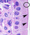
junctional complex/terminal bar

64
Q

Function of Zonula Occludens (Tight Junction):

A
  • Maintains selective passage of substances from one side of the epithelium to the other.
65
Q

What is the red arrow pointing to?

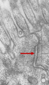
A

zona occludens (tight junction)

66
Q

What are the red arrows pointing to?

A

zona occludens (tight junction)

67
Q

Function of zonula adherens (belt desmosome):

A

Belt-like attachment that anchors cell to cell

68
Q

What is the red arrow pointing to?

A

zonula adherens (belt desmosome)

69
Q

Key molecules in the zonula occludens:

A

Occludin and claudins

(tight junction)

70
Q

Key molecules in the zonula adherens:

A

actin, cadherins, and catenins

(belt desmosome)

71
Q

Key molecules in macula adherens:

A

intermediate filaments, desmoplakin, desmoglein

72
Q

What are the black arrows pointing to?

A

macula adherens

(spot desmosomes)

ladder-like appearance

73
Q

What is this an image of?

A

zonula adherins

(belt desmosome)

74
Q

What is this an image of?

A

zonula occludens

(tight junction)

75
Q

What is this an image of?

A

macula adherens

(spot desmosome)

76
Q

What is this an image of?

A

hemidesmosome

77
Q

Hemidesmosome function:

A

cell to matrix attachment

78
Q

What is this an image of?

A

gap junction between two muscle cells

(connexins)

79
Q

What kind of glands are pictured?

A

exocrine glands

(secrete products into ducts to the surface)

80
Q

What kind of glands are pictured?

A

endocrine glands

(secretes products directly into blood stream via capillaries and lymphatic vessels)