EOR Flashcards
LICHEN PLANUS

Pityriasis rosea

Drug Eruptions
- Erythema Multiform
- Etiology
- Presentation
- Treatment/Prognosis
- SJS/TEN
- Differences
- Treatment
Erythema Multiform:
- Etiology: Herpes Simplex Virus
- Similarities in clinical and histopathologic findings have led to controversy over the distinction between EM and Stevens-Johnson syndrome (SJS), an often drug-induced disorder that may present with cutaneous, targetoid lesions and mucosal erosions. However, there is suggestive evidence that EM with mucous membrane involvement and SJS are different diseases with distinct causes [1]. The term erythema multiforme major should not be used to refer to SJS
SJS/TEN
- According to a widely accepted classification, SJS and TEN are considered a disease continuum and are distinguished chiefly by severity, based upon the percentage of body surface affected by blisters and erosions (table 1) [2,3]:
●SJS is the less severe condition, in which skin detachment is <10 percent of the body surface (picture 1A-C).
●TEN involves detachment of >30 percent of the body surface area (BSA) (picture 2A-D).
●SJS/TEN overlap describes patients with skin detachment of 10 to 30 percent of BSA.
We will use the term “SJS/TEN” to refer collectively to SJS, TEN, and SJS/TEN overlap syndrome.

MEASLES (Rubeola)

IMMUNOGLOBULIN A VASCULITIS

KAWASAKI DISEASE

Laryngeotracheitis
- Etiology: Pathogen?
- Dx:
- Tx

Necrotising Entericolitis


Candidal Rash
Staphylococcal scalded skin syndrome (SSSS)

Patellofemoral Pain syndrome

MARFAN SYNDROME

Legg-Calvé-Perthes disease

Intestinal Malrotation with MIDGUT VOLVULOUS

HAND, FOOT, MOUTH
What is the pathogen?

neurofibromatosis type 1
The boy has both café-au-lait macules and cognitive disability, raising concern for neurofibromatosis type 1, or von Recklinghausen’s disease. Neurofibromatosis is a neurocutaneous disorder caused by a mutation on NF1 on chromosome 17. Cutaneous findings of neurofibromatosis type 1 include café-au-lait macules, axillary and inguinal freckling, Lisch nodules, and neurofibromas. Neurofibromas are peripheral nerve sheath tumors that may be focal and cutaneous or plexiform. Cutaneous neurofibromas are small, sessile or pedunculated lesions that move with the skin. They are non-tender and occur primarily on the trunk. Cutaneous neurofibromas most commonly develop during adolescence, although subtle lesions may be noted earlier in angled light. The number of neurofibromas on an affected individual may range from a few to thousands. Plexiform neurofibromas are present in half of patients with neurofibromatosis and cause a very different clinical picture. They may be superficial, deep, or both. The entangled nerves can become large, complex, and often disfiguring due to associated overgrowth of surrounding tissues. Deep plexiform neurofibromas may compress internal structures, including the airway and spinal cord. Individuals with neurofibromatosis are also at increased risk of bony abnormalities, including bony dysplasia, pseudoarthroses, scoliosis, osteoporosis, and short stature. Additionally, affected individuals are predisposed to a variety of tumors, including optic nerve gliomas and soft tissue sarcomas such as rhabdomyosarcoma, malignant peripheral nerve sheath tumors, and glomus tumors.

Most common pathogens causing Osteomyelitis?
- MC
- Neonates
- Sickle Cell

Meckel Diverticulum

Neonatal cephalic pustulosis

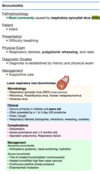
Pertussis- Whooping Cough
- Pathogen
- Presentation
- Treatmnet

Diaphragmatic Hernia

Gastroschisis vs omphalocele
Dx: Clinical
Tx: Silo

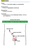
Extrophy of the Bladder

Biliary Atresia
Dx:
- US = No Ducts
- Hitascan after 7 days of phenobarbitol