ENT Flashcards
What are the 3 main chambers of the eye?
- Vitreous chamber
- Anterior chamber
- Cornea to iris
- Filled with aqueous humor
- Aqueous humor drains from anterior chamber thru Schlemm’s canal into venous system
- Posterior chamber
- Iris to lens and ciliary process

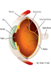
What are the 3 layers of the eye globe?
- Sclera (white part)- outermost layer
- Tough, fibrous
- Cornea- visual fx
- Tissue where cornea meets sclera is limbis
- Curvature of cornea is where we have visual power
- Uveal tract- middle
- Choroid- layer of blood vessels- bleeding of choroid can cause expulsive hemorrhage
- Iris- controls light entry (changes size of pupil)
- SNS stim → dilate pupil (iris muscle contracts)
- PSNS stim → myosis (constrict) → pupil constrict (iris sphincter muscle contract)
- Ciliary body- produces aqueous humor
- Retina (innermost layer) – nerve tissue cont. w/ optic nerve . no capillaries. Completely dependent on choroid layer to provide o2 and nourishment to retina

What is the vitreous humor?
Conjunctiva?
Cornea?
Aqueos humor?
Schlemm’s canal?
What is the pupil formed by?
What is a lens?
- Vitreous humor → center of the eye
- Attachment to BV and optic nerve
- Traction to gel → retinal detachment
- Conjunctiva is clear membrane that covers the sclera in the front of the eye.
- Cornea (part of the sclera) is a strong clear bulge located at the front of the eye.
- Aqueous humor is a watery substance located in the anterior chamber of the eye.
-
Schlemm’s canal:
- channel in the eye that collects aqueous humor from the anterior chamber and delivers it into the bloodstream (drainage system)
- Pupil is formed by the iris
- Lens of the eye is a flexible unit that consists of layers of tissue enclosed in a tough capsule
What are the 6 extraocular muscles of the eye?
6 muscles –
- superior rectus
- inferior rectus
- medial rectus
- inferior oblique muscle
- (1-4 innervated by oculomotor nerve- CN III)
- lateral rectus muscle
- innervated by abducens nerve- CNVI.
- superior oblique muscle
- innervated by trochlear nerve- CN IV.
Sit in cone behind eye
All surround optic nerve, artery, vein, ciliary ganglion

Describe the motor innervation of the eye?
-
Cranial Nerve III- Oculomotor
- innervates the superior rectus, medial rectus, inferior rectus, and inferior oblique eye muscles
- Eye movements
- Pupil constriction
- Opens eyelid
- PSNS fibers
- innervates the superior rectus, medial rectus, inferior rectus, and inferior oblique eye muscles
-
Cranial Nerve IV- Trochlear
- innervates the superior oblique muscle
- Moves eye down and outward
- innervates the superior oblique muscle
-
Cranial Nerve VI- Abducens
- innervates the lateral rectus muscle
- Moves eye lateral
- innervates the lateral rectus muscle
Sympathetic fibers from carotid plexus travel through ciliary ganglion → innervate dilator muscles
- Ex: block ciliary ganglion → fixed/dilated pupil
PSNS- CN III
Describe the sensory innervation for the eye.
-
TRIGEMINAL nerve (CN V) – touch/pain
-
Ophthalmic branch (VI): 1st branch
- Innervates the upper eyelid, conjunctiva, and cornea
- Nosociliary branch (of opthalmic nerve)
- sensory to medial canthus, lacrimal sac, and ciliary ganglion
- Ciliary ganglion → provides sensory to cornea, iris, and ciliary body
- sensory to medial canthus, lacrimal sac, and ciliary ganglion
- Also regulates OCR (oculocardiac reflex)
- Nosociliary branch (of opthalmic nerve)
- Innervates the upper eyelid, conjunctiva, and cornea
-
Maxillary branch (V2): 2nd branch of 5
- Innervates lower lids
-
Ophthalmic branch (VI): 1st branch
-
Facial nerve: VII- Exits skull in stylomastoid foramen
- Supplies motor innervation to orbicularis muscle via zygomatic branch
- Ex: block CN VII → cant squeeze lid
- Innervates the lower lid
- Ex: block CN VII → cant squeeze lid
- Supplies motor innervation to orbicularis muscle via zygomatic branch
What is the function of the optic nerve?
- Function:
- Vision
- Carries neural signals from retina
- Part of the optic chiasm (fibrous sheeth)- contains…
- nerve
- artery
- vein
- sympathetic nerves
- Consequences to INJURY → total blindness
What is normal intraocular pressure?
- Normal: ~ 16 mmHg (+/- 5)
-
Abnormal: > 25 mmHg
- Must maintain normal IOP to maintain normal curvature of cornea
- IO perfusion pressure related to CPP
- (MAP – IOP → how eye regulates perfusion)
- Ex: High IOP → impairs BF to optic nerve (fx effected)
How is aqueous humor produced?
- Posterior chamber:
- 2/3 produced by ciliary body (~80-90%)
- → then actively moved from posterior chamber to anterior chamber by an active sodium pump mechanism.
- Active Na pump (AKA → Na-K ATPase carbonic anahydrase enzyme)
- Passive filtration: 1/3 (~20%)
- comes from passive filtration through vessels in iris. across ciliary epithelium
- Aqueous fluid is produced at a rate of 2 uL/min.
How is aqueous humor eliminated?
- Fluid drains out of eye through trabecular meshwork (spongy tissue) → - into canal of Schlemm’s and the episcleral venous system (in anterior chamber) → eventually ending up at SVC and RA.
- Drainage system
- Trabecular meshwork → eventually go back into central circulation
- Anything affecting flow → cause increase IOP
- Drainage system
What can happen if elimination of aqueous humor is impaired?
- Open angle glaucoma (OAG): Sclerosis of trabecular meshwork
- chronic elevation
- Closed angle glaucoma (CAG): Obstruction of Aqueous drainage from closure of anterior chamber angle
- CAG Causes:
- Iris swelling
- Anterior displacement
- CAG Causes:
- Pts already w/ narrow angle predisposed to acute increase IOP (PAIN & emergency)
What determines intraocular pressure?
- A measurement of the fluid pressure inside eye
- The globe is a relatively noncompliant compartment and the volume of the internal structures is fixed, except for aqueous fluid and choroidal blood volume.
- The quantity of these two factors regulates intraocular pressure.
- The globe is a relatively noncompliant compartment and the volume of the internal structures is fixed, except for aqueous fluid and choroidal blood volume.
-
Determined by:
- Production of aqueous humor
- Drainage of aqueous humor
- Changes in choroidal blood volume or pressure
- Extraocular muscle tone
- Normal IOP: 10-20 mmHg (~ 16 and +/- 5)
- >25 mmHg, you have problem
What increases IOP?
- Drugs:
- Ketamine (?) → theoretically causes issues but does not directly
- Succinylcholine (increases by 8-10 mmHg)
- d/t
- decrease in Aqueous humor outflow
- increase in choroidal BV
- increase in CVP
- d/t
- Other:
- MOST SIG INCREASE → Laryngoscopy & Emergence
- Sympathetic Blunting
- Less manipulation of AW the better (LMA less increase than DL)
- Position changes
- Coughing, valsalva maneuver, straining, vomiting, HTN, injection of local anesthesia, laryngoscopy, hypercarbia/hypoventilation/ hypoxia, lid pressure, eye compression, forceful lid squeeze (increases to 70 mmHg), pupil mydriasis (dilation)
- Increases up to 30-40 mmHg
- MOST SIG INCREASE → Laryngoscopy & Emergence
What can decrease IOP?
- Drugs:
- Most anesthetic drugs:
- VA (excluding N2O → no effect)
- Propofol
- Etomidate
- Opioids
- NDMR
- hypertonic solutions
- Dextran
- Mannitol
- Most anesthetic drugs:
- Other: Hypotension, hypothermia, hyperventilation, pupil miosis (constriction)
What are some anesthetics that have no effect on IOP?
- N2O
- Versed
- ~NDNMB
What is acetazolamide?
MOA, S/E?
Carbonic anhydrase inhibitor (topical for eye)- brand Diamox
- Treatment for glaucoma
- Induces:
- decreases IOP*
- diuresis
- reduces aqueous humor production
-
K+ depletion (+/-)
- want preop labs
- SE:
- Confusion
- Drowsiness
- Low K
- Low Na
- Acidosis
- Polyuria
What is echothiophate?
- Tx for glaucoma
- Irreversible cholinesterase inhibitor
- Produces:
- miosis
- Produces:
- Systemic absorption may cause total body inhibition of plasma cholinesterase
-
CAUTION:
- Prolong effects of:
- Succinylcholine
- Mivacuronium
- Toxicity w/ Ester LAs
- Prolong effects of:
-
Very long DOA:
- Need to stop 4-6 weeks preoperatively
-
CAUTION:
Why is it important to know eye drops pt is on?
**Nasal mucosa/conjunctival capillaries connected to eyes → causing systemic absorption
-
Systemic effects worse in: (greater toxicity risk)
- Children
- Elderly
- Preop: Know what drops pt on
Phenylephrine eye drops? s/e?
- Produces mydriasis
- Associated w/:
- severe HTN
- Arrythmias
- Adverse CV events
- Very high [] (1 drop = 5 mg)
What are clycloplegics?
Atropine and Cyclopentolate
- Pupil mydriasis (dilation)
- Systemically absorbed
- SE: See anticholinergic symptoms
- Dry mouth
- Dry skin
- Fever
- Agitation
- disorientation, psychotic reactions
- Central anticholinergic symptoms
- SE: See anticholinergic symptoms
Effect of acetylcholine in eye drops?
- Produces miosis
- Cholinergic agonist
- Causes:
- Bradycardia
- acute bronchospasm
- hypotension
What is timolol?
- Tx of glaucoma
- B1/2 antagonist
- Produces:
- Miosis
- reduction of aqueous humor production
- SE:
- Bradycardia
- Bronchospasm
- CHF exacerbation
- heart block
- Issues w/ nursing infants
NSAID use in eye drops?
- Ketorolac and Diclofenac
- Used for inflammation
Scopalamine effect on eyes?
- Mydriasis (dilation)
- Can causes central anticholinergic syndrome