ENT Flashcards
Which nerve causes referred pain to the ear in sore throats/cold etc?
Glossopharyngeal nerve
Which of the three ossicles is in contact with the tympanic membrane?
Malleus
Which structure would be affected if the patient had an infection of the middle ear?
Cochlea
Which nerve does the shoulder shrug?
Accessory Nerve
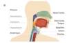
Name these structures
What is a trismus?
Trismus is a motor disturbance of the trigeminal nerve.
In particular there is spasm of the masticatory muscles, with difficulty in opening the mouth - risus sardonicus or ‘lockjaw’ to the layperson
What are the different types of otitis externa?
Acute suppurative
Chronic suppurative
Serous / secretory
What are the causes of otitis media (acute supporative)?
- Common cold
- Acute tonsillitis
- Influenza
- Coryza - profuse discharge of the nose - of measles, scarlet fever, whooping cough.
What are the organisms for otitis media (acute supporative)?
URTI bacteria
Bacterial causes include:
- Strep pneumonia - 30%
- H. influenzae - 20%
- Moraxella catarrhalis - 20%
- Group A Strep and Staph aureus - 5%
Viral causes (less than 10%)
What are the symptoms of otitis media (acute supporative)?
Symptoms
- Earache, usually throbbing and severe
- Pyrexia up to 40 degrees - child may be flushed
- Otorrhoea will often be blood-stained - profuse and mucoid at first, later becomes thick and yellow
- Mucoid discharge: signifies tympanic membrane perforation, after which pain subsides
Signs
- Conductive deafness is always present, and may be accompanied by tinnitus
- Tympanic membrane signs depend on the stage of infection
- Loss of lustre
- Break-up of the light reflex
- Redness
- Impaired mobility of tympanic membrane
What is the diagnosis of otitis media (acute supporative)?
- Acoustic reflectometry
- Pneumatic otoscopy
- Portable tympanometry
- Professional tympanometry
What is the management of otitis media (acute suppurative)?
-
Discussion and reassurance about the natural course of the illness
- in children, 80% recover in around three days without antibiotics
-
Pain relief
- Paracetamol and ibuprofen have been shown to reduce earache
-
Antibitoics
- First choice: amoxicillin for 5 to 7 days
- Penicillin allergy: clarithromycin for 5 to 7 days (but erythromycin is preferred if pregnant)
- Second choice: co-amoxiclav
- If perforated
- See children in 4 weeks and keep ear dry
- Prescribe antibitoics if still looks infected
When should you consider antibiotics in otitis media (acute supporative)?
- Symptoms lasting more than 4 days or not improving
- Systemically unwell but not requiring admission
- Immunocompromise or high risk of complications secondary to significant heart, lung, kidney, liver, or neuromuscular disease
- Younger than 2 years with bilateral otitis media
- Otitis media with perforation and/or discharge in the canal
What is otitis media (secretory)? (glue ear)
‘Secretory otitis media’, ‘otitis media with effusion (OME)’, or `glue ear’, is the accumulation of serous or mucoid fluid (but not mucopurulent fluid) in the middle ear cavity without signs and symptoms of an acute infection
What is the cause of otitis media (secretory)?
The aetiology of secretory otitis media is not fully understood.
What are the symptoms of otitis media (secretory)?
The highest incidence of glue ear is between the ages 2 and 5.
- Hearing impairment may be the only symptom
- Significant hearing loss may be observed in OME which is bilateral and has lasted for more than one month
- Speech or language development delay
- Behavioural problems
- Lack of concentration or attention
- Being withdrawn
- Ear rubbing, irritability or sleep disturbances in infants
What are the investigations for otitis media (secretory)?
Examine the ear with an otoscope
There are no signs of an acute inflammation
Evidence of middle ear effusion include:
- Abnormal colour of the tympanic membrane e.g. - yellow, amber, or blue
- Loss of light refelx
- Opacification of the membrane (except due to scarring)
What is the treatment for otitis media (secretory)?
Conservative
In children, 50% of cases will resolve spontaneously within 6 weeks.
Medical
Medical treatments used for this condition include decongestants and antibiotics
Surgical treatment
Adenoidectomy - if adenoid enlargement with post-nasal obstruction is present, the adenoids are removed
Myringotomy and grommet insertion
What is otitis externa?
Otitis externa is a diffuse inflammation of the skin lining the external auditory meatus.
Who are at risk of getting otitis externa?
Mediterranean ear” describes otitis externa which arises as a result of holidaying in hot climates where the patient tends to sweat and bathe more frequently.
commonly swimmers are more susceptible; hence the term “swimmer’s ear”
What are the causes of otitis externa?
What is the most common bug for otitis externa?
-
Infection:
Bacteria
The vast majority of cases (98%) the cause is bacterial
– Pseudomonas aeruginosa and Staphylococcus aureus, Other possible organisms include proteus and E.coli
Fungi
Candida, aspergillus -
Allergy
Eczema; contact allergy to cosmetics, shampoos -
Iatrogenic
Frequent ear syringing, especially when it causes trauma
What are the symptoms of otits externa?
Symptoms are of rapid onset (generally within 48 hours)
Scanty discharge
There are no mucous secreting glands in the external ear; profuse discharge suggests middle ear disease
Bacterial infection
Typically associated with scant white purulent discharge, which occasionally can be thick
Otalgia
Itch
Feeling of fullnessor hearing loss
What is needed for the dignosis of otitis externa?
- Rapid onset (generally within 48 hours) in the past 3 weeks
AND…
2. Symptoms of ear canal inflammation, which include:
Otalgia (often severe), itching, or fullness,
WITH OR WITHOUT hearing loss or jaw pain,
AND…
3. Signs of ear canal inflammation, which include:
tenderness of the tragus, pinna, or both
OR diffuse ear canal edema, erythema, or both
WITH OR WITHOUT otorrhea, regional lymphadenitis, tympanic membrane erythema, or cellulitis of the pinna and adjacent skin
What is the management of otitis externa?
Analgesia
Eardrops
e.g. containing antibiotic and anti-inflammatory:
Gentisone-HC contains gentamicin and hydrocortisone
Oral antibiotics
May occasionally be prescribed with topical treatment. Use flucloxacillin (if not penicillin allergic) unless pseudomonas is suspected when ciprofloxacin (or aminoglycoside) should be used
WHAT IS MALIGNANT OTITIS EXTERNA?
- Uncommon type of otitis externa that is found in immunocompromised individuals (90% cases found in diabetics)
- most commonly caused by Pseudomonas aeruginosa
- Infection commences in the soft tissues of the external auditory meatus, then progresses to involve the soft tissues and into the bony ear canal
- Progresses to temporal bone osteomyelitis
What are the key features in a history for malignant otitis externa?
- Diabetes (90%) or immunosuppression (illness or treatment-related)
- Severe, unrelenting, deep-seated otalgia
- Temporal headaches
- Purulent otorrhea
- Possibly dysphagia, hoarseness, and/or facial nerve dysfunction
What is the diagnosis of malignant otitis externa?
A CT scan is typically done
What is the management of malignant otitis externa?
- non-resolving otitis externa with worsening pain should be referred urgently to ENT
- Intravenous antibiotics that cover pseudomonal infections
What is leucoplakia?
Leucoplakia is a premalignant, white coloured thickening of the oral mucosa that cannot readily be scraped off. In the mouth the cheeks and the tongue are most often affected.
What are the causes of leucoplakia?
Sore teeth
Smoking
Spirits
Sepsis
Syphilis
How is leucoplakia different from candida?
Candida can be scraped off the tongue
What is the management for leucoplakia?
If leucoplakia is severe or extensive then the patient should be referred for dermatological opinion and biopsy.
If dysplasia is severe then this may necessitate surgical removal. Grafting may then be required.
What is a cystic hygroma?
This is a congenital lesion comprised of lymph-filled spaces which arise from an embryonic remnant of the jugular lymph sac.
It is not a true cyst but rather a lymphatic hamartoma which forms multilocular cyst-like spaces.
What syndromes are cystic hygromas associated to?
Cystic hygromas of the neck in early fetal life are responsible for the webbed neck in Turner’s and Noonan’s syndromes.
Are are the features of a cystic hygroma?
Most occur in the base of the neck
Both in the anterior and the posterior triangle, and may extend up to the jaw, over the anterior chest wall, and down into the axilla. Rarely, it occurs alone in the axilla, pelvis, or groin.
It presents as a soft, fluctuant and highly transilluminable lump just beneath the skin. It may be lobular and usually is painless. It contains clear fluid and may be of any size.
What is the management for cystic hygromas?
-
Surgery
Surgical excision of all affected tissue, but this can be difficult because of infiltration of vital neck structures. -
Sclerosing agents e.g. doxycycline
Can be injected directly into the cystic hygroma.
What is a branchial cyst?
This arises from embryonic remnants of the second branchial cleft in the neck.
What are the symptoms of a branchial cyst?
The cyst may enlarge during an upper respiratory tract infection and then persist.
In the acute stage, it may be tender.
What is the location of a branchial cyst?
It is most common in young adults where it presents as a smooth swelling in front of the anterior border of the sternomastoid at the junction of its upper and middle thirds.
The position is characteristic.
What are some investigations for branchial cyst?
The cyst may enlarge during an upper respiratory tract infection and then persist.
In the acute stage, it may be tender.
On examination
- It is usually fluctuant but does not transilluminate.
- It does not move on swallowing.
- Other enlarged lymph nodes are absent
- Contains cholesterol
What is the treatment for a branchial cyst?
Treatment is by excision.
Care must be taken not to damage the carotid vessels and internal jugular vein which usually lie deep to the swelling
What is a thyroglossal cyst?
The thyroid gland develops from the lower portion of the thyroglossal duct, which begins at the foramen caecum at the base of the tongue.
If a portion of this duct remains patent it can form a cyst - a thyroglossal cyst.
What is the location of thyroglossal cyst?
What age do they occur?
The cysts are usually found between the isthmus of the thyroid gland and the hyoid cartilage, or just above the hyoid cartilage.
They occur at any age but the majority are seen in patients between 15 and 30 years of age
What are the symptoms of a thyroglossal cyst?
Presentation is usually, as a painless, smooth, cystic, midline swelling in the region of the hyoid bone.
A thyroglossal cyst may become symptomatic if it becomes inflamed; resulting in pain and swelling.
What is seen on examination of a thyroglossal cyst?
On examination, the cyst rises as the patient swallows or protrudes their tongue because of its attachment to the tongue via the tract of thyroid descent.
What is the treatment of a thyroglossal cyst?
Often, it presents with infection requiring drainage prior to excision.
Where does pharyngeal cacner happen?
The nasopharynx (behind the nose)
The oropharynx, which includes the under surface of the soft palate, the base of the tongue and the tonsils
The hypopharynx (bottom part of the throat)
What are the symptoms associated with each location of pharyngeal cancer?
Nasopharynx
Most likely to cause a lump in the neck, but may also cause nasal obstruction, deafness and post-nasal discharge
Oropharynx
Common symptoms are a persistent sore throat, a lump in the mouth or throat, and otalgia
Hypopharynx
Problems with swallowing and ear pain are common symptoms and hoarseness is not uncommon
What are the risk factors for pharyngeal cancer?
The majority of upper airway tract (UAT) cancers are triggered by alcohol and tobacco, which together probably account for three-quarters of cases
What are the investigations for pharyngeal cancer?
- Ultrasound
- Fine-needle aspiration cytology or core biopsy for people with a neck lump that is suspected of being cancer of the upper aerodigestive tract
What is epistaxis?
Bleeding from the nose
What are the different classifications of epistaxis?
Where do these occur?
-
Anterior bleeding
More common - seen in around 90% of cases
Occurs from Keisselbach’s plexus in a site known as the Little’s area
2. Posterior bleeding
Present in around 10% of cases
Woodruff plexus - Behind the posterior part of the middle turbinate
The posterior superior roof of the nasal cavity
What are the risk factors for epistaxis?
-
A hot, dry indoor climate
This causes the delicate nasal skin to crack -
Deviated nasal septum
This alters airflow pattern and causes the skin on one side of the nasal septum to become dry -
Colds and allergies
Upper respiratory tract infections and allergies increase the risk of bleeding.due to inflammation - Irritant chemical exposure
-
Medical causes
Kidney failure, thrombocytopenia,hypertension, hereditary bleeding disorders (e.g.hemophilia) - Anticoagulants, nonsteroidal anti-inflammatory drugs (NSAIDs), like aspirin and ibuprofen
What are some local causes of epistaxis?
- Chronic sinusitis
- Irritants (cigarette smoke)
- Medications (e.g. topical corticosteroids)
- Rhinitis
- Trauma
- Nasal vessel degeneration
- Septal deviation and perforation
- Vascular malformation (e.g. telangiectasia
- Neoplastic
What are some systemic causes of epistaxis?
-
Medication
Warfarin
Aspirin and other NSAIDS -
Haematological disorders
Hereditary haemorrhagic telangiectasia
Bleeding diatheses
Leukaemia - Liver disease (cirrhosis)
What are the investigations for epistaxis?
Examination
Nasal passage should be done with a Thudicum nasal speculum under adequate lighting to identify bleeding points
Laboratory studies are indicated in
- Patients with severe hemorrhage - full blood count, coagulation studies, blood typing and screening for possible transfusion
- Patients taking warfarin - full blood count, coagulation studies
- Patients with systemic conditions - investigations for hepatic or renal dysfunction
What is the treatment for epistaxis?
- First aid measures
-
Cautery -using a silver nitrate stick
Used in epistaxis that is refractory to first aid measures and if the bleeding site can be identified -
Nasal packing
If bleeding continues despite cautery methods or if the bleeding point cannot be seen
If nasal packing fails refer the patient to secondary care for further management
What is a red flag for recurrent nosebleeds?
Unilateral epistaxis - nasal cancer
What is a cholesteatoma?
- Are skin or stratified keratinising squamous epithelium growing in the middle ear
- They are a greasy-looking mass or accumulation of debris that is seen in a retraction pocket or perforation
- They often take the form of a cyst or pouch that sheds layers of old skin