EKG Flashcards
Graphical recording of summated electrical activities of heart by surface electrodes?
electrocardiogram
machine that records ECG
electrocardiograph
how do you make a electrocardigraphic lead?
Positive electrode from positive pole and negative electrode from negative pole will pick up electrical activities produced by heart
how many leads are there in total?
12
in what two major groups are leads divided into?
Limb Leads
Unipolar Chest Leads/Precardial Leads
what are the 2 groups of limb leads?
3 are bipolar limb leads
3 are augmented unipolar limb leads
what are the 3 are bipolar limb leads?
Lead I, Lead II, Lead III
what are the 3 are augmented unipolar limb leads?
aVR, aVL, aVF
what do augmented unipolar limb leads do?
Augment electrical potential to match electrical potential of bipolar limbs
limb leads will look at electrical activity of the heart from what view?
frontal plane
Unipolar Chest Leads/Precardial Leads will look at the electrical activity of the heart from what view?
horizontal plane view
what are the Unipolar Chest Leads/Precardial Leads ?
V1
V2
V3
V4
V5
where are the Unipolar Chest Leads/Precardial Leads placed on the body?
o V1: 4th ICS, right sternal border
o V2: 4th ICS, left sternal border
o V3: between V2 and V4
o V4: 5th ICS, midclavicular line
o V5: anterior axillary line
where do you place limb leads (normal and augmented) in the body and at what angle?
o Lead 1: right arm is negative; left arm is positive; angle of orientation is 0 degrees.
o Lead 2: right arm is negative; left leg is positive; angle of orientation is 60 degrees.
o Lead 3: left arm is negative; right leg is positive; angle of orientation is 120 degrees.
o aVL: left arm is positive, all other limbs are negative; angle of orientation is -30 degrees
o aVR: right arm is positive, all other limbs are negative; angle of orientation is -150 degrees
o aVF: legs are positive, all other limbs are negative; angle of orientation: 90 degrees
what is the Anatomical Group of V1 and V2?
Septal
what is the Anatomical Group of V3 and V4?
Anterior
what is the Anatomical Group of Lead 1, aVL, V5, V6?
lateral
what is the anatomical group of Lead II, Lead III, aVf ?
Inferior
what is the anatomical group of aVR ?
none
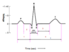
what is P wave?
when SA node generates impulse – spreads to atria,
what is QRS?
what is Q?
what is R?
what is S?
depolarization of ventricles
Q = septal depolarization
R = depolarization of apex
S = depolarization of base of heart
Which ion conductance is maximum during QRS?
sodium
After ventricular depolarization, ventricles will remain how? why?
depolarized before they get repolarized; no potential is recorded; corresponds to plateau phase of ventricular action potential.
what is T wave?
Ventricular repolarization