ECGs Flashcards
(23 cards)

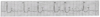
Multi Focal PVC
- this indicates that there is more than one irritable focus, and each focus produces its
own QRS morphology (shape)
- when examining the above example, the first and last PVCs look the same, indicating
that the impulse originated from the same ventricular focus. These QRSs are positively
deflected and the T waves are negatively deflected
- the second PVC looks different because it originated from a different focus. It has a
negative QRS followed by a positive T wave

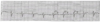
Ventricular Bigeminy
- this reflects more irritability because the PVCs are occurring more frequently
- this occurs when every 2nd beat is a PVC
- when examining the ventricular bigeminy on figure 7-12 above, every 2nd beat is a
PVC
- the sinus beats have P waves, narrow QRSs and T waves that follow the narrow
QRSs
- quickly after the normal T waves, a premature ventricular beat arises. We know they
are PVCs because the QRS is wide and the T waves are attached to these QRSs and
deflect in the opposite deflection (the QRSs are positive and the T waves are negative)

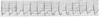
Paired PVCs (sometimes called couplets)
- this example shows 2 PVCs in a row (as well as a single PVC). These all look the
same because they originated from the same focus
- paired PVCs indicate that the single focus is quite irritable and generates two
consecutive premature impulses
missing lesson 4 part c

Run of PVCs
- when 3 or more PVCs occur consecutively, more irritability is obvious
- in this example, a narrow QRS is followed by three PVCs in arrow
- three consecutive PVCs are sometimes called triplets or a salvo of PVCs

Run of PVCs (cont’d)
- this example shows 4 PVCs in a row, and these are mutifocal
- this rhythm indicates more irritability because not only are there 4 consecutive PVCs,
but these beats also arise from different foci
- the very first beat at the start of the strip is normal and narrow
- this narrow QRS is then followed by the 4 consecutive PVCs
- the 1st and 3rd of these PVCs have negative QRSs and attached positive T waves
- the 2nd and 4th PVCs have positive QRSs followed by attached negatively deflected T
waves
- some might refer to this as VT
- halfway through the strip, the baseline sinus rhythm resumes

R on T PVC
- these PVCs are often the most serious and dangerous pattern of ventricular irritability
- it is called ‘R on T’, but technically it means ‘QRS on T’
- the PVC is so early that it strikes on the T wave of the preceding normal beat
- so this PVC occurs just as the ventricles are repolarizing
- if the ventricles do not have the opportunity to fully repolarize prior to the next
depolarization, the rhythm can deteriorate into VT
- figure 7-19 above shows 4 normal beats each with a depressed ST segment and a
small positive T wave
- the 5th beat on this strip strikes right on top of the T wave, so this impulse occurred just
as the ventricles were relaxing (the T wave represents ventricular repolarization)
- this means the ventricles cannot fully repolarize because they have just received
another impulse to depolarize
- the end result is a sinister and ominous VT

SINUS ARRHYTHMIA

SINUS BRADYCARDIA

SINUS TACHYCARDIA
Distinguishable Features
- HR > 100 (AR and VR are the same)
- all other findings are normal

SA BLOCK & SA ARREST (SINUS PAUSE)
Distinguishable Features
- entire PQRST missing amid the baseline rhythm
- all other findings are normal

PREMATURE ATRIAL CONTRACTION (PAC)
Distinguishable Features
- the beat with the PAC is earlier than expected (premature)
- the PAC has a P wave that is abnormally shaped and differs from all the other P
waves that originate from the SA node (different site of origin = different looking
P wave)
- the premature P might be difficult to see
- it can be “lost” in the T wave of the beat preceding the PAC
- the prematurity of the beat shortens the patient’s normal RR interval, causing
an irregularity in the rhythm

ATRIAL FLUTTER
Distinguishable Features
- VR may be fast or slow (varies on the degree of block)
- P waves no longer exist (atria are not contracting, they are fluttering)
- the Ps are replaced by flutter waves that appear saw-toothed or resemble
picket fences
- there are no P waves, therefore PR intervals cannot be calculated
- the QRSs are normal as conduction beyond the AV node is not affected
ATRIAL FIBRILLATION
Distinguishable Features
- the HR varies (depending on whether it is controlled or uncontrolled)
- the ventricular rhythm is always irregular
- the P waves are absent (the atria are quivering, not contracting)
- because of chaotic atrial activity, only a fibrillatory line is seen where Ps would
normally exist
- no P waves, therefore no PR intervals can be measured

PAROXYSMAL ATRIAL TACHYCARDIA (PAT)
Distinguishable Features
- HR is 150-250
- the rhythm is always regular (impulses are initiated with a regular rhythm)
- the P waves may not be visible if the HR is too fast
- if Ps are not visible, the PR intervals cannot be measured
- QRS complexes are usually normal (narrow) as conduction below the AV node
and within the ventricles is not usually affected

PREMATURE JUNCTIONAL CONTRACTION (PJC)
- P is either inverted (with shortened PR), buried, or follows the QRS

JUNCTIONAL ESCAPE RHYTHM
- HR is 40-60 (the junction initiates 40-60 impulses / minute
- the rhythm is regular (the junctional pacemaker fires at a regular rate)
- P waves are either inverted, after the QRS or absent/buried in the QRS
- PR intervals <0.12 seconds (if P waves occur prior to the QRSs)
ACCELERATED JUNCTIONAL RHYTHM
- HR is 60-100
- the rhythm is regular (the junction fires impulses with a regular pattern)
- P waves are either inverted, buried or follow the QRS complex
- the PR interval is < 0.12 seconds (if a P wave precedes the QRS)
- QRS is narrow, due to normal conduction beyond the AV junction
JUNCTIONAL TACHYCARDIA
- HR is 100-200
- the rhythm is regular (junctional rhythms are regular)
- P waves are either inverted, buried or follow the QRS complex
- the PR interval is < 0.12 seconds (if a P wave precedes the QRS)
- QRS is narrow, due to normal conduction beyond the AV junction

FIRST DEGREE HEART BLOCK
- the rhythm is regular
- P waves are normal and there is a P wave preceding each QRS complex
- PR intervals are constant (always the same length), but prolonged (> 0.20 sec)
because impulses are delayed by the AV node
- QRS complexes are narrow (there is no disturbance beyond the AV junction)

SECOND DEGREE BLOCK, TYPE I (WENCKEBACH)
- the AR is that of the SA node
- the VR is slower than the AR (because some Ps don’t reach the ventricles)
- the rhythm is always irregular
- the P waves are normal in configuration, but there are more P waves than QRS complexes
because some impulses are blocked at the AV node and do not reach the ventricles, so
the ventricles do not contract
- the PR interval is variable
- the PR interval progressively lengthens until an impulse is completely blocked at the AV
node and does not reach the ventricles, which produces a missing QRS complex
- then, a new PR interval sequence of lengthening begins again
- QRS complexes are narrow if conduction within the ventricles is normal

SECOND DEGREE HEART BLOCK, TYPE II (MOBITZ II)
- the AR is that of the SA node
- the VR is 2, 3, 4 times slower than the AR
- the rhythm is regular because the block occurs at regular intervals
- the P waves are normal, but there are 2, 3 or 4 more P waves than QRSs
- the PR intervals are constant (always the same length)
- PR interval may be normal in length or can be prolonged, but it’s constant
- PR intervals only exist with every 2nd, 3rd, 4th ventricular beat (where Ps exist)
- the QRS complexes are usually narrow, but can be slightly widened
- the width of the QRS basically determines the location of the block
- if the QRS complex is narrow, the block is at the AV nodal area
- if the QRS complex is widened, the block is sub-junctional, and can more easily
advance to 3rd degree block
- therefore, the wider the QRS complex, the more serious the block

THIRD DEGREE HEART BLOCK or COMPLETE HEART BLOCK (CHB)
- the AR is that of the SA node (60-100) and the VR is 20-40
- both atrial and ventricular rhythms are regular, but independent of each other
- the P waves are normal and occur regularly, but there are more P waves than
QRS complexes (some Ps may be hidden in QRS complexes)
- the PR interval is variable and totally erratic
- because the atria and ventricles have independent pacemakers, there is no relationship
between the Ps and the QRSs, and therefore a variable PR interval is present
- QRS complexes are usually wide and distorted, but can be relatively narrow if the
ventricular impulses originate nearer to the AV node
