DVM 4 Specific Flashcards

1
Q

9 week old Male Labrador

Hx: Vomiting and anorexia for 2 days

HR 144 RR 36 Temp 39.7C NAD on abdo palpation

* Why decreased serosal detail?

* The gas within the GIT– which parts contain gas? Are the gas filled portions of the GIT over distended?

* The fluid filled portions– which parts contain fluid? Are they over distended?

* Which other organs can you ID? Are these within normal limits?

* What are your recommendations for further investigation and treatment of Baughie?

A

Should not exceed 1.6 times the height of the centre of the body of L5 at its narrowest point.

SI/L5 ratio of 1.7 for diagnosis of intestinal obstruction (sens and spec only 66%)

2
Q

Layla 4 yo FS JRT

Vomiting and diarrhoea for 3 days

HR 120 MM pink to very pink CRT 1 sec, tacky

Pulses normal and synchcronous, auscultation

What are the types of ileus?- Which type is present in Layla?

How might you further investigate her diarrhoea and vomiting?

A
3
Q

Oscar 12 yo MN DLH

Hx: Vocalised and collapsed at home

CE: Systolic heart murmur, increased RR. Pale gums, tense abdomen with mass lesion palpable cranially.

Large irregularly margined mass within the cranioventral abdomen– which is the most likely organ of origin for this mass?

The mineralised opacity caudal to this mass- what is it called?

A

2-3 x the length of L2 for cats.

2.5-3.5 the length of L2 for dogs.

Hepatocellular Carcinoma

4
Q

Pip

1 yo MN Ragdoll

Trodden by a horse

HR 138 RR 32 MM pale, lethargic, distended painful abdomen

DDX for decreased intraserosal detail?

What is most likely?

What structures can you see?

Are there any MSK abn?

What procedure has been performed? Describe how you would perform this study.

Can you namet he phases of the excretory urogram?

Is there a rupture of the urinary tract? What is the most likely cause of Pips loss of serosal detail?

A

Excretory urogram is used to visualize the kidneys and ureters while the other contrast studies are used to visualise the lower urinary tract.

AKA IV Pyelogram. An iodinated contrast agent such as iohexol is injected IV. Contrast agent is rapidly concentrated and excreted by the kidneys.

Nephrogram phase- the renal cortex and medulla opacity– rate of renal opacification and wash out of the contrast agent provides a rough indication of the filtraion ability of the kidneys.

The contrast agent is excreted by the nephrons and accumulates within the renal pelvis- the pyelogram phase. The size and shape of the renal
pelvis can be assessed

Cystogram- Almost immediately, the contrast agent will pass down the ureters, and into the
urinary bladder, producing a ‘cystogram’.

5
Q

1 yo MN Burmese

5 week history of weight loss and reduced appetite

CE: Abdo palpation: pain on palpation with thickened intestinal loops/poss descending colon palpable. Estimate poss < 5% dehydrated.

DDX decreased intraserosal detail? Most likely?

Rule for assessing gastric distension?

Oval, comma shaped and C shapred SI throughout the mid abdomen? What is the rule for SI overdistension in the cat?

The distended loop of intestine containing fragmented gas opacities within the cranioventral abdomen on the lateral projection within the right cranial abdomen on the VD projection– do think this is SI or LI?

A

Stomach– no greater than 3 ICS– fundus typically 2 x wide as pylorus

6
Q

11 yo FS Maltese Terrier

Not quite right for a couple days now, vomiting

HR RR WNL- pendulous abdomen, not painful on palpation. V+ and diarrhoea

DDX intraabdominal decreased serosal detail? Most likely?

Hepatomegaly- what are the rules for assessing liver ? DDX?

Gas distended SI…

Fragmented mineral opacity in the descending colon….

A
7
Q

8 yo F GSD

Vomiting and inappetance

INcreased HR and RR, temp 40.5C, tense abdomen on palpation

Large fluid filled tubular structures within the mid to caudal ventral abdomen? What do you think this is most likely to be?

What is the circular soft tissue opacity seen on VD within the left abdomen at the level of the 5th vertebra?

A
8
Q

8 yo M Staffy

Previous history of two laparotomies- Owners concerned re: another FB

HR 130 RR 38.6C

No pain on abdo palp

A
9
Q

12 yo MN Labradoodle

Acute v+, lethargy, and profound weakness, but concurrent history of chronic vomiting and weight loss

HR 160 with injected MM

panting

Relaxed abdomen– firm area cranio ventrally– mass?? Cannot rule out tail of spleen

Gas filled stomach– rule for distension?

Over distended SI– what is the rule to assess for presence of mechanical SI obstruction?

The over distended SI loop within the mid ventral abdomen containing multiple fragmented gas opacities… what do you think this is most likely to be?

Ventral spondylosis L2-3

What is your plan for Austen

A
10
Q

10 yo MN Himalayan Cat

hx: Constipation

CE: 240 HR, RR 30, Pale MM, tense abdo, bladder distended, difficulty passing catheter

Why do they look different?

What procedure?

What contrast agent?

What is the dose rate?

Can you ID the reason he has having trouble urinating?

A
11
Q

Janis 13 yo Bull Terrier

Presenting: Dull, showing signs of depression, tachycardic, very tense on abdo palpation

The decreased intra-abdominal serosal detail centrally- what are the differential diagnoses for decreased intra-abdominal serosal detail? What is most likely?

Abnormal fragmented gas opacities in the ventral abdo- What are the DDX for pneumoperitoneum? Do you think it is free gas within the abdomen or within an organ? If you think it is arising from an organ, which do you think is the most likely organ of origin for these fragmented gas opacities?

A
12
Q

5 yo M JRT “Spike”

Stranguria since last night, vomited twice

CE: Panting and agitated, tense on abdominal palpation, no other abnormalities on abdo palp

A
13
Q

Vanessa 4 yr old F Doberman

Whelped 6 live and 2 dead puppies 24 hours prior to presentation. Noted to be straining again on morning of presentation. Bitch quiet and has reduced appetite.

HR 124 RR 36 MM tacky T 39.4C

A
14
Q

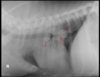
14 yo M King Charles Spaniel

Presenting complaint: Purulent saliva from mouth, malodorous breath. Enlarged submandibular LNs. Grade V systolic heart murmur, normal resp rate/ sounds. Thoracic radiographs taken prior to GA for dental.

CE: 160 HR, RR 40, Temp 38.5C. Grade V Systolic heart murmur.

Rules for assessing cardiac silhouette?

VD/DV RG do the caudal lobar veins lie medial or lateral the arteries?

Is this dog in Left sided CHF?

Are you happy to go ahead with the dental?

A
15
Q

Will you see anaemia with haemorrhage? With a severe coagulopathy, what are possible ddx?

A

* Yes + possible hypovolaemic shock

* Anticoagulant rodenticide toxicity, hepatic failure, DIC, malabsorptive diseases

16
Q

When might you see mild thrombocytopenia?

A

Secondary from blood loss/ increased utilisation, DIC

17
Q

What is a heart murmur? Why would you see a heart murmur with anemia? What else could cause a heart murmur?

A

* Heart sounds produced when blood flows across one of the heart valves that is loud enough to be heard with a stethoscope– vast majority are due to turbulent blood flow brought on by high velocity blood flow

* the body has a lower than normal number of RBCs therefore the blood is thinner and flows faster than normal

* Structural abnormality (e.g. valve leaflet), fever, hyperthyroidism

18
Q

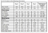
In a dog with anticoagulant rodenticide toxicity, what kind of blood transfusion product would be ideal?

A

Fresh whole blood for the red cells, clotting factors, proteins, and platelets (lack of vitamin K reductase does not allow the activation of vitamin K and therefore lack of clotting— so fresh whole blood with stop bleeding by providing clotting factors that have been lost)

** Max is also hypovolemic and hypoxic shock from severe anemia– so supplementation of red cells is essential

19
Q

What are possible complications from a transfusion?

A

* Acute immunological transfusion reaction- acute hemolytic reaction (type II hypersensitivity reaction), febrile non hemolytic reaction, non-hemolytic immunological reaction (type I hypersensitivity)

* delayed immunological transfusion reaction- delayed hemolytic reaction, post transfusion purpura (internal bleeding)

* acute non immunological tranfusion reactions- bacterial contamination of transfused bloods, hemolysis from damaged RBC transfusion, hyperkalaemia/hypocalcemia/hypomagnesemia after massive transfusions, hypothermia, circulatory overload

20
Q

What are some things you can do to prevent transfusion reactions?

A

* blood typing (especially cats), cross matching major and minor (especially if second transfusion), test infusion 0.25 m/kg/h over the first 5-10 minutes, use of an in line blood filter, monitor patient parameters every 15-30 minutes during transfusion for acute reactions (demeanor, temperature, pulse rate and quality, BP, RR and character, MM colour, plasma and urine colour), ensure transfusion complete within 4-6 hours to preven bacterial growth, proper collection and storage of blood, appropriate blood donor screening

21
Q

Dog with anticoagulant rodenticide poisoning, what other treatments over than an infusion?

A

* Vitamin K1 supplementation

* strict cage rest to prevent further hemorrhage

* oxygen supplementation

* Intravenous fluids

22
Q

What are X-rays?

A

A form of electromagnetic radiation

EM radiations of frequencies higher than UV light have enough energy to remove an outer-shell electron from an atom and are therefore called ionising radiation (X-rays, gamma rays, and cosmic rays are ionising radiation). When ionising radiation interacts with atoms it causes the ejection of an electron and creates an ion pair which has the potential to damage cells.

** They can produce damage to molecules and induce temporary or permanent cell damage, causing problems such as mutations or cancer

24
Q
A
25
Q

What are some unusual properties of ionising radiation?

A

* You cannot see, smell, touch or taste it

* Penetrates all matter

* We do not develop tolerance or immunity to radiation

* There is little difference between how individuals react to a given dose

* Dose is cumulative

* Delayed effects: May take years to see the effects

26
What does it mean that effects of radiation are stochastic or deterministic?
\* Stochastic- increased risk with increased dose-- the dose of radiation you receive, a higher dose of radiation will increase the probability of a stochastic effect e.g. a genetic mutation leading to cancer-- higher radiation dose or exposure will increase the chance of cancer developing (this is the one that is important in vet med) \*\* no threshold below which the risk of the effect will cease & effects are cumulative over a lifetime \* Deterministic- inevitable at high doses-- in contrast with stochastic effects, the severity of the effect, not the probability, increases with increasing dose. Exposure below a certain threshold will not result in the effect (THERE IS A THRESHOLD). e.g. radiation induced skin burns, uncommon however in medicine
27
What is the direct effect of ionising radiation? Indirect effect?
\* direct effects may damage DNA, protein or other macromolecules \* Indirect effects create free radicles within cytoplasm and these free radical damage macromolecules \*\* Cell may either repair itself or if DNA damage has occurred a mutation may result, causing cell death, cancer, or birth defects
28
In Victoria, the use of radiation comes under what department? And which Act? What does it state?
\* Department of Health which administers The Radiation Act 2005-- species that business must have a management licence to possess a radiation source and conduct a radiation practice & individuals must have a use licence
29
What is ARPANSA?
\* A national governing body -- Australian Radiation Protection and Nuclear Safety Agency-- responsibility for protecting the health and safety of the people and environment from the harmful effects of ionising radiation. They publish a series of codes and standards then implement in the form of legislation. RPS 17: Radiation Protection in Veterinary Medicine (2009) \*\* Also provide "Personal Radiation Monitoring Service"-- monitors and records occupational exposure to radiation
30
What are the three basic methods of monitoring for radiation exposure?
\* Thermoluminescent Dosemeter (TLD)- electrons int he crystal structure of the TLD card are excited to high energy levels as a result of irradiation and are trapped in the crystal structure. By heating the material, electrons return to their ground state and light is emitted. The amount of ligh is proportional to the radiation dose that the TLD material has received. Filters are used to allow only certain radiation to pass. \* Film badges- degree of darkening can be measured and will indicate the absorbed dose. But dose to film does not equal dose to tissue. \* pocket dosimeters-- different to TLDs and film badges... pocket dosimeters can be read instantaneously-- useful when high exposures are expected.
31
What tissues are more susceptible to radiation?
Rapidly dividing tissue
32
What is the recommended effective dose limit for occupational exposure? In a single year? What is the avg annual dose for veterinarians?
20 mSv per year, averaged over consecutive years \* 50 mSv in a single year \*\* Avg annual dose for veterinarians is around 16 microSv = 0.016 mSv
33
What is the ALARA principle?
\* As Low As Reasonably Achievable \*\* Is the radiograph necessary for the patient and management of the case? Would ultrasound or a blood test answer the clinical question? \* Minimize retaking radiographs, minimize manual restraint, under no circumstances should any part of your body be within the primary x-ray beam
34
What are the three types of radiation encountered in medical practice?
\* Useful beam \* Leakage radiation \* Scatter radiation
35
How can radiation exposure be minimised ?
Modifying 1. Time (take good radiographs the first time around) 2. Distance from the radiation source-- the energy of the scatter radiation will decrease as distance from the radiation source increases, according to the inverse square law... e.g. so by doubling the distance from the radiation source, you will reduce your radiation exposure by a factor of 4. (avoid manual restraint, use ancillary positioning devices such as sand bags, stand well back) 3. Shielding- concrete walls, PPE
36
How thick should lead PPE be?
Lead equivalent of 0.25 mm will shield scatter radiation with energy of up to 100 keV (electron volt)
37
How are x-rays generated? What elements are necessary for x- ray production?
\* Generated in the x-ray tube by firing a high energy electron beam at a tungsten target 1. A source of electrons (cathode) 2. An obstacle- free path for the passage of high speed electrons 3. A target (the anode) in which the electrons can interact, releasing energy in the form of x-rays 4. a method of accelerating the electrons- electrical potential difference applied between the cathode and anode
38
What should the local radiation management plan follow?
ARPANSA RPS 17 + ALARA by observing time distance and shielding principles
40
What is the cathode? What is it made of?
\* cathode acts as a source of electrons \* it is of a tungsten coiled filament mounted within a molybdenum focusing cup. Tungsten has a high melting point and a high atomic number. When a current is run through the cathode filament, it heats up and releases electrons.
41
What is the anode? What is it made of?
\* when an electrical potential difference is applied across the x-ray tube, the anode has a positive charge relative to the cathode and attracts the electrons \* When electrons collide with the anode, 99% of the kinetic electron energy is converted to heat and only 1% gets converted into x-rays Because of the heat produced anodes have a tungsten target which is resistant to heat. This is mounted on a copper base which helps conduct heat away from the target. (Either rotating-- more powerful x-ray machines-- or stationary-- dental and portable-- copper base)
42
What is the glass envelope?
\* Contains the cathode and anode within the vacuum. The x-ray tube has a small exit window through which the x-ray photons escape. The tube is surrounded by oil which helps dissipate the heat and acts as an electrical insulator.
43
What is meant by quantity in regards to properties of the x-ray beam? Quality? Intensity?
Quantity= the number of x-ray photons produced Quality- the energy of the x-ray photons, hence their penetrating power Intensity- the amount of x radiation per unit area. The intensity is determined both by the number of photons and the energy of each photon
44
What does the quantity of electrons produced at the cathod filament dependent on?
The electrical current of the cathode circuit. This is an adjustable parameter that the x-ray machine operate can control, known as the Milliamperage (mA) setting. Increasing mA increases the number of electrons within the electron cloud
45
What does increasing mA do?
Increasing milliamperage (mA) increases the number of electrons within the electron cloud
46
What does the higher the kV mean? What is kVp?
\* In order to accelerate the electron cloud toward the anode and produce x-rays, a potential difference or kilovoltage (kV) is applied between the anode and the cathode. The highter the kV, the greater the acceleration and thus the greater the energy of the electrons-- the greater the energy of electrons-- the greater the resultant x-rays which EQUALS HIGHER QUALITY X-rays \*\* kVp- kilovoltage peak- the maximum voltage available at the kV setting and is an adjustable paramter that the x-ray machine operator can control
47
What is the meant by the exposure time?
\* The potential difference between the anode and cathode is only applied for a very short period of time (milliseconds)-- the exposure time is an adjustable parameter that the x-ray operator can control. A long exposure time allows may electrons to impact with the anode, while a short exposure time allows fewer electrons to impact with the anode
48
What is the quantity of x-rays produced a function of? What determines the quality of x-rays produced?
both the mA setting and the time setting (s) \*\* mA determines the number of electrons produced, while time determines the number of electrons that hit the anode \*\* some x-ray machines allow the operator to select the mA and s independently \*\* kVp setting determines the energy of the electrons and therefore the energy of the resultant x-rays and thus the quality or penetrating power of the x-rays
49
How does the radiographer ensure the x-ray beam has the appropriate intensity?
Intensity = quality and quantity of x-ray photons \* this is so that enough x-ray photons penetrate the patient and interact with the x-ray detector to form the image \* The x-ray operator achieves this by manipulating the kVp, mA, and second settings on the machine to be appropriate for the anatomic area that is being radiographed
50
What is the focal spot? What does the size focal spot influence?
The area of the target with which the electrons collide... the size of the focal spot influences the detail (edge definition) of the image and also affects the heat loading capacity of the machine. e.g. a stationary anode is limited to having a larger focal spot in order to accomodate higher temperatures. The focal spot of a rotating anode can be smaller due to the larger surface area over which electrons impact the target and superior ability of a rotating anode to dissipate heat. Common focal spot sizes in veterinary medicine are 1-2 mm^2
51
What is the collimator?
The device to limit the size and shape of the primary x-ray beam \* To minimize scattered radiation, the primary beam should be confined to a size and shape that will just cover the region of diagnostic interest
52
What are the benefits to using the collimator to limit the x-ray beam to the area of interest?
Less scatter radiation is produced which improves image contrast and image quality and radiation safety...
53
What is full wave rectification?
\* Converting the AC into a direct current (DC) without losing any electricity-- full wave rectification creates a nearly constant electrical potential across the x-ray tube as both halves of the alternative voltage are used to produce x-rays \* A consideratlbe portion of the exposure time is still lost however while the voltage is in the valley between the two pulses
54
What is a three phase generator?
Present in most modern machines- produce an almost constant electrical potential difference between the anode and the cathode, which results in: 1. More power available to the x-ray tube per unit time and therefore for shorter exposure 2. Intensity of the x radiation generated is higher 3. Radiation quality is greater because it contains less low energy x-rays 4. Tube utilisation is more efficient because the target is not subjected to bombardment of low energy electrons, which only create heat \*\* main disadvantage is the increased cost of the machine relative to more simply rectification machines
55
What are the three main parts of the x-ray machine generator?
\* The control panel- allows the operator to select appropriate setting for the voltage (kVp), tube current (mA), and the time of exposure (sec) \* The transformer assemby- step up or step down transformers to increase or decrease the voltage as required (which is needed to boil electrons from the filament and to accelerate these electrons from the cathode to the anode) \* The rectification circuit- incoming mains electricity is alternating current (AC), when x-ray exposure is made and a potential difference is applied across the x-ray tube, direct current (DC) is required. Most efficient systems produce constant positive lectrical potential, cheaper units the DC is more variable and only reaches peak voltage (kVp) for a small proportion of the time
56
What are the two stages that accomplish the exposure?
\* Preparation- press and hold the button on the hand piece half way down; the cathode is heated by the cathode current and the anode starts rotating \* Exposure- when the system is ready, the operator fully depresses the button; this causes the potential voltage to occur for the set exposure time
59
What is the tube rating? What is it based on?
Each x-ray machine has a tube rating expressed in kilowatts-- the tube rating dictates themaximum combinations of kVp, mA and time that can be safely used without over-loading the tube Based on: 1. Focal spot size 2. Target angle 3. Anode speed 4. Electrical current
60
How is a radiographic image formed? What is attenuation?
Differences in attenuation between different components of the object to be radiographed. Attenuation is the proportion of x-rays that are stopped/ absorbed by a given thickness of matter
61
What 3 things happen when x-ray hits matter? What determines which one predominates?
1. Transmission- the x-ray photon passes straight through 2. Photoelectric effect- x-ray photons can be absorbed 3. Compton effect- x-ray photons can be scattered \*\* Which one of these interactions predominates is determined by: - energy of the x-ray beam (kVp) - atomic number of the absorber. Remember more interactions occur with matter that has a high atomic number - thickness and density of the part being radiographed. It makes sense that thicker or denser objects will stop more x-rays.
62
What is the photoelectric effect?
PE results in complete absorption of an x-ray photn by an atom and the ejection of an orbital electron, which produces a positive ion. Largely responsible for radiographic contrast. The main way x-rays are attenuated at 40-120 kV. The primary interaction between x-rays and bone. \*\* Probability of the PE depends on: 1. Energy of the x-ray photon (must be above threshold kV) 2. Atomic number of the absorber (elements with a higher atomic number are more likely to have PE absorption)
63
When is PE more likely to occur?
In low energy x-rays (50 to 70 kVp) \* high Z material (more in iodine, barium and lead than in oxygen, carbon or hydrogen) \*\* PE explains why better contrast is achieved with low kVp and why iodine and barium (used in radiographic contrast studies) stop more x-rays than organic matter \*\* soft tissue is made up of hydrogen, carbon, nitrogen, and oxygen-- avg atomic number of 7.. vs. bone which is mostly calcium (atomic number 20)
64
How do you increase subject contrast?
Decrease the kVp setting
65
Why is PPE made from lead?
Atomic number is Z82-- materials made from elements with a high atomic number are good absorbers of x-rays
66
What is screen speed?
Part of a cassette. An important characteristic of intensifying screens. Faster screens requires less radiation (lower mAs, usually achieved with shorter exposure time)-- at the expense of detail however.
67
What is the compton effect? Why is it not useful in diagnostic radiology? What influences the likelihood of the compton effect?
Responsible for almost all the scatter radiation produced in diagnostic radiology No useful function in diagnostic radiation because 1. Scattered radiation poses a safety threat 2. Decreases contrast of the radiographic images (e.g. the difference between black and white on the radiograph is less obvious) \*\* Likelihood of the compton effect 1. Very dense absorbers are more likely to produce compton scatter 2. large volumes of irradiated tissue are more likely to produce compton scatter-- thick body aprts e.g. fat dog abdomen, horse back or shoulder; or poorly collimated image will produce more scatter 3. High kVp settings thuse higher energy x-ray photons are more likely to produce compton scatter
68
How do you absorb scatter radiation before it hits the detector?
Use a grid They should always be used when the patient thickness is greater than 10 cm because more scatter is produced in thicker body parts Placed between the patient and the detector- so under the patient or under the table between the patient and the cassette
69
What is the relationship between optical density of a radiograph to the exposure needed to produce that density?
Sigmoidal \* Ideally, most of the radiographic image should be exposed in the linear portion of the curve. If the film is exposed at the shoulder or toe of the sigmoidal curve, the image will appear overexposed (too dark) or under exposed (too light) respectively
70
How does a film screen x-ray work? What is the most commonly used x-ray films?
\* x-ray film consists of a polyester base coated on both sides with a gelatin emulsion containing silver halide crystals. When visible light or x-rays interact with silver halide crystals- an invisible image is formed then processed to be made visible-- by exposing the silver halide to an x-ray photon or light photon which activates the crystals which precipitate silver... which aggregates producing darkenened areas \*\* Most commonly used x-ray films are double emulsion- having emulsion on both sides of the base (single emulsion can be used with pocket pets)
71
What are cassettes?
Cassettes are used in traditional film-screen and computed radiology (can be reusable) \* rigid lightproof containers that hold both the intensifying screens and the radiographic film. Double emulsion film is used in cassettes with two screenes which therefore results in two images on either side of the film
73
What is an intensifying screen? Why must the x-ray film match the intensifying screens of a cassette?
\* Intensifying screens in a cassette convert x-ray photon energy into light photons, which expose the x-ray film. The use of film with intensifying screens (film-screen) is a more efficient detection system than x-ray film alone and allows a reduction in radiation exposure up to 100 times. \* X-ray films are only sensitive to one colour of light and different screen types give off different colours
74
What is optical density ? What is it primarily controlled by?
Measure of the blackness of film-- primarily controlled by mAs
76
What is film speed?
Term used to describe how sensitive the film is to x-ray or light photons. Film that has a high speed rating is very sensitive to photons, an adequate optical density can be achieved with lower radiation exposure. The trade off for fast fil is that it has slightly lower detail/ spatial resolution compared to slower film.
77
What is film latitude? What is film latitude's relationship to film contrast?
Exposure range over which acceptable optical densities are produced. Film with a wide latitude will accept a significant variation in exposure factors or processing without exhibiting any great chance in optical film density. Most x-ray film has WIDE LATITUDE making it relatively easy to avoid under or over exposure... narrow latitude film or high contrast film is less forgiving with exposure factors-- FILM LATITUDE VARIES INVERSELY with FILM CONTRAST
78
CR advantages and disadvantages?
79
What speed film would you want to use for cat's manus?
Slow speed 100 to 200- higher in detail, small body part, very little movement so longer exposure times okay to use
80
Advantages and Disadvantages of DDR
81
What speed film would you use for a fat dog's abomen?
Fast speed- 400 to 600 speed
82
What film speed would you want for a horse's carpus?
400 speed- fast system. Move a lot so short exposure time
83
What are the steps in film processing?
1. The Developer- chemical solution that converts the latent image on the film to a visible image by reducing exposed silver halide crystals to black metallic silver. \*\* Time and temperature dependent (chemical reaction occurs at a greater rate at higher temperatures)-- usually at 20C about 3 minutes to develop 2. The rings- x-ray film retains developer in the gelatin and if this were transferred to a fixer the alkaline developer would neutralize the acid fixer-- so rinsing stops the developing processes and prevents carry over the fixer 3. The Fixer- removes unexposed silver halide crystals. Fixation also hardens the gelatin coating so that it can be dried without damaging the film surface. Fixed fro twice the development time to ensure maximum hardening of the emulsion. An incompletely fixed film takes on a milky or cloudy appearance. 4. Wash Bath- removes the remaining silver complexes and excess fixer. (20-30 minutes) 5. Dry the film. Hanging.
84
What are the limitations to traditional film- screen?
Image can have good contrast or good latitude but not both \* And once the film has been exposed to the x-rays, the contrast and latitude can't be adjusted
85
What are the three steps of Computed Radiography?
1. Image acquisition- x-ray photons strike a reusable storage phosphor imaging plate (IP)-- placed in a plate reader where it is scanned by a laster beam which releases the stored latent image as visible light. Then the IP plate is erased for reuse by scanning the plate with an intense white light 2. Image processing- using algorithms the computer evaluates and manipulates the digital data 3. Image display- the user can then apply post processing techniques such as edge enhancement, magnification, adjustment of contrast range and dynamic range
87
How does direct digital radiography work?
\* Direct systems use an electronic detector made of amorphous silicon or selenium or a charged couple device to capture the image. The detector is attached to the computer system via a cable. The signal is transmitted to the computer and reassembled into a digital image.
89
What is the major advantage of digital imagine?
Due to the wider latitude, the number of retakes due to poor exposure are reduced BUT although digital image contrast can be manipulated by viewing software, subject contrast is still influenced by collimation, scatter and kVp settings
90
What are the six basic steps in making a technique chart?
91
What is quantum mottle?
An underexposed digital image will have a grainy appearance Quantum mottle is caused by the discrete nature of an x-ray photon; underexposure results in an insufficient number of x-ray photons reaching the detector to fully form the image \*\*\* think of black paint spots outlining something
92
What does radiographic contrast depend on? When is high contrast preferred? When is low contrast preferred?
Radiographic contrast depends on: 1. Subject contrast- due to attenuation of the x-ray beam in different tissues; low kVp will produce high subject contrast as long as the kVp is high enough to penetrate the tissue (higher kVp = more compton scattering) 2. Film contrast- homogeneity in size of silver halide crystals within the film emulsion, over or under exposure-- technical factors, over or under deveopment will push film contrast to the toe or shoulderof the characteristic curve and produce low contrast image 3. Fog and Scatter- inadvertent exposure of the x-ray film to light High contrast is preferred for bone images; low contrast is preferred for chest and abdominal images
93
How does spatial resolution of digital systems compare to film-screen systems? Does this matter?
\* Spatial resolution of digital systems is not as good as film-screen systems, but it is not a clinical limitation
94
Effect of kVp, mA and exposure time on the radiographic optical density and contrast
95
What are the four steps to processing the digital image that occur automatically?
1. Creation of a histogram- maps optical density vs. frequency which allows mathematical manipulation of the raw data- histogram is compared to an expected histogram for the anatomic area selected to be radiographed 2. Data adjusted for under or over exposure- histogram shifts left or right to compensate 3. Enhancement of image contrast, using a look up table 4. Edge enhancement or smoothing- complex mathematical function is applied to the raw data to allow processing in the spatial frequency domain-- boundaries such as bone margins can be smoothed or enhanced
96
What is PACS?
Archiving and distribution of digital images
97
What are the features of radiographic image quality?
\* Optical density, radiographic contrast (subject contrast, film contrast, fog and scatter, digital image contrast), detail (spatial resolution, edge definition), technical faults
98
How can optical density be manipulated?
By adjusting mAs (mA or seconds) setting
99
How does a film with high optical density appear? Why would this occur? What should you do?
Too dark over-all due to overexposed (too much radiation reached the film-- likely because mAs setting was too high) or over-developed. \*\* may also occur if film-focal distance is too short \*\* If too dark then reduce mAs +/- increase the film focal distance
100
Why does under exposure occur? What should you do?
\* check kVp settings-- have to be high enough to produce x-rays that will penetrate the patient \* most common cause of under- exposure is too low mAs setting... therefore increase the mAs and maybe increase the kVp
101
What is a technique chart?
Helps us determine the best x-ray machine settings (kVp, mA, seconds) \* technique charts are specific to the x-ray machine, film-screen speed, film to focal distance, whether a grid is used \*\* measure the thickness of the area you intend to radiograph and read off the technique chart what the ideal kVp and mAs setting should be
105
What are the four factors that determine the degree of attenuation of an x-ray beam as it passes through matter?
1. Thickness of the absorber 2. The density 3. Atomic number 4. Energy of the radiation
107
How can you reduce the production of scatter? How can you reduce the impact scatter has on the xray image?
\* Reduce production of scatter by 1. Selecting lower kVP (still has to be high enugh to penetrate the patient) 2. restricting the collimator field to only the area of interest \* Reduce impact of scatter on the x-ray image - using a grid when you radiograph body parts thicker than 10 cm - having a small air gap between the patient and the cassette 3. Reduce scatter radiation dose to the operator - PPE - distance
108
What is the biggest contributor to contrast of digital images?
The image processing algorithm and windowing of the image by viewing software (still impacted by subject contrast and scatter radiation)
109
What are the five basic radiographic opacities?
110
What are the two main components of image detail?
Spatial resolution and edge definition
111
What is spatial resolution? How does film screen compare to digital? How is it determined?
\* determined by the x-ray detector-- ability to distinguish between two adjacent contrasting objects \*\* gilm- screen has better spatial resolution than digital... but advantages of edge-enhancement, superior latitude and ability to zoom images outweighs the disadvantage of lower spatial resolution \*\* Spatial resolution of film-screen is determined by the size of the halide crystals of the film emulsion and the size of the phosphor crystals of the intensifying screen \*\* Spatial resolution of digital radiography is determined by the size of the detector elements and size of the pixel matrix used to store the image
112
What is penumbra? What determines?
Edge definition geometric factors that determine: 1. Focal spot size (small focal spot produces a shart image, think of hand and iphone) 2. Focal spot to film distance (as large as possible is important to improve image sharpness and reduce magnification) 3. Object to film distance (close to the film as possible to improve image sharpness and reduce magnification)
113
Main causes of a blurry image?How can this be minimized?
Motion of the patient or cassette \* Minimized by shortening exposure time, sedation and anaesthesia \*\* Cassette motion a problem in equine radiography where cassettes may be positioned by a cassette holder \* blurry images can also be caused by geometric factors taht increase the penumbra
114
What is B-mode ultrasound imaging?
115
What is M-mode ultrasound?
116
What is Colour Doppler?
117
What is spectral doppler?
118
What is distortion resulting from?
Unequal magnification of different parts of the same object
119
What is forshortening?
\* object being radiographed must also be parallel to the film
120
What three things might happen when ultrasound pulses interact with the patient?
1. Transmission into deeper tissues 2. Refraction (bending) of sound as it passes from a tissue of one density to a tissue of a different density 3. Attenuation of sound, which is a reduction in amplitude or intensity of the pressure wave
121
What is parallax error?
Since the x-ray beam is divergent the periphery of radiographic images have different projections-- e.g. spine radiographs
122
What is a B-mode ultrasound image composed of? What does the brightness of each x correspond to?
123
What is acoustic impedance?
\* Sound reflection or transmission characteristics of a tissue, largely dependent on the density of the tissue and speed of sound within the tissue. Because we assume constant speed of sound in soft tissue, density is the most important component in this equation \*\* It is not exact value of acoustic impedence that matters but the difference in acoustic impedence between two tissues \*\* At boundaries between soft tissue and boen or soft tissue and air there are large differences in acoustic impedance resulting in little sound transmission and the image is displayed as a shadow-- THIS IS WHY WE CLIP ANIMALS FUR, clean the skin and apply coupling gel-- this removes air between the skin and transducer
124
WHat is the preferred imaging modality to assess bone or lung?
125
What does the ultrasound transducer do?
\* converts electrical energy to acoustic energy and acoustic energy to electrical signals \* consist of piezoelectric crystals aligned within the footprint of the transducer and when a voltage is applied the crytal vibrates and produces a sound-- each one has a unique frequency at which is vibrates and will produce sound of an inherent frequency. The tranducer transmits sound pulses for 1% of the time and listens for returning echoes 99% of the time. \*\* The operator can select the main frequency band that is used for imaging, selecting higher frequencies for imaging superficial structures and lower frequencies for deeper
126
127
What is the phased array?
128
What is an artefact?
\* Structure or feature not normally present in an image, but visible as a result of an external agent or action-- fake out e. g. film screen dark marks caused by pressure on the film or white marks-- dust or hair int eh cassette which prevents light from the intensifying screen from reaching the film e. g. grid cut off appears as light areas caused by misalignment or inappropriate placement of a focussed grid e. g. milk/ opaque image- typically a problem with manual processing- incomplete fixation leaving behind silver halide crystals OR brown image-- incomplete washing leaving fixer behind which oxidises over time OR linear roller marks with automatic processors with dirty rollers e. g. digital artefacts- grainy image = underexposure; double image- double exposure with both images displayed due to wide latitude radiography; moire artefact- bands or lines from ; white lines on a CR image- dirty plate readers; misapplication of the algorithm- poor collimation and beam centering can result in mis-application of the algorithm and poor image contrast resolution; uberschwinger- excessive edge enhancement appears as a thin black rim around high denisty object such as bone margins or metal implants and may mimic bone lysis
129
What is axial resolution?
Dependent on the spatial pulse length
130
What is lateral resolution?
131
How do contrast agents work? What are the two main categories?
Work by having different physical density and atomic number hence different radiogrpahic density than normal patient tissue. \* Positive and negative contrast agents
132
how do negative contrast agents appear? Examples of agents? Examples of studies? Risk?
\* Negative contrast agents are gaseous and do not effectively absorb x-rays so appear radiolucent (dark) \*\* room air, carbon dioxide, nitrous oxide \*\* used to inflate organs e.g. GIT- pneumogastrogram or pneumocolon, or urinary tract- pneumocystogram \*\* Risk of fatal air embolism if any gas leaks into the vascular space e.g. damaged mucosa from necrotising haemorrhageic cystitis-- to minimize risk of death from air embolism when performing pneumocystogram place patient in left lateral recumbency and use CO2 or NO which are more soluble than room air (would be reabsorbed in the right ventricle otherwise could become trapped in pulmonary artery or travel and occlude pulmonary circulation)
133
What are positive contrast agents? Examples of agents? Which agents used where?
\* Positive contrast agents have a higher atomic number than soft tissue, thus are an efficient absorber of x-rays and appear more radiopaque \* iodine- Z= 53; barium Z= 56 \*\* Barium used in the GIT-- irritant so not IV-- excreted by the GIT \*\* Iodinated contrast agents are ionic or non-ionic. Ionic agents are markedly hyperosmolar relative to blood so are associated with adverse reactions such as hypotension, nephrotoxicity and thrombosis-- excreted by the kidneys, biliary tract, GIT, saliva \*\* All iodinated agents can trigger allergic reactions \* Iodinated agents-- angiography (IV), cytography (urinary tract), myleography (spinal cord) \*\* Myelography should only be performed with non-ionic!!
134
What are the conventional hanging protocols?
\* Dorsoventral OR ventrodorsal radiograph of the body- head at the top and the patient's right on the viewers left side \* Lateral radiograph- animal standing with the head to the viewers left and dorsal (for the body) or proximal (for the limbs) to the top of the screen \* Craniocaudal or dorsopalmar/ dorsoplantar- proximal is to the top and the lateral aspect displayed to the viewers left (no matter whether you are looking at the left or right limb)
136
Indications for ultrasound
\* internal structure of organs- size, shape, margins \* dynamic evaluation of blood flow-- which can provide some information on organ function e.g. echocardiography where blood flow and changes to the heart size over the cardiac cycle can be measured \* more sensitive at detecting pathology, low specificity for identifying a specific disease \* GIT e.g. heptobiliary disease, pancreatitis, vascular anamolies such as portosystemic shunts
137
Patient preparation?
\* Hair coat clipped, skin cleaned and alcohol applied to defat the skin, ultrasound gel applied to provide good acoustic coupling (transmission of sound) to reduce artefact and allow the ultrasound probe to slide over the skin
138
Special considerations for echocardiography ?
Sedation will affect cardiac function, so preferably an unsedated animal
139
Special considerations with scintigraphy?
140
Special considerations for abdominal ultrasound? Musculoskeletal ultrasound?
\* Abdominal- animals should be fasted \* musculoskeletal ultrasound- may benefit from using a stand off pad since structures are superficial and often hard
141
What is the Hounsfield scale?
The operator can choose what part of the scale to assign the central colour gray
144
What is an MRI sequence?
146
Safety issues with MRI?
148
What is duplex doppler ultrasonography?
Simultaenous use of real time B mode imaging and spectral doppler.
149
What is sound? What is the relationship of wavelength and frequency at a given velocity? What is the range of frequencies of ultrasound? What is the assumed constant speed of sound for ultrasound and where is that number from?
\* Sound is a pressure wave with properties of wavelength, frequency, velocity and amplitude \* For a given velocity, wavelength and frequency are inversely related e.g. high frequency = short wavelength. \* ultrasound has frequencies between 2 and 20 MHz \* Ultrasound assume constant speed of sound at 1540 m/sec (which is the average speed of sound in soft tissues)
151
What does sound attenuation depend on? What kind of transducer do we use for superficial structures? Deep structures?
\* Sound attenuation depends on the distance travelled and teh frequency of sound \* High frequency tranducer for superficial \* low frequency to image deeper structures
152
What is Poiseulle's Law? How does it relate clinically?
Physical law that governs the pressure drop in a gluid flowing through a long cylindical pipe. Clinically you can think about how it governs the pressure needed to push a volumetric rate of fluids e.g. in ml/hr through your tube (catheter) \*\* both length and radius have an effect on catheter flow but the radius effect is much greater than length. Double the length, you double the pressure it takes to get fluid through at the same rate.... Catheters have a high resistance to flow. 18 gauge are the biggest we use in peripheral veins.
153
What gauge through rabbit ear veins, bird wings and tiny kittens? What gauge through cats and very small dogs? Large cats and medium sized dogs? Larger sized dogs (heeler upwards)?
160
What should you consider when selecting a transducer?
\* consider frequency of the transducer and the footprint or shape of the field of view of the transducer
163
how is the best spatial resolution achieved?
Imagine with a high frequency transducer and imaging within the focal zone
164
When are low dynamic range setting used for? When are high dynamic range setting used for? What is true for both?
Low dynamic range settings are used for echocardiography \* High dynamic range for abdominal ultrasonography \* You will have the best perception of contrast resolution when imaging with a low gain setting in a darkened room
165
What is nuclear scintigraphy?
Allows imaging of the distribution of radiopharmaceuticals within the body using a gamma camera. It allows imaging of both structure and function
166
What is the most commonly used radionuclide in vet med?
Technetium- as it decays, the nucleus is converted to a more stable state by emitting gamma radiation which is detected by a gamma camera. It has a half life of 6 hours-- long enough for most imaging procedures to take place but short enoug hthat the radioactivity of the patient rapidly decays, typically reaching background levels within 24 hours... thus minimizing radiation dosage to the patient, staff and owners
167
Static vs. dynamic nuclear scintigraphy
\* Static mode- provides anatomic information about the distribution of the radiopharmaceutical at one point in time, allowing determination of organ size, shape, position and pattern of distribution that may be specific for a disease state e.g. bone scintigraphy-- shows areas of active bone turnover as seen with physes, stress fractures, infection or bone neoplasia \*\* dynamic mode provides functional information allowing measurement of rate of accumulation or removal or the radiopharmaceutical within an organ-- example is portal scintigraphy where a series of images is acquired over a four minute period allowing mapping of the passage of pertechnetate through the portal circulation, liver and heart
168
What is the most appropriate description of the radiographic appearance of a positive contrast agent in comparison to soft tissue?
Appear more radiopaque-- higher atomic number
169
What is used in bone scintigraphy?
Technetium methylene diphosphonate (MDP)- taken up by areas of active bone turnover \*\* bone scintigraphy is much more sensitive than radiography at detecting bone lesions but is relatively non-specific
170
What is thyroid scintigraphy?
Technetium pertechnetate-- trapped and concentrated by thyroid follicular cells, mimicking biological behavior of iodide \*\* useful to evaluate hyperthyroidism-- determines whether cervical masses are thyroidal or non-thyroidal in origin... differentiates between bilateral and unilateral disease, detection of ectopic thyroid tissue, and functional metastic lesions
171
What is portal scintigraphy used for?
Sensitive and specific for diagnosis of portosystemic shunts-- even quantification of the severity of the shunt-- Technetium is administered into either the colon or into the spleen where it is taken up by portal circulation
173
What is computed tomography?
Cross sectonal imaging modality based on x-rays. It gives excellent contrast resolution compared with conventional radiography. Produces high detailed cross sectional images with no superimposition of overlying structures. \*\* Helical scanners are the preferred type of machine-- newer helical CT scanners have multiple rows of detector arrays-- multi-slice CT scanners have very rapid acquisition times
175
Indications for a CT examination
\* Superimposition of structures in radiography prevents identification of lesions \* Improved soft tissue contrast is required \* lesions are suspected but not demonstrated with conventional modalities such as radiology or ultrasound \*\* e.g. brain and spinal imaging (MRI is best however but CT is more sensitive than MRI at detecting early brain haemorrhage with trauma), nasal disease to avoid superimposition, Tympanic bulla (changes seen with otitis media for example), musculoskeletal imagine (bone loss, complex trauma e.g. pelvic fractures provides 3D reconstructions), thoracic imaging (pulmonary metastases), abdominal imaging, foreign bodies and draining sinuses (localisation of abscesses and foreign bodies)
176
Excretory urography- to look for ectopic ureters and tumors and to evaluate kidney function
177
Urethrocystogram
178
What is magnetic resonance imaging?
\* imaging of hydrogen atoms throughout the body-- measures how hydrogen atoms are influenced by radiofrequency pulses \* Most common source is water \* MR images are based on the distribution of water and the interaction of water with its surrounding molecules \* MRI exploits the property of hydrogen as a magnetic dipole-- the rotating positive charge of the hydrogen atom induces a magnetic field-- thus hydrogen atoms have a magnetic moment (direction and strength of their magnetic field)-- with a strong magnetic field-- the magnetic moments of the hydrogen atoms align with the external magnetic field
180
What is better about MRI? Problem with MRI?
\* superior contrast resolution-- for soft tissue especially neural or musculoskeletal tissue... sensitive to the detection of early lesions \*\* takes a long time so susceptible to motion artifact from respiration or peristalsis \*\* most pathologies involve increased water content (tumours, inflammation, oedema, infection) and MRI is highly sensitive at detecting most diseases
182
DDX for abdominal pain?
ischemia, inflamm, distension, traction-- FB obstruction, pancreatitis, obstructive neoplasm, biliary obstruction, etc.
183
When does a patient need rehydration? Maintenance?
\* Rehydration in response to e.g. interstitial dehydration due to increased losses from vomiting and diarrhoea, coinciding with reduced intake (inappetance) \* Maintenance-- e.g. if he doesn't eat/drink
184
In a 25 kg dog. TBW? ICF? ECF? If the animal has a PCV of 50%, what is the total intravascular blood volume of the dog?
TBW = 60% of 25 kg = 15 L ICF = 40% of 25 kg = 10 L ECF intravascular = 1.25 L ECF interstitial = 3.75 L \*\* If PCV if 50%-- the other 50% is water (plasma). The IV volume of water (plasma) is 1.25 L, so that means that PCV is also 1.25 L... therefore a total blood volume of 2.5 L (2.5 L, 10% of body weight)-- clinically important in resuscitative fluid therapy where one sequentially boluses portions of blood volume at a time until targeted resuscitation goals are met.
185
What would your fluid therapy plan be (type, amount, over what time, by which route)?
\* Isotonic: LRS (Hartmann's) or 0.9% NaCl because he is not acidemic already \* Amount: - deficit = 25 kg \* 0.5 \* 1000 = 1250 mL, 52 ml/ hr + maintenance... maintenance= ((25\*30) + 70)/24= 34 ml/hr. 86.2 ml/hr + ongoing losses... 28 ml/hr on going losses?? 114 ml/hr
186
Why do many animals appear brighter at the vets?
\* Stress response, catecholamine/ cortisol release
187
B- mode
190
Describe what would happen to the body water (how it would shift between compartments) in a patient in which you infuse a hypertonic fluid such as mannitol 25% or hypertonic saline 7%?
\*\* water would shift into the IV compartment from both the interstitial and intracellular compartments-- then as normal serum sodium concentration is re-established the balance between the ICF and ECF will normalize
191
What is normal serum osmolality in cats? Dogs? What substances contribute to serum osmolality?
290- 310 mOsm/l in dogs; 300-310 in cats \* Serum osmolality = 2 (Na+K)+ glucose+ urea (in mmol/L)
192
Spectral doppler
193
Phased array
194
What is osmolality?
\* Final osmolality is the sum of the number of mmols solutes present in the bag. Irrespective of their size or charge, it is the simple sum
195
What is a balanced fluid? What are others?
\* LRS is balanced as it contains an electrolyte composition similar to serum \* 5% dextrose in water, 0.9% NaCl, 0.45% with 2.5% dextrose all non-balanced
196
Calculate how many mls 50% dextrose you would add to a 1 L bag of LRS to make a 2.5% solution? What is the resultant osmolality?
Goal is 2.5% dextrose in 1000 mL. \* to convert any percentage to mg/ml just add a zero... so 2.5% solution is 25 mg/ml \* If you want a 2.5% solution, you need 25 mg/ml and since there are only 1000 ml in the bag of LRS, you would need 25,000 mg dextrose in 1 L total solution. Since 50% dextrose - 500 mg/mL, you would add 50 mL dextrose to your bag of LRS after removing 50 ml of the LRS \*\* The osmolality of 50% dextrose is 2780 mOsm/L, so 2.78 mOsm/ml... since you are starting with a bag of Hartmann's which has 272 mOsm in it, and you took out 50 mls, you are left with a bag of 245 mOsm in it (272-(50\*0.272))= 253 mOsm. Then you will add back in 139 mOsm from the dextrose (2.78 mOsm/ml x 50 ml = 139 mOsm), so 258 + 139 - 397 mOsm
197
Before approving a veterinary radiation procedure, the veterinarian must take into account what three factors?
Need, safety for the animal, other options that do not involve radiation
198
Which of the following best describes the radiation effects experienced during exposures made for diagnostic radiology?
DNA damage
199
What ancillary positioning devices may be used?
Examples: sandbags, radioluscent pads, cassette holders, positioning troughs, other- compression bands
200
In Victoria, the Radiation Act 2005 states that in order to operate an x-ray machine...
must have a user license
201
What form of radiation x-rays take?
Ionizing electromagnetic radiation
202
As specified by ARPANSA (The Australian Radiation Protection and Nuclear Safety Agency), the occupational radiation effective dose limit is:
20 mSv per annum over a period of 5 years
203
What principles do we follow to minimise radiation exposure
Time (as few x-rays as possible), distance, and shielding
204
What is the radiographic constrast? Subject contrast?
Number of shades of gray in an image \* Subject contrast- different tissues absorb a different amount of rays
205
What is an artefact?
Something that is not real, NOT a microchip
207
How do you achieve the best results in regards to geometric factors?
\* select the smaller focal spot when possible (as the focal spot becomes larger the penumbra is increased because blurring of the edges) \* Keep FFD as large as possible (about 100 cm)-- focal spot to film distance (closer the x-ray tube is to the film, the greater the image magnification and greater the penumbra)- improve image sharpness and reduces magnification \* keep the OFD as small as possible (object to film distance)-- improves image sharpness and reduces magnification
208
Example of positive contrast agents? Is Helium a common example of a negative contrast agent?
\* Positive contrast agents: barium and iodinated contrast agents \* Helium is NOT a negative contrast agent
209
Mechanism of action of a postive radiographic contrast agent?
High Atomic number is an efficient absorber of photons
210
What is the main route of excretion of barium? Main route of excretion of iodinated contrast agents?
\* Barium is mainly excreted through faeces \* Iodinated contrast agents are mainly excreted through urine
211
What is the major difference between an ionic and non-ionic contrast agent?
Ionic agents have high osmolality which have adverse reactions-- nauseau and vomiting. \* The iodinated anion is really the only part of the molecule that is important for radiographic purposes, both the anion and cation are osmotically active and when they dissociate in solution, tend to thave osmolalities double that of equivalent non-dissociating non-ionic compounds
212
Excretory urogram, what is the most appropriate contrast agent to use?
Iodinated- non-ionic agents reduce the incidence of contrast media induce nephrotoxicity (CMN) in excretory urograms
213
What is the most appropriate contrast agent for a myelogram?
Non-ionic iodinated-- seizures, hyperthermia, prolonged recovery from anaesthesia, and intensification of pre-existing neural signs are postmyelographic effects commonly reported in dogs. Neurotoxicity of contrast media as well can occur-- direct chemotoxic effects on neural tissue and their hyperosmolality
216
Appropriate choice of contrast agent for performing a contrast study in a patient with suspected gastric wall mass
Barium
217
Appropriate medium to evaluate ureters in a young cat with abdominal trauma
Excretory urogram
218
Most appropriate diagnostic imaging test to evaluate kidneys with chronic renal failure?
Ultrasound
219
Most appropriate agent to use in a patient suspected to have an oesophageal FB and oesophageal perforation?
Non-ionic iodinated
220
What potential complication may occur when performing a pneumocystogram in a patient with haematuria?
Air emboli
221
What is an example of an adverse reaction that may occur following intravaneous injection of iodinated contrast media?
Renal failure
222
What is the major cause of hypotension observed following intravenous injection of iodinated contrast agent?
Osmolality
223
Prior to performing an excretory urogram, which of the following options lists the most appropriate tests to evaluate the risk of an adverse reaction to IV injection of an iodinated contrast agent?
Biochemistry and urinalysis
224
True or false: Perivascular injection of an ionic iodinated contrast agent may lead to sloughing of the skin?
True
226
Which of the following statements is true of diagnostic ultrasound? A. The ultrasound machine assumes sound travels faster in fat than in the liver B. Diagnostic ultrasound uses sound frequencies of between 1 and 10 Hz C. The wavelength of sound is inversely proportional to the frequency D. Ultrasound probes continuously produce sound and receive echoes to form the B-mode image
C. The wavelength of sound is inversely proportional to the frequency
227
What circumstances will produce an ultrasound image with the best possible spatial resolution? \* high frequency transducer \* imaging with a low gain setting \* low frequency transducer \* imaging within the focal zone \* high dynamic range
\* High frequency transducer, imaging within the focal zone
228
Physical properties that influence the rate at which sound is attenuated as it passes through the body?
Irregular surfaces and different tissue interfaces (NOT frequency and distance travelled-- they are not physical properties)
229
Most appropriate use of transducer to use when imaging structures deep within the abdomen
Low frequency
232
Purpose of applying gel to skin?
Better sound transmission so it can glide
233
In B-mode ultrasound imaging, what does the brightness of a pixel on the display indicate?
Amplitude of each returning echo, low intensity echoes are black and high intensity echoes are white
234
T or F: Ultrasound is very specific for identifying what disease is present?
False
235
236
237
238
What is a morgan line?
Femoral head/neck exostoses - may be a thin line of enthesophytes on the caudal aspect of the femoral head (Morgan line or “caudolateral curvilinear osteophyte”…even though it’s really an enthesophyte)
239
240
241
What are the most common sites of IVD herniations-- type I in large dogs?
T11-12, T12-13, T13-L1 and L1-L2. The intercapital ligament prevents extrusion of the nucleus pulposus in the cranial and midthoracic IVDs.
242
243
anisokaryosis within the one cell Spindle appearance-- meschenchymal cells seen individually and in small bundles \* Spindyloid with pale round to oval nuclei with some binucleation \* Soft tissue sarcoma- locally aggressive but rarely metastasize, concerned it may be more aggressive. Histopathology is required for grading
245
6 yo MN Golden Retriever Hx: would not get up from his bed this morning and not interested in food. Has been quiet the past few days. PE: HR 112, RR 20, Pale MMs. CRT \< 2 seconds. T 39.6C. Decreased Hb, Decreased Ht, Decreased RCC, Increased MCV, Increased MCH, Decreased MCHC, Neutrophilia\*, Monocytosis\*, NRBC- 17, Increased reticulocytes, Total bilirubin increased 1. Why has the WBC been corrected? 2. Analyser count accurate? No. 3. Morphological abn? 4. Error flag for PLT count? 5. Characterise the anaemia? 7. Most likely cause of haematology abnormalities? 8. Potential causes of mild elevation in ALP?
1. Why has the WBC been corrected? Due to the presence of NRBC using the formula: Corrected WBC= (WBCx100)/ (100+NRBC) 2. Is the analyser differential count accurate? NO. Bands are included with normal nuetrophils, high monocytes 3. Morphological abn? Leukocytes- left shift and toxic change. Occasional activated lymphocytes (after they bind to an antigen-- they are activated). Erythrocytes- agglutination, spherocytosis, anisocytosis, macrocytosis, polychromasia. 4. Error flag for PLT count? Clumping 5. Characterise the anaemia? Moderate macrocytic hypochromic anaemia that is markedly regenerative. 6. Macrocopic agglutination in blood tube 7. Most likely cause of haematology abn? IMHA would account for the regenerative anaemia with normal protein, spherocytosis and agglutination. The metarubricytosis is likely secondary to the regenerative response. 8. ALP- sources of ALP include hepatocytes and biliary epithelial cells, bone, gut, placenta, and corticosteroid induction in dogs. The elevation in this case could reflect mild cholestasis or corticosteroid induction (endogenous or exogenous). IMHA tests-- : CBC, Biochemistry, Blood Gases, PCV/TP, Blood Smear, Saline auto-agglutination, (Direct Coomb’s test), Blood Pressure, Coagulation profile, urinalysis (haemoglobinuria- intravascular haemolysis OR bilirubinuria), May be second line, though highly suspicious: infectious titers/ PCR testing: Idexx panel– tick/vector canine comprehensive Real PCR panel (Anaplasmosis, Babesia, Mycoplasma - depends on your area), (ELISA for heartworm antigen- depends on where you are) \*\* Essential to look for underlying disease process in order to correct or to R/O if primary IMHA --Coomb’s test- agglutination of RBCs after the addition of an antibody mixture to the patient’s blood. Antibodies in the mixture will bind to Ig or Complement proteins as they are bound to the RBCs. Serial dilutions of the antibody mixture are added to the patient’s blood to quantify the degree of agglutination. False positives and false negatives may be frequently observed if post transfusion or other disease process.
246
What is toxic change in a neutrophil?
Morphological abnormalities acquired during maturation under conditions that intensely stimulate neutrophilc production and shorten the maturation time in the boen marrow. This accelerated maturation occurs secondary to cytokine stimulation, which is usually in reponse to inflammation. Animals recovering from bone marrow injury or who are administered hematopoietic cytokines (e.g. granulocyte-colony stimulating factor or G-CSF) can also show accelerated maturation in neutrophils or toxic change. On the standard hemogram, the presence of toxic change is reported under the WBC exam section and is subjectively graded as mild, moderate and marked. Most of the toxic changes reflect asynchrony of maturation between the nucleus and cytoplasm.
247
EDTA \* Routine haematology in mammals \* Routine fluid cytology \* Endogenous ACTH, Cyclosporin, PCR tests, genetic tests, Catecholamines, Lead \* NOT suitable for most biochemistry assays \* NOT suitable for culture \* Na/K EDTA irreversibly chelates calcium preventing clotting
248
Heparin \* Routine biochemistry \* Ammonia \* Routine haematology and biochemistry in avians and reptiles \* NOT suitable for most serology \*\* Sodium heparin inhibits clotting through activation of antithrombin III
249
Serum \* Routine biochemistry \* Drugs (gel tubes may be unsuitable) \* Serology \* Hormones \* Protein electrophoresis \* Sterile plain tubes for fluid culture and cytology (NOT SST or gel tubes) \*\* Gel aids separation from cells from serum
250
Fluoride Oxalate \* Glucose \* L-lactate \* Not suitable for routine biochemistry or haematology \*\* Sodium Fluoride/ Potassium Oxalate-- stops erythrocyte utilisation of plasma glucose. Binds calcium to prevent coagulation.
251
Citrate \* Coagulation testing e.g. PT, APTT, Fibrinogen, FVIII, FIX, VWF \* Haematology (need to correct for dilution) \* Blood products \*\* Sodium citrate inhibits coagulation through binding of calcium, must be filled with correct blood volume
252
253
13 yo female neutered DSH Hx: Vomiting daily for the last 5 days, diarrhoea last few weeks. Weight loss. PE: HR 148, RR 20. Mild splenomegaly and discomfort on abdominal palpation. High monocytes, low platelets High urea, hypocalcaemia, hypoalbuminaemia 1. What is unual about the WBC scattergram? 2. Monocytosis actually present? 3. Accurate count? No. 4. What morphologic abn. can you ID in the leukocytes and erythrocytes? 6. Hypocalcaemia-- significant in this case? 7. Tests for further workup? 8. Most likely cause?
1. Scattergram- broad monocyte cloud which is merging with the eosinophil cloud. Slight merging of the neutrophil and lymphocyte clouds. Suggest the differential list is likely to be inaccurate. 2. May be large round cells with variable numbers of deeply purple large cytoplasmic granules. Mast cells. Due to merging of eosinophil cloud may also be counting the eosinophils as monocytes. 3. No-- abn-- band neutrophils, eosinophils, mast cells, basophils 4. Leukocytes- left shift and mild toxic change. Occasional neutrophils containing purple cytoplasmic granules (likely phagocytosed mast cell granules). Rare activated lymphocyte. Erythrocytes- rouleaux, mild anisocytosis. 6. Hypocalcaemia likely to reflect hypoalbuminaemia (reduced protein bound fraction). Can further assess my measuring ionized Calcium. hypoalbuminaemia could reflect GI loss, renal loss, or reduced synthesis due to inflammation (negative acute phase inflammation) 7. Tests to further workup-- Abdo U/S to further assess liver and spleen, and FNA/biopsy of any suspect lesion. Check for cutaneous masses and GIT bleeding (GIT ulceration can occur secondary to MCTs due to histamine release). Ionized calcium. 8. Most likely visceral or disseminated MCT (splenic or multiple organs) if no cutaneous massess present-- depression, anorexia, weight loss, vomiting. More commonn in cats compared to dogs.
254
6 yo female neutered Beagle with shifting lameness. You notice she has swollen carpi and hocks. The dog also has a mild fever and a grade 2/6 heart murmur. 1. Primary DDX 2. What tests would you consider and what samples should be collected for these tests?
6 yo female neutered Beagle with shifting lameness. You notice she has swollen carpi and hocks. The dog also has a mild fever and a grade 2/6 heart murmur. 1. Primary DDX 2. What tests would you consider and what samples should be collected for these tests? 1. Immune mediated polyarthritis- inflammatory, erosive (Grey Hounds, Mycoplasma?), Rheumatoid or non-erosive (idiopathic, diag. of exclusion, secondary to remote infection, GI disease, Neoplasia (type 4, rare), SLE (body attacks nucleated cells, why it is diffuse), drugs- sulfonamide), Breed related-- secondary to renal failure and amyloidosis.... may be accompanied by glomerulonephritis, anaemia, thrombocytopaenia, leukopaenia, retinitis, myositis, aseptic meningitis. Another DDX- Coagulopathy (heart murmur). Multiple Myeloma. 2. Arthocentesis- Markedly elevated cell count (4-350 x 10^9/L, mostly neutrophils)-- submit for culture, negative result does not rule out septic arthritis (Mycoplasma), FOUR JOINTS. Infectious arthritis v. autoimmune. Joint fluid is really hard to grow bacteria. (joint tap—plain, serum, EDTA—not EDTA for culture—one drop—smear—then swab into jelly material—discuss with a pathologist on how to best use) \* Antibody- Anti ANAs- plain tube. Serum testing. \* Synovial biopsy especially if difficulty obtaining joint fluid \* Blood culture- expensive bottles and expire—surgical prep (under GA). Send away for culture. 2-3 samples. Good for multiple resistant infections if you’ve tried multiple antibiotics with no luck. \* Radiography of joints-- mineralisation of periarticular tendons and ligaments-- after several weeks of erosive forms \* Osteosarcoma- FNA (18 G needle, 70-80% of the time diagnostic or Radiographs can help)
257
Terry 8 yo MN Staffy Hx: Weight loss and diarrhoea despite good appetite. Slowing down last few months. PE: HR 94, RR 16. Pink MMs, CRT \< 2 seconds. T 37.8C High WBC. High LYM, Low Eosinophils. 1. DDX for lymphocytosis? 2. Significant numbers of granular lymphocytes. Describe the morphology. 4. Eosinopenia... why does the scattergram indicate this is incorrect? 6. Most likely cause of haematological abn? 7. What other tests?
1. Neoplasia: Chronic lymphocytic leukaemia can occur in an animal of any age... Thymomas have also been associated with peripheral lymphocytosis. Infectious agents: Ehrlichia canis- granular lymphocytosis in the bloood, not in AUS Endocrine: Hypoadrenocorticism can result in lymphocytosis, but this is usually mild. Physiologic: Excitement can result in lymphocytosis, usually mild and occus more commonly in young animals, cats, and horses. 2. Significant numbers of granular lymphocytes. Describe the morphology. Granular lymphocytes are intermediate lymphocytes with round nuclei of clumped chromatin, occasionally showing with a faint nucleolus and abundant pale cytoplasm, often containing a cluster of fine pink-purple granules. These lymphocytes are not usually foudn in the circulation in such high numbers and are therefore classed as atypical. 4. Eosinopenia... why does the scattergram indicate this is incorrect? Neutrophiil and eosinophil clouds are not well separated. 6. Most likely cause of haematological abnormalities? HIgh number of these cells and their monomorphic morphology strongly supports a granular lymphocyte lymphoic leukaemia or a stage V granular lymphocyte lymphoma. In dogs, cells with this morphology are often of splenic rather than marrow origin. Their size, clumped chromatin and indistinct nucleoli suggests this is more likely to be a chronic lymphocytic leukaemia rather than an acute lymphoic leukaemia. The cytoplasmic granules support either T cell or NK cell origin. 7. What others tests?
258
11 yo MN wire haired Fox Terrier Hx: Two possible seizures today where he collapsed and appeared unaware, no prior episodes. Fully vaccinated. PE: HR 148 bpm with grade II/VI systolic murmur, RR 32 bpm, pale MM, CRT 1.5 seconds, T 38.3C. Neuro exam unremarkable. Low RCC, Low Hb, Low PCV, High MCV, Low MCHC (reticulocytes are large cells, immature, less Hb), Neutrophilia, NRBC, Reticulocytosis, high CK 1. Is the anaemia regenerative or non-regenerative? 2. What RBC morphologic changes can you ID on the blood smear? 3. What is the likely mechanism for the anaemia? 4. What are the potential causes for this disorder?
1. Is the anaemia regenerative or non-regenerative? Regeneratie-- moderate macrocytic hypochromic anaemia that is mildly regenerative as there is a reticulocytosis and polychromasia is evident on the blood smear. 2. What RBC morphologic changes can you ID on the blood smear? Eccentrocytes (up to 25% of erythrocytes), occasional large Heinz bodies, polychromasia, anisocytosis, macrocytosis, pyknocytes (resemble spherocytes but with a small membrane tag) 3. What is the likely mechanism for the anaemia? Oxidative injury causing haemolytic anaemia. 4. What are the potential causes for this disorder?
259
11 yo FN Labrador Retriever Hx: Presented for removal an of eyelid mass which has been irritating her for a few weeks PE: Overweight. Generalised lymphadenopathy. Small mass on left upper eyelid (suspected Meiobomian gland adenoma) Low RCC, Low Hb, Low Hct, Low MCHC, Markedly High WCC, Neutrophilia\*, Lymphocytosis\*, Monocytosis\*, Low glucose, High ALT, High ALP, High GGT 1. What is abn about the histogram? 2. What are the potential causes for a marked lymphocytosis? Which is more likely in this case and why? 3. What are the potential mechanisms for thrombocytopaenia? Which is most likely? 4. Why might Kelly have hepatopathy? 5. Potential causes for hypoglycaemia? 6. What are the potential causes of anaemia? Characterise from smear. 7. How would you further assess this case?
1. What is abn about the histogram? Intermediate to large atypical mononuclear cells present. Round nuclei of granular chromatin with 1-3 variably distinct nucleoli and moderate amount of deep basophilic cytoplasm, resembling intermediate to large lymphocytes. Occasional mitotic figures are evident along with frequent basket cells. It is likely that many of these atypical cells are being counted as monocytes and neutrophils based on their large size. 2. What are the potential causes for a marked lymphocytosis? Which is more likely in this case and why? Stage V Lymphoma- most likely given the peripheral lymphadenopathy and magnitude of lymphocytosis \* Lymphoid leukemia- in this case lymphadenopathy suggests LN origin (ie lymphoma) is more likely than bone marrow origin (ie leukemia) \* Myeloid Leukaemia- both myeloid and lymphoid leukaemias can have similar cytomorphology \* Inflammation- not at this magnitude of lymphocytosis \* Physiologic- not at this magnitude of lymphocytosis and unlikely in an adult dog \* Endocrine- hypoadrenocorticism- not at this magnitude of lymphocytosis 3. What are the potential mechanisms for thrombocytopaenia? Which is most likely? Reduced bone marrow production due to myelophthisis- most likely given the marrow is likely heavily infiltrated with the lymphoma Sequestration- seen with splenomegaly and splenic masses DIC- can be a complication of lymphoma IMTP- can be a complication of lymphoma but less likely as they often present with much lower counts and mucosal bleeding (petechiation) 4. Why might Kelly have hepatopathy? Likely reflects infiltration of the liver with neoplastic cells 5. Potential causes for hypoglycaemia? Paraneoplastic syndrome, Storage artefact- utilisation of glucose by erythrocytes and leukocytes, Hypoadrenocorticism, Hepatic disease- other functional parameters are normal so less likely (ie cholesterol, albumin, urea) 6. What are the potential causes of anaemia? Characterise from smear. \* Mild polychromasia and thus anaemia is likely mildly regenerative and hence could reflect: - blood loss e.g. mucosal bleeding secondary to thrombocytopenia- often not seen until PLT count \< 30 x 10^9/L - blood loss- coagulopathy secondary to liver disease (unlikely to be hepatic failure but consider PT/APTT to further assess) - Haemolysis- IMHA can be triggered by lymphoma- no evidence on the smear - Haemolysis- microangiopathic e.g. DIC- no evidence of shear injury on the smear but worthwhile to check PT and APTT and if available D-dimers (fibrin degradation product, a small protein fragment present in the blood after a blood clot is degraded by fibrinolysis. It is so named because it contains two D fragments of the fibrin protein joined by a cross link) or FDPs (fibrin degradation product- components of blood produced by clot degeneration) given the thrombocytopenia \*\* Reticulocyte count manual or automated in order to assess if regenerative 7. How would you further assess this case?
261
8 yo MN Blue Heeler Hx: Lethargy over past few weeks. Intermittent pasty dark diarrhoea. Mild weight loss. PE: Pale MMs, HR 132 with grade 1/6 systolic murmur, RR 24, Thin BCS 3/9. T 38.1C. Mild abdominal discomfort on palpation. 1. Evaluate the blood smear. What erythrocyte morphologic abn can you ID? 2. What is RDW-SD and how does this help us evaluate an anaemia? 3. Describe the anaemia. DDX? 4. Compare the patient RBC/PLT scattergram with the normal dog scattergram. What does Bluey's scattergram suggest? 5. What is PDW and MPV? What the increases in these values reflect? 6. What are your DDX for the azotaemia? How could you further evaluate this patient?
1. Evaluate the blood smear. What erythrocyte morphologic abn can you ID? Microcytes, hypochromasia, polychromasia, keratocytes, schistocytes, dacrocytes, rare spherocytes 2. What is RDW-SD and how does this help us evaluate an anaemia? RDW-SD= red cell distribution width std deviation (a measure of erythrocyte anisocytosis). An increase could reflect macrocytosis, microcytosis or both. The low MCV with high RDW-SD and reticulocytosis suggests both microcytosis and macrocytosis are likely in this case. 3. Describe the anaemia. DDX? Moderate microcytic hypochromic regen. anaemia. Marked reticulocytosis-- regenerative. Haemorrhage or haemolysis. The low protein suggests haemorrhage is much more likely. Microcytosis is seen with iron deficiency, or with portosystemic shunts, and can also be normal in some Asiatic breeds eg Akita, Chow Chow, Shiba Inu Given the regen. anaemia is microcytic and there are GIT signs than iron deficiency due to chronic GIT haemorrhage is most likely. The schistocytes, keratocytes, etc. support microvascular damage to erythrocytes which is likely to reflect increased cell fragility due to iron deficiency in this case. 4. Compare the patient RBC/PLT scattergram with the normal dog scattergram. What does Bluey's scattergram suggest? The erythrocytes and reticulocytes have merged with the platelets in the scattergram. The reason the populations have merged is that the microcytosis and presence of macroplatelets has meant that the erythrocytes and platelets are similar in size. 5. What is PDW and MPV? What the increases in these values reflect? PDW= PLT distro width (a measure of platelet anisocytosis) MPV= mean PLT volume IN this case the high PDW and MPV suggest macroplatelets are present 6. What are your DDX for the azotaemia? How could you further evaluate this patient? There is a mild increase in urea only with the creatinine being low normal. This suggests prerenal azotaemia is most likely. Potential causes include dehydration, increased protein breakdown, hypovolaemia. GIT haemorrhage leads to increased protein in the GIT and mimics the effect of a high protein diet. IN this case the GIT bleeding is likely the cause of the high urea. Dehydration is usually associated with increased Hct, increased TP, and increased Na and Cl. In this case Na and Cl are normal. Looking at Hct and TP in this case for evidence of dehydration is problemative as the bleeding is likely making these values low.
262
6 yo FN DLH Cat Hx: Vomited numerous times overnight and now depressed and weak PE: HR 99 bpm, RR 24 bpm, T 37.3C. Pain on abdo palpation. Small soft bladder. 1. Eval the leukocyte scattergram for Misty vs. normal cat. 2. The analyser flagged the PLT results. Eval PLT histogram-- what are the potential causes for the elevation from the baseline?
1. Eval the leukocyte scattergram for Misty vs. normal cat. There is merging of the lymphocyte and neutrophiilc clouds and thus the differential may be inaccurate. THis is usually due to the presence of immature neutrophils (bands and neutrophils with toxic change which leads to higher fluorescence of these cells). This is confirmed on the smear as there is a significant left shift and toxic change, consistent with a marked inflammatory response. 2. The analyser flagged the PLT results. Eval PLT histogram-- what are the potential causes for the elevation from the baseline? The elevation from baseline indicates overlap of the PLT and RBC histograms. This could reflect the presence of macroplatelets, microcytic erythrocytes or platelet clumping. The MCV is normal, only occasional macroplatelets are evident and small PLT clumps are present in the feathered edge-- the PLT clumping is likely a result of the abn histogram.
268
1 yo Dobermann dog entire with epistaxis 1. Questions to ask the owner 2. Primary DDX 3. Tests would you consider and samples to be collected
6 yo female neutered Beagle with shifting lameness. You notice she has swollen carpi and hocks. The dog also has a mild fever and a grade 2/6 heart murmur. 1. Primary DDX 2. What tests would you consider and what samples should be collected for these tests? 1. Immune mediated polyarthritis- inflammatory, erosive (Grey Hounds, Mycoplasma?), Rheumatoid or non-erosive (idiopathic, diag. of exclusion, secondary to remote infection, GI disease, Neoplasia (type 4, rare), SLE (body attacks nucleated cells, why it is diffuse), drugs- sulfonamide), Breed related-- secondary to renal failure and amyloidosis.... may be accompanied by glomerulonephritis, anaemia, thrombocytopaenia, leukopaenia, retinitis, myositis, aseptic meningitis. Another DDX- Coagulopathy (heart murmur). Multiple Myeloma. 2. Arthocentesis- Markedly elevated cell count (4-350 x 10^9/L, mostly neutrophils)-- submit for culture, negative result does not rule out septic arthritis (Mycoplasma), FOUR JOINTS. Infectious arthritis v. autoimmune. Joint fluid is really hard to grow bacteria. (joint tap—plain, serum, EDTA—not EDTA for culture—one drop—smear—then swab into jelly material—discuss with a pathologist on how to best use) \* Antibody- Anti ANAs- plain tube. Serum testing. \* Synovial biopsy especially if difficulty obtaining joint fluid \* Blood culture- expensive bottles and expire—surgical prep (under GA). Send away for culture. 2-3 samples. Good for multiple resistant infections if you’ve tried multiple antibiotics with no luck. \* Radiography of joints-- mineralisation of periarticular tendons and ligaments-- after several weeks of erosive forms \* Osteosarcoma- FNA (18 G needle, 70-80% of the time diagnostic or Radiographs can help)
270
6 yo neutered female Springer Spaniel with malaena, pale MM, lethargy 1. Questions to ask the owner? 2. Potential causes of malaena? 3. Further tests?
1. Questions to ask the owner? 2. Potential causes of malaena? Ulcers in GIT, tumors or oesophagus or stomach, infections, FB in GIT, Disorders involving inflammation of the intestinal system, kidney failure, drug toxicity, diet with raw food 3. Further tests? Low RCC, Low Hb, Low Ht, High Urea, Low PLT (9!), Neutrophiilia, Lymphopaenia, Monocytosis, Reticulocytosis, Low TSS, Low TP \*\* consistent with GI bleed-- stress leukogram. \*\* Ensure not associated with coagulopathy-- Coagulation panel \* Confirm GI bleeding exists \* Is the cause ulcer or numerous erosions that cannot be resected? \* Determine underlying cause? Abdominal radiographs generally do not help diagnose GI ulceration, but they can help exclude GI obstruction, intussusceptions, and peritonitis. Abdominal ultrasonography may show abnormalities in GI wall thickness or presence of a mass, but its primary utility is the identification of non-GI lesions. Endoscopy allows visualization of the esophagus, stomach, duodenum, and colon and identification of mucosal lesions and ulcers. Endoscopy also allows for fine-needle aspirates of lesions or collection of biopsy samples, although full-thickness surgical biopsies may be required to identify infiltrative disease and tumors. Ulcerated areas should be biopsied only on the periphery to avoid perforation. Gastric fluid can be tested for pH to help diagnose hypersecretory states. \*\* Blood smear-- microcytic, hypochromic anaemia (iron deficiency anaemia), takes time to develop so may be normocytic, normochromic. BUN to Creatinine ratio has been reported with GI haemorrhage especially with upper GI haemorrhage... not always the case. \*\* Faecal occult blood test can be performed. False positive can occur due to eating of meat or certain types of bacteria within the GIT. Meat free diet for 72 hours before faecal occult blood test. \*\* ACTH, CBC, biochem, imaging and endoscopy to determine underlying... coag profile may identify rodenticide toxicity or clotting factor deficiencies)
271
275
2 yo Male Cairn Terrier Hx: Two week history of intermittent anorexia and vomiting. Has had a number of seizures in the past two days. Has just recovered from a seizure. PE: HR 120, CRT 2 seconds, RR 24, T 37.8. Quiet and responsive Low RCC, Low Hb, Low Hct, Increased RDW-SD, low PDW (platelet distro width), low MPV (MPV is higher when there is destruction of platelets), NRBC, ALP increased, increased CK 1. Is the mild anaemia regenerative or non-regenerative? What erythrocyte morphologic abnormalities can you ID in the blood smear? 2. What are the potential causes for the increased nucleated RBCs? 3. Why has the WBC count been corrected? 4. What further tests?
1. Is the mild anaemia regenerative or non-regenerative? What erythrocyte morphologic abnormalities can you ID in the blood smear? The mild normocytic normochromic anaemia is non regenerative as there is no polychromasia/ reticulocytosis. There is metarubricytosis (immature erythrocytes and their release into the blood-- called polychromasia-- due to lead toxicity, phosphofructokinase deficiency, anaemia) and basophilic stippling. 2. What are the potential causes for the increased nRBCs? \* As there is no polychromasia/ reticulocytosis this is not part of the regenerative response thus is termed inappropriate metarubricytosis. Potential causes include lead poisoning, heat stroke/ hyperthermia, splenic disease, sepsis, inflammation, dyserythropoeisis e.g. erythroid leukaemia, myelodysplastic syndrome (a group of cancers in which immature blood cells in the bone marrow do not mature and therefore do not become healthy blood cells). 3. Why has the WBC count been corrected? Analysers utilising impedance counts include the NRBCs as leukocytes. We can correct the WBC count with: Corrected WBC= (WBC x 100)/ (100+NRBC) 4. What further tests?
276
* D. Clenbuterol (Tocolytic) Beta2 adrenergic receptor agonists- acts on vascular smooth muscle, myometrium, and the bronchial tree * Uterine pushing-under hormonal control * Abdominal pushing- under reflex, skeletal muscles * You must give an epidural and tocolytic if you want to stop-- not common to have to give both-- so the epidural stops her straining. Don't reach for tocolytic too quick because you can use her uterine pushing for her to help after your reposition. * Don't use xylazine- uterotonic- harder to get the uterus up to your surgical site. She may also sit down. (same with Ace, PGF2alpha- uterotonic)
277
b. ocular menace reflex- takes time to develop (learned response) and can't see the response, dark in there
278
D. Radial Nerve paralysis- rolling on the front foot, drag their foot and throw their front limb forward with their shoulder-- damage with any short of trauma to the shoulder-- recumbency can damage the radial nerve as well from doing the forelimb splits (damage the brachial plexus)
279
Sciatic Nerve Can end up with peroneal (upside down P) or tibial nerve damage (upside down T)
280
* Long acting corticosteroids used to induce parturition impair the secretion of proteins that are critical to normal cellular and hormonal immune responses, an effect that is strongly linked with changes in the composition of the WBCs. The resulting immune suppression is still profound at the time of parturition * Increased incidence of RFMs when induced to calve (long acting produce lower incidence of RFMs compared to short acting corticosteroids or prostaglandins) * Increased incidence of mastitis especially coliform mastitis endotoxaemia, peracute metritis, septicaemia, enteritis * Photosensitivity reaction can occur in the non-pigmented skin and teats of induced cows, in some cases can be severe enough to preclude milking. CS used to induce parturition MAY lead to hepatic damage * Premature calves reduced chances of survival however most studies report that when calving is induced using short acting CS or PGs within two weeks of term, little difference is seen in the number of stillbirths, calf vigour or calf mortality compared with natural calves except when Long acting CS’ have been previously used (gestation 283 days, 9.3 months) * Blood in milk is probably the least likely….
281
c) Penethamate hydrochloride- APVMA - register drugs in AUS \*\* Care with using intramammary antibiotics as if the milk is gluggy may not penetrate teat canals well-- may need systemic antibiotics even if she is not systemically unwell
282
a) insufficient fibre in the herd's diet- increases acetate and butyrate- milk production is less but butterfat concentration is higher (vs. higher energy (concentrates) means more proprionate-- more glucose, lactose, more volume)-- milk production will go down but quality goes up-- most farmers are paid on butterfat content (b- insufficient energy in a cows diet-- butterfat and milk concentration will actually go up, c- excessive VFAs (acetate, butyrate, proprionate (further broken down into lactose-- draws water into udder and increases volume of milk, ketones, glucose) fed to the cows-- product of rumen digestion, cows make VFAs, e- inadequate rumen bypass protein in the concentrate ration-- bacteria break down protein and use it themselves but by products of amino acids which is how the cow gets protein, d- excess ionophores in the concentrate ration would kill cattle just like horses)
283
c) Phenylbutazone- look it up in MIMs
284
e) Eimeria zuernii Severely affected cattle develop thin, bloody diarrhea that may continue for \>1 wk, or thin feces with streaks or clots of blood, shreds of epithelium, and mucus. They may develop a fever; become anorectic, depressed, and dehydrated; and lose weight. Tenesmus is common because the most severe enteritis is confined to the large intestine, although pathogenic coccidia of cattle can damage the mucosa of the lower small intestine, cecum, and colon.
285
d) Fibrinogen
286
a) Plain tube
287
288
289
b) Staph epidermidis
290
e) Anthrax
291
d) 6 days-- more than 60 hours
292
b) Progesterone
293
e) Pasture bloat
294
b) 1.0 ml
295
e) Flaccidity of the tail
296
WHAT IS BACTOSCAN AND THERMODURIC?
Bactoscan- up to 200,000 premium milk with bonus 1 cent per litre, \> 400,000 penalized 10%, \>600,000 for more than a couple of weeks 30 cent penalty (depends on the company) \* Thermoduric- 600, OK
300
What is the 3 week submission rate? What is the conception rate?
3 week submission rate dependent on heat detection and oestrous
301
Aims for repro for individual cows
302
Seasonal herd aims for repro performance
303
What is the NRR?
304
Common causes of repro problems in dairy cows
305
Maximizing conception rate and service rate
normal heifer replacement rate- 20-25%
306
What is proximal regional anaesthesia? Which vertebrae?
307
Which side is blocked with the paravertebral technique?
308
What is the distal paravertebral block?
Distal paravertebral block Indicated for same as proximal paravertebral block * The dorsal and ventral branches of the spinal nerves T13, L1 and L2 are desensitized at distal ends of L-1, L-2 and L-4. * A 7.5-cm, 18-gauge needle is inserted ventral to tips of respective transverse processes in cows - approx 10-20 ml of a 2% lidocaine solution injected in a fan-shaped infiltration pattern. * Needle completely withdrawn and reinserted dorsal to transverse process, where the cutaneous branch of dorsal branch is injected with about 5 ml of the analgesic. * Procedure repeated for the second and fourth lumbar transverse processes. 10-20 ml 2% lidocaine is used per site and onset and duration similar to proximal technique.
309
Advantages of regional anaesthesia inverted L?
310
Advantages and disadvantages of proximal paravertebral, distal paravertebral, infiltration anaesthesia, and inverted L or 7 block?
329
When do you have a mastitis problem?
\> 5% new cases every month 17 cases/120 milkings/ period of time \* 2-3 squirts of milk-- if you still have changes in the milk 3-4 squirts in, she has mastitis
330
5 Major Pathogens in Mastitis
Strep uberis, Strep agalatia, strep dysgalactia, Staph aureus, Coliforms (e. coli, Klebsiella)
331
332
Put in the order the Australian domestic consumption (kg/year/person)? Pigs, Poultry, Beef, Small Ruminants
Poultry (41.7) Beef (34) Pigs (25.5) Small Ruminants (10.4)
333
T or F: All carcasses are swabbed for bacteria (under the E. coli and Salmonella Monitoring program) and tested for pesticide and veterinary medicine residues (under the National Residue Survey).
False: Carcasses are randomly selected for testing under these programs
334
Functions of veterinarians in the meat production and processing sector relate to seven (7) areas. These are 1. Protection of human health 2. Maintenance of export markets 3. Enhancement of consumer confidence 4. Assuring animal welfare 5. \_\_\_\_\_\_\_\_\_\_\_\_\_\_\_\_\_\_\_\_\_\_\_\_\_\_\_\_\_\_\_\_\_\_\_\_ 6. Improvement of meat quality and profitability 7. Management of processing staff
Surveillance for notifiable diseases and biosecurity.
335
The principle document that governs international agreement on what constitutes safe food is called the
Code Alimentarius
336
On-going changes to Australia’s meat inspection system reflect the need to Response **Your answer** **Choice** **Correct?** **Score** **Feedback** **Correct answer** keep pace with evolving international standards and processes for trade 0 account for reductions in prevalence of the classical meat safety diseases and use a more risk-based system of inspection 0 move to a more self-regulatory (as opposed to Government-inspected) approach 0 all of the above 0
All of the above
337
The organisation responsible for the Sanitary & Phytosanitary (SPS) Agreement is
World Trade Organisation (WTO)
338
The organisation responsible for setting maximum residue limits in Australia is
Australian Pesticides and Veterinary Medicines Authority (APVMA)
339
Electrical stunning Lairage Trimming Evisceration Post-mortem carcass inspection Match these five key stations in a sheep abattoir to the order they occur:
1. Lairage 2. Electrical Stunning 3. Evisceration 4. Post-mortem carcass inspection 5. Trimming
340
What is a weasand? Sticking? Pluck? Bung?
Weasand- oesophagus Stick- throat cut Bung- rectum Pluck- heart and lungs
341
Potential food safety threats originate in six different areas. Here are five; can you remember the other one (from the virtual beef abattoir)?: 1. Microbial contamination 2. General disease 3. Pharmaceuticals 4. Hormones 5. Pesticides and environmental chemicals 6. \_\_\_\_\_\_\_\_\_\_\_\_\_\_\_\_\_\_\_\_\_\_\_\_\_\_\_\_
Foreign materials
342
What are the five freedoms?
[1. Freedom from hunger and thirst](http://www.cve.edu.au/abattoirresource/five-freedoms-continued#collapseOne) In order to decrease the amount of faecal contamination and gut spillage during carcass dressing, animals presented to abattoirs are kept off food prior to slaughter. However, transport hauliers and abattoir management have to be aware of certain species and age-related standards regarding time off feed. For example, bobby calves cannot go longer than 18 hours since their last feed. Water should be available at all times to all species, and animals should be able to drink within an hour of arrival at the abattoir. [2. Freedom from discomfort](http://www.cve.edu.au/abattoirresource/five-freedoms-continued#collapseTwo) [3. Freedom from pain, injury and disease](http://www.cve.edu.au/abattoirresource/five-freedoms-continued#collapseThree) [4. Freedom to express normal behaviour](http://www.cve.edu.au/abattoirresource/five-freedoms-continued#collapseFour) [5. Freedom fear and distress](http://www.cve.edu.au/abattoirresource/five-freedoms-continued#collapseFive)
343
The current *Industry Animal Welfare Standards for Livestock Processing Establishments* relate to six areas. Five are listed here – can you remember the other one? 1. Management procedures and planning 2. Design and maintenance of facilities and equipment 3. \_\_\_\_\_\_\_\_\_\_\_\_\_\_\_\_\_\_\_\_\_\_\_\_\_\_\_\_\_\_\_\_\_\_\_\_\_\_\_\_\_\_\_\_\_\_\_\_ 4. Management and humane destruction of weak, ill or injured livestock 5. Management of livestock to minimise stress and injuries 6. Humane slaughter procedures
Staff competency
344
Correct stunning Response **Your answer** **Choice** **Correct?** **Score** **Feedback** **Correct answer** results in animals experiencing no pain on slaughter 0 ensures large livestock are adequately restrained 0 results in better meat quality 0 all of the above 0
All of the above
345
T or F: Head-to-body electrical stunning is not suitable for Halal processing.
True ## Footnote This statement is true. Head-to-body electrical stunning causes cardiac arrest resulting in death prior to slaughter. According to Halal rules, this is not permitted.
346
There has been controversy about the humaneness of gas stunning (CAS) because __________________ is not instantaneous.
insensibility
347
T or F: Stunning is not used in the slaughter of animals for Halal meat in Australia.
False
348
Restraint devices for slaughter without stunning which involve inverting the animal onto their backs are considered inappropriate by Australian standards because Response **Your answer** **Choice** **Correct?** **Score** **Feedback** **Correct answer** activates the sympathetic nervous system 0 delays the onset of unconsciousness following neck incision 0 impairs heart action and impedes blood flow 0 all of the above 0
All of the above
349
There are six areas monitored and used to underpin auditing in the Industry Animal Welfare Standards. These are: 1. Electronic prodder use 2. Vocalisation 3. Correct stun first shot – Cattle 4. Bleed rail – Cattle 5. Tong placement – Pigs 6. \_\_\_\_\_\_\_\_\_\_\_\_\_\_\_\_\_\_\_\_\_\_\_\_\_\_\_\_\_\_\_
Slips and falls
350
Within how many hours before slaughter must ante-mortem inspection be performed?
24 hours
351
Animals that fail ante-mortem inspection and are separated out of the slaughter line include those animals that are (add the missing condition): 1. not healthy 2. affected by a physiological condition 3. still in a withholding period 4. not mobile 5. \_\_\_\_\_\_\_\_\_\_\_\_\_\_\_\_\_\_\_\_\_\_\_\_\_\_\_\_\_\_\_\_\_\_\_\_\_\_\_\_\_\_\_\_\_\_\_\_
Excessively dirty
352
The Authorised Officer must view all animals on both sides and from the front and rear in these two states: 1. At rest in an undisturbed state 2. \_\_\_\_\_\_\_\_\_\_\_\_\_\_\_\_\_\_\_\_\_\_\_\_\_\_\_\_\_\_\_\_\_\_\_\_\_\_\_\_\_\_\_\_\_\_\_\_
In motion
353
Animal whose condition is not easily or quickly determined Animal that is affected by a disease that would respond to treatment Animal cannot move unassisted Animal that is excessively soiled Animal with clinical mastitis
Animal whose condition is not easily or quickly determined- retained for further inspection Animal that is affected by a disease that would respond to treatment- returned for further treatment Animal cannot move unassisted- emergency slaughter Animal that is excessively soiled- rejected from slaughter Animal with clinical mastitis- restricted slaughter
354
Post-mortem inspection is carried out for four main reasons. Add the missing reason. 1. to ensure the quality and integrity of the product 2. because it is a legal requirement, as mandated by federal and international governing agencies. 3. to protect animal welfare by identifying evidence of in-humane handling (e.g. multiple stunning, bruising) 4. \_\_\_\_\_\_\_\_\_\_\_\_\_\_\_\_\_\_\_\_\_\_\_\_\_\_\_\_\_\_\_\_\_\_\_\_
To protect the health of the consumer
355
In post-mortem inspection, the inspection of the head is not a requirement for export of cattle, as it is with sheep.
This statement is false. Inspection of the head is not a requirement for export of sheep, as it is with cattle.
356
Post-mortem inspection involves five methods. Four are listed below. What is missing? 1. Visual 2. Olfactory (smell) 3. \_\_\_\_\_\_\_\_\_\_\_\_\_\_\_\_\_\_\_ 4. Incision 5. Sampling and analysis
Palpation
357
It is important to note that animals passed for unconditional slaughter may still have internal pathology that was not detectable on ante-mortem inspection. Such carcasses may require ________________ or even condemnation.
Trimming
358
A carcass that is fit for human consumption, subject to conditions, is
Diverted to retain rail for trimming
359
Johne’s disease Contagious ecthyma Acute arthritis Generalized bruising Hydatids
Johne’s disease- intestines and mesentery condemned Contagious ecthyma- affected carcass parts condemned Acute arthritis- carcass and all parts condemned Generalized bruising- carcass and all its parts condemned for human consumption (but OK for animal food) Hydatids- affected organs condemned
360
The typical presentation for a horse with a bony sequestrum is
* Sequestrum- excessive granulation tissue
361
What is the most common neoplasia affecting the equine penis?
SCC
362
A horse that you castrated earlier in the day now has small intestine hanging from the scrotum. Your initial management should be ...
Protect it and transport it
363
An 8-year old Arabian mare used for endurance riding presents to you for assessment of the day after a 100 km race. On close examination, you are unable to appreciate an abnormalities of the horse's gastrointestinal tract. However, the horse is reluctant to move, has firm quadricep muscles on palpation, and is passing dark red urine. Which of the following is the MOST likely diagnosis?
* Exertional rhabdo
364
Palomino
365
An increase in blood lactate concentration in a horse with abdominal pain strongly suggests the presence of devitalised intestine.
* False
366
367
You are monitoring a 500 kg horse that has been hospitalised for an elective arthroscopy. The horse is receiving only dry feed (hay, chaff/ and grain) and the weather is mild. Approximately, how much water do you expect that this horse should drink daily?
18-37 liters/day
368
You need to sedate a flighty 3-year-old Thoroughbred colt so it will stand still for a series of prepurchase radiographs. You have access to "Dormosedan" which contains detomidine 10mg/ml. You estimate the weight of the colt to be 500 kg. How much "dormosedan" will you give it intravenously?
0.5 mL
369
From the following list, select the 2 (two) clinical signs that might be expected with anterior enteritis (duodeno-jejunitis)?
* AE: Fever and nasogastric reflux
370
A 300 kg pony is to be treated with 2.2 mg/kg of phenylbutazone orally. You have access to green "Butin" paste that contains 200 mg/mL of phenylbutazone. How much "Butin" paste will you administer to this pony?
371
How much chaff and hay would you expect an idle 10 year old gelding weighing 500 kg to eat daily when in hospital for a routine elective procedure?
* 5kg feed
372
Bay
373
Synovial fluid analysis is most useful for evaluating which of the following diseases?
* septic arthritis
374
From the following list, select the two (2) that you would expect to present with sudden onset, severe and unrelenting abdominal pain?
* SI strangulation from pedunculated lipoma, volvulus
375
You are presented with a 15-year old Quarter Horse gelding that has a two-week history of a persistent cough and a bilateral mucopurulent nasal discharge. The horse is housed outdoors 24 hours a day with constant access to a large round bale of hay. There are 3 other horses in his paddock, none of which are affected. On physical examination, temperature and heart rate are normal, respiratory rate is 44 breaths/minute and you notice some mild-moderate abdominal effort on expiration and nostril flare. Plasma biochemistry analysis, a complete blood count and fibrinogen concentration are normal. Of the following, which is the MOST likely diagnosis?
RAO
376
Buckskin
377
The most informative radiographic views of the carpus of a thoroughbred racehorse with mid-carpal joint effusion are:
Skyline
378
What is the main aim of any cruciate ligament repair surgical technique? A. To return limb to 100% normal function B. To reduce pain associated with instability C. To delay progression of DJD by stablising the joint
ANSWER = C. DJD is almost always already present in the stifle at the time of diagnosis as the process of inflammation in the joint starts the cartilage degeneration. All cruciate stabilising surgeries can do is slow down this progression. The joint can never be 100% normal but the pain from an unstable joint and the risk of meniscal tears can be reduced. Answer A and B incorrect – a joint that has been damaged can never be 100% normal again and although stabilisation of the joint will reduce pain associated with the condition, the main function is long term management to delay the progression of DJD
379
Halstead's principles
1. [Gentle] tissue handling 2. Meticulous [haemostasis] 3. Preserve [blood] supply 4. Maintain strict [aseptic] technique 5. Minimum [tension] across tissues 6. Accurate [apposition] of tissues 7. Eliminate [dead] space
380
After performing and single enterotomy on the anti-mesenteric border of the jejunum to remove a linear foreign body in a cat, what is the best method of closure of the enterotomy site?
The submucosa has all the collagen and healing potential, so apposition of this layer is most important for primary healing, a continuous pattern allows even tension if performed correctly and tightens at the site during distension. Omentum can be used to augment the enterotomy closure (acts as drainage).
381
What characteristics of the meniscus make it vulnerable to injury when the cruciate ligament is damaged?
The caudal pole of the medial meniscus is most commonly injured when the cranial cruciate ligament is damaged, since it is less mobile due the attachment to the collateral ligament and an unstable joint leads to increased shear force from internal rotation and cranial translation of the tibia.
382
When performing an exploratory laparotomy, the skin incision is made then the subcutaneous tissue is incised to expose the linea alba. In dogs the subcutaneous tissue is connected intimately with the linea alba and exposure can be difficult. What is the best method to use to improve visibility and ensure accurate entry into the abdomen?
Blunt dissection can be performed in a longitudinal and lateral plane to expose the linea alba using metzembaum scissors. If necesaary subcutaneous tissue overlying the linea alba can be carefully resected to improve visualisation. Correct – this gives the best exposure of the linea alba and improves accuracy for the abdominal incision. Any dead space created can be closed by suturing the subcutaneous layer.
383
During and exploratory laparotomy for a 9 year old dog that had a gastric foreign body, you find a 2cm nodule in the spleen. What is the best course of action?
Perform a complete splenectomy and submit for histopathology The spleen is highly vascular and has a very thin capsule that makes it almost impossible to suture, therefore haemorrhage is a significant problem, so a complete splenectomy is preferred over biopsy or partial splenectomy. Answer A is incorrect as cytology often will not give a definitive diagnosis, a tissue sample is preferred
384
lease sequence the following spinal cord function from the first one to disappear (1st) to the last one to disappear (4th) when a dog or a cat has a compressive spinal cord disease? Urinary/faecal continence [a] Proprioception [b] Deep Pain sensation [c] Voluntary motor function [d]
conscious proprioception is the first clinical sign to change, followed by voluntary motor function, urinary/faecal continence and finally deep pain perception.
385
During an exploratory laparotomy for a linear foreign body in a cat, you find there is an area of perforation at the mesenteric border of the duodenal flexure distally and you decide an intestinal resection and anastomosis is required. What is the best method of exposure at this location?
Transection of the duodenocolic ligament allows easier manipulation and access of this segment, as it acts as an anchor point making exteriorisation difficult.
386
The 5 year old, 30kg German shepherd dog that presented for RHL lameness has thickening (effusion and medial buttress) of the right stifle on palpation. He is very tense in the consultation room and tibial thrust/cranial drawer are both negative. What other physical examination method can be used to assess if stifle pain is present?
Stifle hyperextension often causes significant reaction when stifle pain (and likely cruciate disease) is present. Hyperextension causes tensioning of the gastrocnemius tendon, producing cranial thrust with cranial tibial translation
387
What length of intestine can be resected before complications such as short bowel syndrome, become a problem?
If less than 70% of the intestine is resected long term complications are minimal
388
§5 Yr.o. Female (N) crossbreed §Vomiting 5 days – haematemesis §Diarrhoea (recent) §Anorexia §Dry mm membranes, CRT = 4s §Weak pulse, HR = 140 bpm §Mild abdominal pain \*\* DDX? \*\* Diagnostic tests?
Differential Diagnoses? úDietary indiscretion úPancreatitis úIntestinal obstruction úGastric ulceration úHepatitis úPeritonitis úOther ‘acute abdominal’ crisis? úHypoadrenocorticism úDKA §Which diagnostic test(s) would you do next? § úAbdominal radiographs úAbdominal ultrasound úAbdominocentesis §How would you treat the patient? § úFluid therapy (0.9% NaCl indicated by electrolytes) plus potassium chloride úExploratory laparotomy
389
§6 Yr old M(N) Border Collie §Vomiting & Diarrhoea (intermittent, 6 months) § §Anorexia, Lethargy §Drinking ↑ § §Pale membranes §CRT = 3s §Dehydration (7%) §HR = 62/min, RR = 15/min \*\* DDX? \*\* Most likely? §What are the strangest findings? § úBradycardia in this seriously ill dog (you ‘expect’ tachycardia…) úEosinophilia in this seriously ill dog (this is not typical of stress) ú
Anaemia úDecreased production úDestruction úLoss úSequestration Low cholesterol úLiver disease úDecreased intake Hypoglycaemia úArtefact úInsulin overdose úInsulin-producing tumour úAdvanced liver failure úSepsis úHypoadrenocorticism Azotaemia úRenal úPre-renal Hyperproteinaemia úDehydration úHyperglobulinaemia (not in this case) § Reduced Na: K ratio úHypoadrenocorticism úRenal and urinary tract disease (low sodium) úSevere liver disease úSevere GI disease Incl. whipworm úAcidosis úChronic heart failure úSevere muscle injury (crush) \*\* Renal disease and hypoA §How would you confirm a diagnosis of hypoadrenocorticism? úACTH stimulation test \*\* no response to ACTH rejection= hypoA mmediate §Intravenous fluid therapy with 0.9% NaCl à restores blood volume and decreases potassium immediately §Replacement of adrenal steroids úHydrocortisone or úDexamethasone or úDOCP §Long-term Management úDOCP (Desoxycorticosterone Pivalate) plus prednisolone/cortate, or úFludrocortisone plus prednisolone/cortate
390
§7 Yr.o. male Golden Retriever §Anorexia §Increased thirst (mild) §Pale faeces § §Relatively bright §Icterus (= jaundice) \*\* What causes jaundice? \*\* DDX? \*\* What tests should you consider before surgery and why?
1. Pre-hepatic causes • Haemolysis 2. Hepatic causes * Injury to hepatocytes * Hepatitis * Neoplasia • 3. Post hepatic causes • Bile duct obstruction Post-hepatic disease seems most likely (clinically “well”, AP very high but ALT marginally increased only, and acholic faeces) §What are the main DDx? Bile duct cancer Cholelithiasis Pancreatic disease (mass, pancreatitis) Duodenal mass §How would you investigate further? Abdominal ultrasound Abdominal radiology Exploratory laparotomy \*\* What test should you consider BEFORE surgery? (and why?) Clotting status ! Bile duct obstruction à reduced fat absorption à reduced vitamin K absorption à reduced clotting factors 2, 7, 9 & 10
391
§14 year old neutered male DSH §Weight loss §Possible increase in thirst § §BCS 2/9 §Pale mucous membranes §Tachycardia, cardiac murmur §Abdominal mass \*\*DDX? \*\* Ionized calcium = normal What are the significant laboratory changes? §Non-regenerative anaemia §Neutropenia §Lymphopenia §Thrombocytopenia What do these suggest to you? úBone marrow disorder (myeloproliferative, neoplastic, toxic, fibrosis, infectious) úHypersplenism
§Abdominal mass úNeoplasia, lymph node, intestinal foreign body, organomegaly (spleen), granuloma §Weight loss with increased appetite úHyperthyroidism, EPI, malabsorption due to intestinal disease, diabetes mellitus, cardiac cachexia §Tachycardia, cardiac murmur úCardiac disease (congenital or acquired-such as hypertrophic cardiomyopathy) úAnaemia §Possible polyuria and polydipsia úRenal insufficiency, hyperthyroidism, diabetes mellitus, hepatic insufficiency, hypercalcaemia §Pale mucous membranes úAnaemia Decreased production Destruction Loss Sequestration úCardiac disease §FeLV and FIV negative §Imaging úNormal thoracic radiographs úHepatomegaly, splenopathy, and multiple small nodules (likely lymph nodes) on abdominal ultrasound
392
§3 yo M(N) Siamese §1 month history of coughing and lethargy §Acute onset of dyspnoea §Physical Exam: úAnxious but responsive úMM pink & moist, CRT \< 1 sec úHR = PR = 180 bpm úOpen mouth breathing, RR 50 with increased expiratory effort and expiratory wheezes úNo abnormalities abdominal or lymph node palpation, T 38.8 deg C §Problem list úExpiratory dyspnoea (tachypnoea) úCoughing (1 month) úAnxious - acute úLethargy (1 month) \*\* What diagnostic test?
§Minimise stress and handling §Oxygen (oxygen cage \> flow by \> facemask) §Get IV access §? Should you try thoracocentesis NO §Bronchodilator YES- which one? úTerbutaline IV or SC úSalbutamol / albuterol by inhalation úAminophylline (IM) úNorepinephrine IV (IM, SC) §? Furosemide IV §? Glucocorticoids Dexamethasone IV (IM, SC) Prednisolone sodium succinate IV §?Nitroglycerine topically What diagnostic tests would you do NEXT? §Thoracic radiographs úWhen? §Blood tests- pick the most useful úComplete blood count úBiochemistry úHW testing úBlood gas úFIV/FeLV úToxoplasma titre úCoronavirus titres §Faecal examination + Baermann §Lung cytology úHow? §Echocardiogram §Laboratory data §Peripheral eosinophilia úInflammatory/allergic úParaneoplastic úParasitic úSystemic eosinophilic disease §What is your interpretation? §Diffuse bronchial pattern, hyperinflation, flattening and caudal displacement of diaphragm §What does this suggest? §Suggests inflammation of airways §DDx asthma, lymphoma, lungworm, less likely pneumonia, FIP, fungal §What would you do next? §On lung cytology \> 50% of cells are eosinophils; no aetiological agents are seen §What other test would you do on this samples? Culture, specify Mycoplasma. This sample was negative §What are your DDx now? §Feline bronchial disease §Heart worm (Dirofilaria immitis) §Parasites: Aelurostrongylus abstrusus, Capillaria aerophila, Paragonimus spp. §Fungal disease §Hypereosinophillic syndrome §What is the most likely diagnosis? Feline Bronchial Disease §Acute setting úOxygen úMinimise stress / handling úIV or inhaled bronchodilators ú+/- IV glucocorticoids §Chronic setting úGlucocorticoids – PO, inhaled úBronchodilators – PO, inhaled úRx secondary infections
393
§6 year old FN Bichon Frise §Acute onset of collapse §PE : ú Depressed but responsive, unable to stand ú5/9 BCS úMM pale pink and moist úCRT \< 1 sec, HR = PR = 180 bpm úGrade 2/6 left apical systolic cardiac murmur úRR 40/min úNormal abdomen, T 37.9˚C §Collapse (unable to stand) úMetabolic (↑↓K, ↓glucose) úHaematological (↑↓ PCV) úCardiovascular (CHF, arrhythmia) úRespiratory úNeuromuscular (CNS, myopathy, myasthenia gravis, IVDD) úOrthopaedic (bilateral cruciate rupture) úEndocrine (hypoadrenocorticism, hypothyroidism, pheochromocytoma) §MM pale- anaemia, high sympathetic tone: reduced cardiac output with poor perfusion §Grade 2/6 left apical systolic murmur- anaemia (haemic murmur), mitral regurg (endocardiosis, endocarditis, DCM) §Tachycardia- §Anaemia / hypoxia, CHF, shock, hypotension, sepsis, anxiety / fear, pain, high sympathetic tone, tachyarrhythmia §Tachypnoea §Overweight §Mentally dull
§Severe regenerative anaemia úRed cell destruction (inc. IMHA, onion or Zn toxicity, microangiopathic haemolytic anaemia, DIC), red cell loss (haemorrhage) §Spherocytes úRed cell destruction as above §Microscopic auto-agglutination úRed cell auto-antibodies §Neutrophilia with left shift úNon specific bone marrow stimulation, infection §Monocytosis úStress response, inflammation, immune mediated disease, haemolysis §Immune mediated haemolytic anaemia úPrimary – idiopathic úSecondary – infectious, neoplasia, drug, Zn FB §What questions do you have for the owners? úPrior drug history (including vaccines, over the counter and preventative health-care) úTravel history - ticks úToxicity ? Including foreign bodies, onions etc úPrior disease úPrior transfusions §Search for an underlying cause úThoracic radiographs úAbdominal ultrasound ú+/- Infectious disease panels (geographically dependant) E.g. HW, Babesia canis or Gibsoni, Ehrlichia spp., Rickettsia ú+/- BM – if no response to Rx, for BM disorder ú+/- PT/APTT (ACT), FDP, fibrinogen, AT III for DIC §+/- Cross match / blood type úOnly if has had prior transfusion §Initial treatment plan for idiopathic (primary) IMHA úpRBC transfusion (or whole RBC transfusion if not available) úPrednisolone 1 mg/kg BID PO until normal PCV and and no auto-agglutination or spherocytes, then taper ~ 10-20% every 2-4 weeks while monitoring PCV to lowest effective dose úAzathioprine 1-2 mg/kg SID PO for 1 week, then EOD, then taper., or start at 1-2 mg/kg eod. úLow dose aspirin. Why? úMonitor PCV q 12-24 hr initially úLong (months) treatment duration úPrognosis – fair, variable if discharged
394
§8 year old MN DSH §Pollakiuria 2 days §Stranguria §Inappropriate urination, periuria §Pink urine §Uncomfortable §Hyporexia §Only cat §No changes in household §Indoor and outdoor lifestyle §New cat has moved in next door §Diet: Whiskas dry, fed ad lib. §Defecating normally §Has a single litter tray §6.7 kg, BCS 7/9 §QAR §Small and painful urinary bladder §Otherwise normal May consider neurological and orthopaedic exams – normal Small bladder makes obstruction unlikely, as does otherwise normal physical exam. §Acute onset LUT signs (pollakiuria, stranguria, periuria, pigmenturia, small painful urinary bladder) §Obesity §Hyporexia §Quite
Ultrasound ideal – non invasive, evaluates kidneys and urinary bladder, check for uroliths, mass, wall changes (can’t see entire urethra, usually don’t see ureters). Bladder wall can look more distended / irregular when small Normal wall layering is preserved §After urinalysis and imaging, what is the most likely diagnosis? § §Idiopathic FIC (FLUTD) §Treatment: úPain relief for 3- 5 days (e.g. buprenorphine) or NSAIDs (ensure eating and hydrated) ú+/- smooth muscle relaxants (phenoxybenzamine), +/- skeletal muscle relaxants (prazosin / dantrium) ú+/- anti-anxiety medication (usually if chronic problem), may consider Feliway ® úGood hydration §Prevention: úAddress obesity úRisk factors in this case for FIC – obesity, new cat next door, dry food diet, only 1 litter tray, fed ad lib. úReduce stress, resolve inter-cat conflict, weight loss, increase number and cleanliness of litter trays, increase owner interaction / playing, increase water intake úHill’s c/d §Prognosis úUsually resolves in 1 week ú45% relapse in 6 months, 70% relapse in 1 year úYet with diet – relapse rates reduced by 89% (1.2 incident days / 1,000 days vs. 11.2 incident days / 1,000 days with controls
395
§5 yo M(N) Samoyed §4 week history of sneezing and epistaxis (R sided) §Recently decreased appetite §Had grand mal seizure last night §P Exam: úTPR all normal úSwelling bridge right nose úBlood tinged R sided nasal discharge úNeurological exam normal úPainful around head §Problem list úSeizure Intra-cranial (tumour, fungal, bacterial, viral, idiopathic, inflammatory, vascular) Extra-cranial (metabolic, haematological, toxin) úR sided nasal bridge swelling úNasal discharge - R sided epistaxis úNasal pain These all indicate a primary nasal disease. Other DDx would be coagulopathy but unilateral úInappetance Extension of nasal disease into the brain would be the most logical explanation, but we can’t rule out others just yet
§Blood tests? úBlood work shows dehdyration and leucocytosis úLCAT positive \> 1: 1024 §Cytology? úFungal organisms (cryptococcosis) on cytology §Imaging? úNasal radiographs úHead CT scan úWhy do we need these if we have a diagnosis?
396
Reasons for hypotension in anaesthesia?
\* Meds,hypovolaemia, underlying disease, hypoT, cuff size Check patient, turn iso down, fluid bolus
397
Reasons for hypertension?
\* pain, drugs, stress, underlying disease, fluid overload, cuff size, too light anaesthetic, renal disease, hyperA \* check patient, increase iso, increase propofol or increase methadone
398
What would you consider as a low SPO2. Reasons for low SPO2- what actions can you take to get it back up to acceptable levels?
\* Lung disease \* FB in the tube- mucus plug-- you can often hear it (sound of bag)- cat flu, Bordatella bronchiseptica, suction tube \* Check patient, CRT, reposition, check tubing- make sure not breath holding- give breath, tube in oesophogus? Pre-oxygenate, increase oxygen, not reading properly, decrease iso/ reverse drugs
399
What sort of drug is ACP, what does it do? How logn does ACP last? What are the side effects? Contraindications?
Phenothiazine- alpha 1 adreno receptor antagonist 6-8 hours Hypotension, vasodilation, splenomegaly Contraindication: CV disease, hypothermia (2C decrease in temp), avoid in epileptics, mild anti-emetic, avoid in Boxers, HUskies, C-section as it crosses the placental barrier
400
What sort of drug is methadone? What does it do to our patients? How long does Methadone last and what are the side effects?
Opioid mu receptor 4-5 hours Sedation, bradycardia, nausea, salivation (Cerenia) \* Reversal: Nalaxone, Butorphanol
401
What sort of drug is Atropine? What are its effects? How long does it last and what are the side effects?
Anticholinergic, 10-15 minutes, Tachycardia, increase in BP, Decrease in gastric motility, increase chance or regurg, side effects: anti- SLUD \* Use more eye lube
402
What sort of drug is dexemeditomidine? How long does dexemeditomidine last? What are the effects and side effects?
Alpha 2 agonist, reflex bradycardia, kittens--\> faster, CARE asphyxiation, 45 minutes, Atipamezole
403
What MAP is considered as hypotensive? What would you do if your patient is hypotensive?
\< 60 mmHg, ephedrine 0.1 mL/kg, decrease iso, give fluid (bolus first)
404
How can you tell if your patient is too light?
Palpebral reflex, jaw tone, central eye position, increase HR, increase BP, increased RR
405
How can you tell if your patient is at surgical depth?
Eye position (medial), stable vitals, jaw and palpebral reflex
406
How can you tlel if your patient is too deep?
Bradycardia- dilated pupils, dry eyes, RR decrease, eye position- centrla, decrease in vitals
407
What do you do if your patient is too light?
Increase iso, give some breaths, top up on propofol, 3-5 minutes quicker with non-rebreathing
408
What do you do if your patient is too deep?
\* Decrease iso, give reversal
409
How many fluid boluses can you deliver in the first hour of surgery
3
410
Why would you get pale MMs?
Decrease in perfusion, vasoconstriction, blood loss, anaemia
411
B. Call the exotic disease hotline
412
D. Insufficient chilling of the bulk vat- epithelial and inflammatory cells- not bacterial cells (BMCC)-- somatic cells
413
Oxytocin (IM)- may want it for heifers after first calving
414
True Under the Prevention of Cruelty Act 1986-- 24D- you have the authority under 24A POCTA inspector can decide to destroy an animal if not on the person's dwelling
415
416
A. Administer an epidural injection of lignocaine. B. don't do!! vasodilation, hot water, wash with room temperature water C. Frog leg position D. Face the animal slightly down hill
417
D. Stab those cows with breathing difficulties in the left flank with a knife (do simultaneously with A) A. Get the herd out of the paddock. (would be highest priority if you don't have help) B. Administer bloat oil to the cows with a drench gun C. Call the knackery to remove the dead stock
418
Eimeria zuernii- Coccidiosis Mycobacterium paratuberculosis- JD Streptococcus uberis- Mastitis Clostridium hemolyticum- bacillary haemoglobinuria
419
B. 6 weeks of age (rumen development)
420
Meloxicam- anti-inflammatory Trimedexil- LA Corticosteroid- induction of parturition Cloxacillin- injectable treatment of mastitis Engemycin (Oxytet)- broad spectrum injectable antibiotic
421
D. Horses
422
423
424
425
426
427
428
429
430
431
432
433
434
435
436
437
438
439
440
441
442
443
444
445
446
447
448
449
450
451
452
Which set of results is consistent with upper intestinal obstruction? a) Dog A b) Dog B c) Dog C d) Dog D
453
Which set of results is consistent with diabetes ketoacidosis? a) Dog A b) Dog B c) Dog C d) Dog D
454
455
456
457
458
459
460
461
462
463
464
465
466
467
468
469
470
471
472
473
474
475
476
477
478
479
480
481
482
483
484
485
486
487
A. The ewe is grazing improved pasture of 500 kg dry matter/ha availability-- she would be on 1000 kg dry matter/ha (twin bearing 1200)
488
C. All affected cattle should be given a course of IM injections of penicillin registered for use in cattle.
489
C. Hyperthyroidism and renal insufficiency can be excluded
490
A ten year old German Shepherd from suburban Melbourne presents with progressive abdominal distension over a period of days, and recent onset of dyspnoea. The herat and lung sounds are muffled on physical exam, and fluid obtained on abdominocentesis was classified as a modified transudate. What is the most likely diagnosis? a) Pulmonic stenosis b) Heartworm disease c) Pericardial effusion d) Tricuspid valve dysplasia
d) Tricuspid valve dysplasia
491
Which set of results is consistent with hypoadrenocorticism? a) Dog A b) Dog B c) Dog C d) Dog D
d) Dog D
492
B. Mineralisation within the parenchyma
493
D) Diabetic ketoacidosis
494
B) Collapsing trachea
495
B. ulna carpal bone
496
Anaemia of iron deficiency characterisation
microcytic, hypochromic, potentially severe anaemia with a variable regenerative response
497
Anaemia of chronic disease characterization
Mild to moderate, nonregenerative, normochromic and normocytic \* secondary to chronic inflammation or infection, neoplasia, liver disease, hyperA or hypoA, hypothyroidism
498
Anaemia of onion poisoning characterisation
Heinz bodies and eccentrocytes Strong regenerative response, macrocytosis, aniscocytosis, hypochromasia
499
IMHA anaemia characterisation
Moderate to marked regenerative response, macrocytic, hypochromic
500
HACCP CCPs and CLs
CCPs (Critical Control Point)/ Critical Limits (CLs) Milk pasteurization Down to 5C within 24 hours or at 60C or hotter when it is received, displayed, transported or stored Nitrite (prevents C. botulinum) Meat cooked at 65C for 10 minutes Metal detector
501
Salmonella typhimurium PPE
Gloves, closed toed shoes, lab coat, face and eye protection
502
Most important sample for histopathology confirmation of Bovine Johne's Disease
Tested intwo stages: 1. Screening test of a large sample of mature animals from the herd- HT J PCR or ELISA 2. Test of animals that showed a reaction to the bacteria Follow up faecal culture or tissue culture and histopathological investigation of any reactors---histology- high specificity and sensitivity but requires animal slaughter and immediate collection of samples
503
Staging of Lymphoma
Stage I: Involvement of a single Lymph Nodes (or lymphoid tissue in a single organ) only Stage II: Regional involvement of multiple LNs (either in front half or back half of the body) Stage III: Generalized lymph node enlargement (this typically refers to enlargement of lymph nodes in BOTH the front and back half of the body) Stage IV: Involvement of the liver and/or spleen Stage V: Involvement of blood, bone marrow, central nervous system, or other organs \*\* Usually diagnosed in Stage III Substage a: Where there are no systemic signs of disease (i.e. the patient generally seems healthy and symptom free) Substage b: Where there ARE systemic signs of disease (i.e. the patient demonstrates general symptoms of not feeling well) \*\* substages consistent predictor of whether or not a dog will go into remission \*\* Staging tests (ALL): - chest rads - abdo U/S - full blood panel - bone marrow aspirate - urinalysis
504
Appropriate conservative treatment of IVDD with mild pain (how long to rest, appropriate analgesia)
For both disk extrusions and protrusions-- strict cage rest 4 to 6 weeks duration (two to three walks 5-10 minutes on leash are allowed-- but harness if cervical)-- followed by 4 to 6 weeks of progressive exercise re-introduction \* Pain relief: \* Tramadol 2 to 10 mg/kg at least three times a day \* Gabapentin 10 to 20 mg/kg 3 times per day \* NSAIDs for a few days \*\* Tramadol and Gabapentin first and add some NSAIDs if necessary \*\* Corticosteroids are C/I as they have increased morbidity (risk of GI disorders by 3 and risk of UTI by 5 without any benefit)
505
Urinary incontinence for a UMN bladder
Phenoxybenzamine hydrochloride (alpha adrenergic antagonist) relaxed internal urethral sphincter Diazepam- relaxes the striated fibres of the external urethral sphincter
506
Urinary incontinence LMN bladder
\* Bethanechol- parasympathomimetic drug facilitates the detrusor contraction
507
Yorkie with tetraparesis, reduced reflexes, positive pain, diminished blink/menace-- next step?
Idiopathic Cerebellitis/ Shaker Dog Disease/ White hair shaker syndrome \* Maltese, West highland white terriers, Bichon frise, spitz, samoyed, beagle, dachshund, yorkies, spring spaniels Clinical signs: tremors in all four limbs and head, absent menace response, nystagmus, head tilt, tetraparesis, paraparesis, +/- hypermetria and body swaying, rarely seizures \*\*UNK pathogenesis-- acquired autoimmune disorder affecting neurotransmitter synthesis (dopamine, epinephrine, and norepinephrine) has been hypothesized \* Immunosuppressive doses of CS e.g. prednisolone at 1-2 mg/kg PO BID until clinical remission followed by the lowest dose that controls the signs Concurrent administration of Diazepam 0.5 mg/kg TID for 4 weeks speeds resolution of clinical signs
508
Clinically healthy dog with low thyroid but negative for auto-antibodies-- euthyroid sick syndrome, hypothalamic problem, auto-immune or early atrophy
TT4- if normal- dog probably doesn't have hypoT, if low could be hypoT Free T4 (equilibrium dialysis)- low TSH- high BUT still could be euthyroid sick \* if unsure and want to know.. U/S, scintigraphy, TSH, thyroid biopsy, treatment trial \*\* circulating antithyroglobulin antibodies can detected in as many as half of dogs with hypoT-- autoimmune thyroiditis \* Certain breeds have lower T4-- Grehounds, sight hounds, Alaskan Sled Dogs (intense racing or training) \*euthyroid sick syndrome- any illness can affect thyroid function tests (or drugs- GCs, phenobarbital, sulfonamides, clomipramine and aspirin)
509
Calculate how many mLs of 50% dextrose would you add to 1L bag of LRS to make a 2.5% solution. What is the resultant osmolality? Hint: to convert % to mg/mL, move the decimal point one place to the right e.g. 50% dextrose = 500 mg/mL, 2.5% dextrose = 25 mg/mL
goal is 25 mg/mL.....25,000 mg dextrose in 1000 mL (or 1 L) Since 50% is 500 mg/mL, 25,000/500 = 50 mL-- you need to remove 50 mL first \* Osmolarity of 50% dextrose is 2780 mOsm/L, so 2.78 mOsm/mL. Since you are starting with a bag of Hartman's which has 272 mOsm in it, you took out 50 mLs you are left with a bag with 258 mOsm in it (272- (50\*0.272))= 258 mOsm. Then you add back in 139 mOsm from dextrose [2.78 mOsm/mL x 50 mL= 139 mOsm], so 258+139=397 mOsm.
510
Which product is combined heartworm and intestinal wormer? Interceptor spectrum, HeartGuard Plus, Revolution, Comfortis
HeartGuard Plus
511
What do you give a cow with grass tetany?
4 in 1-- mainly magnesium but also calcium as often hypocalcaemia coincides
512
Daily fluid requirement for maintenance?
513
Treatment of a cow with Ketosis
Oral dextrose or oral propylene glycol or IV glucose
514
Most important step in managing a goat herd with CAEV detected in several age cohort mobs
Snatch rearing/ dirty mob/clean mob
515
Ferret with hyperkeratotic lesions on footpads likely Disease? Pyoderma, bumblefoot, sarcoptic mange, trixacara mange-- unlikely any of these
Canine distemper virus (and nasal planum= pathognomonic)
516
Two most important blood parameters for kidney health in a bird
Uric acid-- consider with clinical signs, protein level, PCV and water intake
517
Which disease less likely to transfer from wild birds to domestic fowl? Mycobacterium avium, Salmonella, Yersinia, Mycoplasma
Mycoplasma
518
Optimal analgesia for a spay perfomed by an inexperienced surgeon \* Bupivicaine v. Lignocaine \* Meloxicam v. Carprofen \* Buprenorphine v. Methadone
\* Bupivacaine v. Lignocaine - Bupivacaine longer duration (up to 8 hours when combined with epinephrine) but more cardiotoxic, lower toxic dose - Lidocaine duration of action dependent on uptake but around 1 hour, 2 hours with epinephrine-- may cause some local irritation and swelling which is esp a problem in a horse \* Meloxicam v. Carprofen (Rimadyl) - Meloxicam: 12-24 hours (up to), onset of effect 1 hour - wash out period of 5-7 days before starting a new NSAID-- LONG half life in dogs - Carprofen longer onset of action-- absorbed faster when administered orally vs. subcut. - 18-24 hours after sub cut in cats - Rimadyl (Carprofen) accepted as safer?? \* Buprenorphine v. Methadone - Bup- partial mu opioid agonist (high affinity for mu receptor but only partial activity therefore moderate analgesia and variable sedation), s/e bradycaria, respiratory depression, etc., slow onset of action (30-40 minutes) and a long duration of action (6-12 hours), especially effective in cats-- considered safe in cats with HCM - Methadone- pure mu opioid agonist, s/e dysphoria, nausea, reduced GIT motility, respiratory depression, and bradycardia, short onset of action (lipid soluble and cross the brain barrier quickly), provides 4-6 hours of analgesia + binds to the NMDA receptor in the CNS where it works as an antagonist which prevents central sensitisation and wind up, two phenomena involved in the development of chronic and neuropathic pain
519
Optimal anaethesia for a seizure patient having an MRI- butorphanol v. methadone, propofol TIVA v. isoflurane, spontaneous v. mechanical ventilation
\* butorphanol v. methadone - methadone: mu receptor agonist, non-competitive inhibitor of NMDA receptors, reduces reuptake of norepinephrine and serotonin which may contribute to its analgesic effects, C/i CHF, hypertension, head injuries, elevated CSF pressures, and geriatric or severely debilitated patients-- can reverse Methadone... 4-6 hours for analgesia - butorphanol: partial opiate agonist/antagonist, C/i liver disease, hypothyroidism or renal insufficiency, Addison's , head trauma, increased CSF pressure or other CNS dysfunction (e.g. coma), geriatric or severely debilitated patients... sedation 2-4 hours; analgesia \< 1 hour \*propofol TIVA v. isoflurane \*spontaneous v. mechanical ventilation - mechanical ventilation would be better than spontaneous in this situation in order to control CO2 levels
520
Spironolactone
Aldosterone antagonist used as a potassium sparing diuretic or for adjunctive treatment for heart failure (controversial in dogs)-- should not be substituted for Furosemide in CHF C/I Addison's, Hyperkalaemia, anuria, acute renal failure, renal impairment \*\* Used in patients not responding to Furosemide and ACE inhibitors, who develop hypokalaemia on other diuretics and are unwilling or unable to supplement with exogenous potassium sources
521
Best test to diagnose hyperA? Best test to monitor hyperA?
522
Interpret test results for monitoring trilostane therapy and advise next management step
Trilostane can treat both PDH and AT. However when no signs of metastasis, surgery is the prefered treatment for AT. Trilostane may control clinical signs, but is not associated with reduction in size of tumour. When treating a larger dog, mitotane treatment may be preferred for economic reasons. \* Similar survival times, but less side effects apparently with Trilostane \* Do not use in animals with hepatopathy or pre-existing renal disease s/e v+ & d+, hyperkalaemia, hypoA, adrenal necrosis (sudden death or hypoA) \*Taken with food-- controls signs of HAC 67-100% of cases within 3-6 months \* Trilostane is a synthetic steroid which competitively and reversibly inhibits the action of an enyme essential to the production of steroid hormones. Peak serum concentration in dogs seen in 1.5; at 18 hours the drug has been completely metabolized. \*\* Monitor-- ACTH stim test 6 monthly - monitoring at 10 days, 1 month, then every 3 months for signs of hypocortisolaemia (addison's) are observed or the dose is adjusted. Blood samples should be obtained 2-6 hours after the trilostane is given. (Trilostane changes the appearance of the adrenals on U/S, they increase in size and become more heterogenous, thus they may be mistaken for adrenal tumours).
523
Maintenance fluids for a 300kg pony
Foals: 80-100 mL/kg/day Adults: 40-60 mL/kg/day 15 L/day \* middle of the range 50\*300
524
The most life threatening electrolyte imbalance in a dog with GDV?
Many dogs have normal K+ levels on admission but within 24 hours hypokalaemia occurs. Often compounded by the administration of IV crystalloid solutions deficient in potassium ions. Potassium supplementation is sometimes warranted.
525
Which fracture force are intramedullary pins best at resting?
Effective at resisting bending forces. Easily overcome by axial (compression/distraction) and rotation forces unless the fracture configuration allows an interlock between the major fragments
526
Which physical test is useul to assess hip dysplasia?
Ortolani sign \* Laxity is the primary abnormality in juvenile hip dysplasia \* Mediolateral translation of the proximal femur relative to osseous portions of the pelvis- positive Ortolani sign \* Assessment of pain during forced extension
527
If you move 2 m away from the x-ray machine, what is the percent reduction in radiation dose?
Energy of the scatter radiation will decrease as distance from the radiation source increases according to the inverse square 25%?
528
RG rule for assessing the size of pulmonary vessels in relation to ribs?
529
Elbow dysplasia-- four parts? treatment?
\* Ununited anconeal process (UAP) \* Fragmentation of the medial coronoid process (FMCP or FCP) \* Osteochrondritis dissecans of the medial humeral condyle (OCD) \* Elbow incongruity \*\* Evolution of treatments: - Lag screw fixation, ulnar osteotomy, then fixation + osteotomy + IM pin
530
531
Elbow dysplasia
532
What are the radiographic signs for free peritoneal gas? Which projections?
There are a few different signs to look for when you suspect free peritoneal gas. Serosal surfaces, especially ones that you would not normally see (liver lobes) become visible from the high contrast between gas and soft tissue. You will also see gas that is not contained within the gastrointestinal tract. These gas bubbles often have odd, angular shapes. Gas also tends to rise to the highest point in the abdomen, which is under the diaphragm in both projections. Look for the diaphragm to be outlined by gas on both sides. It is usually highlighted by gas (lungs) on the cranial side, but the caudal side should have stomach wall or liver adjacent to it. When outlined on both sides by gas, the diaphragm will appear as a very thin, sharp, soft tissue line. The major diagnostic differentials for the distended abdomen in this dog were gastric dilatation–volvulus, hemoabdomen, a ruptured gastrointestinal tract causing pneumoperitoneum, uroperitoneum, and ascites due to right-sided heart failure, hepatic failure, or protein-losing nephropathy/enteropathy. Pneumoperitoneum can generally be diagnosed on lateral and dorsoventral radiographs if there is a substantial amount of gas present, as in this case. Right lateral recumbency is not recommended because gas bubbles will rise to the left side and may be confused with gas in the gastric fundus.
533
Horse gets a penetrating FB going vertically up through its mid frog, which structure is unlikely to be involved (pedal bone, navicular bursa, DDFT, SDFT)?
SDFT inserts much higher up
534
Which opioid is a partial mu agonist?
Buprenorphine Routine pain assessment will be integral to successful use of buprenorphine in the clinical setting. Buprenorphine has a delayed onset of action, and analgesic effects may take 30 to 45 minutes following intravenous or intramuscular injection. The authors also point out that duration of action may be shorter than previously thought and repeated dosing may be required. For this reason, cats should be consistently re-evaluated for pain following buprenorphine administration. Whenever possible, intravenous is the preferred route of administration (0.02 mg/kg), as it will have the fastest speed of onset and superior duration of action compared with other routes. Buccal buprenorphine should be reserved for cats that have received premedication with a full mu agonist such as morphine, methadone, or fentanyl. A multimodal analgesic plan that includes a nonsteroidal anti-inflammatory agent or loco-regional anesthesia should be considered whenever possible.
535
kVp and mAs settings best suited.... minimizing exposure time, contrast v. latitude...
mA- milliamperage second - amount of x-rays that will be produced at the target area. Overall exposure (blackness) is controlled by mAs kVp- kilovoltage peak max value of the applied x-ray tube voltage during x-ray production \*\* Many shades are gray are produced when using high kVp and low mAs -- abdomen and lung \*\* Increasing kVp increases the number of photons produced and the penetration of the x-ray beam-- may cause image to be over-exposed-- too black Preferred Relative Exposures: • Skeletal – High mAs with low kVp • Abdomen – Medium mAs with medium kVp • Thorax – Low mAs with high kVp
536
Best empirical antibiotic choice for pyothorax prior to C&S results coming back (I think it was pyothorax; may have been peritonitis)
In a study of bacteria isolated from pleural fluid from 51 dogs, obligate anaerobes were isolated from 60 per cent of samples, and a mixture of obligate anaerobes and facultative anaerobes from 44 per cent (Walker and others 2000). The most common anaerobes isolated were Peptostreptococcus anaerobius, Bacteroides species and Fusobacterium species, and aerobes isolated were Actinomyces species, Pasteurella species, Escherichia coli and Streptococcus canis. Therefore, empirical antibiotics chosen for canine pyothorax should have activity against anaerobic and facultative bacteria, be distributed into the pleural space and have low toxicity. Walker and others (2000), recommend the beta lactams ceftizoxime, clindamycin and metronidazole. Cytological and histopathological descriptions of pleural samples often report ‘filamentous … \*\* if it was empirical AM tx of septic peritonitis-- beta lactam (e.g. ampicillin) combined with an aminoglycoside or a fluoroquinolone.
537
Best insecticide to use for acute treatment of flystrike in sheep?
Flystrike dressings contain one of a range of chemicals: diazinon, propetamphos and chlorfenvinphos (OPs), cyromazine (IGR), ivermectin (ML), and spinosad (spinosyn). OPs are generally not recommended because of extensive resistance in flies to this group. The IGRs will help to prevent re-strike while the wound heals but will not kill any third instar larvae until they attempt to pupate. Prevention relies mainly on reducing the susceptibility of the sheep, using a range of breeding, surgical, husbandry and chemical measures.  Mulesing is being used less and less as animal welfare pressures accumulate.  The major chemical preventives are the fly-specific IGRs cyromazine and dicyclanil, mostly used as long-wool products.
538
Which parasite-control agent has the narrowest safety margin? (think this was an easy one, as there was an OP listed alongside various better ones)
\*\*Can't find safety index for OPs-- but sounds like Levamisole is worst... OPs and Slicylanilides are bad too, may be tied... OPs for fish SI- 1.7 then they would be worse Salicylanilides sheep: drug of choice for Haemonchus resistance limited but spreading dogs: effective against hookworms but rarely used Imidazothiazoles- Levamisole Activity: • broad spectrum • all GINs & lungworms • NOT effective against inhibited larvae and flatworms • Immunomodulatory effect at lower doses (2-3mg/kg)
539
Legalities in Australia for electric collars for training, debarking, tail docking, ear cropping?
Tail docking & ear cropping: In 2004, tail docking for non-therapeutic reasons was banned across Australia. Since then it has been illegal to dock dogs' tails unless there is a veterinary medical reason for the operation.Aug 23, 2014 Debarking: Owners of dogs who are debarked in NSW or Victoria must show an official council order identifying the dog as a nuisance barker, and sign a statutory declaration. Victoria goes further in requiring the vet to notify the Bureau of Animal Welfare within seven days of each procedure.
540
Haematuria in a cat DDX
\* FLUTD \* Idiopathic cystitis \* UTI--Infections e.g. Leptospirosis \* Neoplasia \* metabolic disorders (e.g. chronic renal disease where uremia results in cystitis \* renal disease
541
Kitten with ocular discharge, salivation, cough, etc… most likely causative agent (herpes vs calici vs chlamydia vs mycoplasma)
Feline Upper Respiratory Tract Infection \* FHV-1 is more virulent than most calicivirus strains but less common.
542
how often does a bitch come into heat?
How often does a female dog come into heat? Most dogs come into heat twice per year, or about every six months, although the interval can vary between breeds, and from dog to dog. Small breed dogs may cycle three times per year, while giant breed dogs may only cycle once every 12-18 months. When young dogs first begin to cycle, it is normal for their cycles to be somewhat irregular. It can take up to 2 years for a female dog to develop regular cycles. There is no season of the year which corresponds to a breeding season for domesticated dogs.
543
Someone wants to inject their stallion with testosterone to enhance breeding performance – what’s the best reason not to? (in terms of the animal; wasn’t about the legalities or anything)
\* used to improve stallion libido-- too much results in a negative feedback on the hypothalamus and pituitary, reducing gonadotrophins (LH and FSH) release and hence stimulation of the testis including sperm production.
544
Young German Shepherd with softserve-style diarrhoea and weight loss, but otherwise normal
IBD (or EPI)
545
Best surgical treatment for an otitis externa dog with completely stenotic horizontal ear canals and no response to medical treatment?
Potential complications –Facial nerve damage –Haemorrhage –Can have some hearing after surgery –Fistulation if tissue left in situ
546
Which PCV/TS combination has the worst prognosis for a horse with sanguinous diarrhoea?
PCV normal 32-52% TP 4.6 - 6.9 g/dL (46-49 g/L) \* Sanguinous diarrhoea- may be frank blood Clostridium perfringens ENTEROCOLITIS IN FOALS AND COLOTYPHLITIS IN ADULTS - a spore forming, gram-positive rod that is an obligate anaerobe Diagnosis: Usually made on the basis of Clostridium perfringens isolation (moderate to large growth), identification of the toxic gene (this would be great to have available in Australia) and of course, clinical signs of enterocolitis in foals and colitis/colotyphlitis in adults – remember that diarrhea can be sanguinous but does not have to be
547
Normal PCV in a dog or cat? Normal TP in a dog or cat?
PCV 35-45% TP 6.5-80 g/dL (65-80 g/L) \* Elevated TP: dehydration, less common- chronic inflammation, neoplasia, infectious disease (e.g. FIP), multiple myeloma \* Lower TP: haemorrhage, malabsorption, liver disease (e.g. lack of production of albumin), GI disease (e.g. protein losing enteropathy), or kidney disease (e.g. protein losing nephropathy)
548
What are the two things fluorescein is useful for in an opthalmic exam?
Damage to the cornea or FB, patency of the nasolacrimal duct
549
Why is atropine useful in managing uveitis in a cat?
mydriatic (atrophine or tropicamide)-- prevents development of posterior synechiae and to provide analgesia via cycloplegia (paralysis of the ciliary muscle) \* Dilation of the pupil should be maintained until clinical improvement of uveitis is observed (1-3 x per day) \* Prolonged pupillary dilation is not of concern as long as the IOP remains below 25 mmHg \* Tropicamide can also be used but requires frequent administration (6-8 hours) duration is not as long as atropine
550
Causes of feline uveitis
551
What Triadan number is a cat's left maxillary 4th premolar?
There isn't one??
552
What Triadan number is a dog's left maxillary 4th premolar?
553
What Triadan number is a horse's left maxillary 4th premolar?
554
Is ethylene glycol a hepatotoxin in dogs?
No... renal toxin Clinical signs are dose- and time-dependent and can be divided into those caused by unmetabolized EG and those caused by its toxic metabolites. The onset of clinical signs is almost immediate and resembles alcohol (ethanol) intoxication. Dogs and cats exhibit vomiting due to GI irritation, polydipsia and polyuria, and neurologic signs (CNS depression, stupor, ataxia, knuckling, decreased withdrawal and righting reflexes). Polydipsia occurs due to osmotic stimulation of the thirst center, and polyuria occurs due to an osmotic diuresis and decreased production and release of antidiuretic hormone. As CNS depression increases in severity, dogs and cats drink less; however, the osmotic diuresis continues and results in dehydration. Dogs may appear to transiently recover from these CNS signs ~12 hr after ingestion. Oliguric acute renal failure usually develops between 12 and 24 hr in cats and between 36 and 72 hr in dogs. Signs include lethargy, anorexia, dehydration, vomiting, diarrhea, oral ulcers, salivation, tachypnea, and possibly seizures or coma. The kidneys are often swollen and painful on abdominal palpation.
555
Poor growth and some diarrhoea in a mob of weaners: best first step in diagnosing/managing?
\*DDX: high worm counts or heavy larval challenge \* Diagnosis: WECs (+/- oocysts), faecal smears and culture, post mortem (ileum, caecum, colon): TWC, gut smears and culture, histo, response to treatment Weaner enteritis: often mixed aetiology: sub-optimal management, nematodes, undernutrition, Se/Vitamin E deficiency, M. ovis
556
Moraxella diagnosed in several sheep in a mob: best mangement step?
\*often secondary in small ruminants \* Spontaneous recovery without treatment in 2-3 weeks \* LA oxytet parenterally or injected subconjunctivally-- not cloxacillin for sheep
557
What do you do if you suspect an exotic disease at an abattoir? Stun to stick interval?
Chief Veterinary Officer, Department of Agriculture and Water Resources (National), Agriculture VIctoria Stun-to-stick intervals The stun-to-stick interval is the time from application of stunning equipment to the start of bleeding. A maximum stun-to-stick interval of 15 seconds is recommended for all species in the field. In the abattoir, all pigs, sheep and goats should also be stuck within 15 seconds. However, on the majority of cattle lines, where the carcase must be hoisted to a bleed area, maximum stun-to-stick intervals of 60 seconds for the penetrative captive-bolt and 30 seconds for the non-penetrative captive-bolt are acceptable. It is essential that equipment is well maintained and that stunning is carried out accurately, using the correct cartridge, to ensure that animals are effectively and irreversibly stunned.
558
Diagnosis of PPID?
Endogenous ACTH assay-- preferred screening test, single blood sample, seasonal variation (autumnal hyperactivity) Other possibilities: \* Dex suppression test Oral (in feed) glucose tolerance test Fasting plasma insulin concentration (\> 20 micoU/mL suggests EMS)
559
Appropriate next step for a grade 3 vaginal laceration in a broodmare?
Third Degree = complete perforation of the vaginal wall and rectum producing a single opening to the rectum and vagina (First Degree Vulval lacerations should be repaired immediately. Second Degree The wound should be surgically repaired after the formation of granulation tissue.) Third Degree Treatment of third degree perineal laceration is always surgical and the aim is to restore normal anatomy. If the foaling injury is less than 3 hours old, immediate repair can be considered but is rarely performed. There is usually extensive bruising and laceration therefore repair should be delayed until bruising has subsided and granulation tissue has formed, usually a minimum of 6–8 weeks after foaling. Preparation for surgery: A laxative diet should be fed. The horse should be placed in stocks and sedated and restrained appropriately. The tail should be wrapped and tied, an epidural administered and the rectum emptied and packed to prevent contamination. The area should be clipped and scrubbed and dorsolateral and ventrolateral retention sutures placed. There are two techniques for repair of a third degree perineal laceration: The Aanes technique is a two-stage repair technique: Stage 1 is the reconstruction of the recto-vestibular shelf (the perineal body is left open). The wound is dissected to 2-3cm past the defect and then the shelf is closed in two layers - a simple continuous pattern is used in the tissue beneath the rectal mucosa to invert it and a six-bite interrupted purse-string suture is used to close the perineal shelf and vaginal mucosa. Stage 2 involves the closure of the perineal body 3–4 weeks later. The site should be infiltrated with local anaesthetic prior to surgery. Excess tissue is removed and the edges of skin are sutured from the anal sphincter to the most ventral point of the incision. To eliminate dead space additional sutures may be placed deep to the perineal body. It is important that no sutures are placed in the anal sphincter. Sutures should be removed after 7-10 days. The Goetze technique is a one-stage operation that involves everting the rectal mucosa into the rectum and vaginal mucosa into the vagina using a purse string suture Endometrial swabs should be taken once the wound has healed to check for the presence of endometritis.
560
What are the four consistent laboratory findings with DKA?
\* Hyperglycaemia, glucosuria, ketonuria, and metabolic acidosis \* CBC, biochem, urinalysis, urine culture, and blood gas analysis
561
What are the classic laboratory findings in hypoadrenocorticism? Diagnosis?
ACTH stimulation test for diagnosis (evaluates the ability of the zona fasciculata and zona reticularis of the adrenal cortex to respond to a maximal stimulus to produce cortisol-- animals that do not have enough functional adrenal cortical tissue to respond appropriately) Na/K \< 27:1 (hyperkalaemia and hyponatraemia) \*\* DDX of hyperK and hypoNa: Addisons, renal and urinary tract disease (anuric renal failure, obstructive uropathy, uroabdomen), severe GI disease, trichuriasis- assosiated pseudohypoadrenocorticism, severe metabolic or respiratory acidosis, peritoneal effusion, chylothorax, CHF, pregnancy, artifacts (erythrocyte lysis in Akitas, marked thrombocytosis, marked leukocytosis) \* Patients with chronic or intermittent GI signs or actue severe GI illness-- check Na/K \* fecal analysis is indicated in patients suspected of having hypoA
562
What is atypical addisons?
Patients with secondary hypoadrenocorticism do not have electrolyte abnormalities. Lack only glucocorticoid activity-- so they generally present with non-specific signs-- chronic or intermittent lethargy, weakness, vomiting, diarrhoea, anorexia, and weight loss.... \* Presence of hypoglycemia (symptomatic or asymptomatic), lymphocytosis, eosinophilia, or an inappropriately normal lymphocyte count in a moderately to severely ill patient should prompt consideration of atypical hypoadrenocorticism \*\* Suspect hypoA in patients with unexplained GI disease/ collapse/ hypoglycemia or other vague illness
563
Laboratory findings associated with GI FBs?
Leukocytosis with a mild left shift Marked leukocytosis or leukopenia with a degenerative left shift can be present in cases of GI perforation and secondary bacterial peritonitis or sepsis \* Wide variety of electrolyte and acid-base changes have been described \* Proximal GI obstruction- hypochloraemia, hypokalaemia, metabolic alkalosis \* Distal GI obstruction- metabolic acidosis \* Hyperlactaemia and hemoconcentration (increased PCV and TS) are frequently identified
564
Sick kitten at the time of vaccination, do you vaccinate?
No
565
Nitrite and Nitrate poisoning in cattle and sheep
Ruminants vulnerable because ruminal flora reduces nitrate to ammonia with nitrite (10 times more toxic than nitrate) as an intermediate product \* acute - methemoglobin formation= anoxia, secondary effects due to vasodilatory action of the nitrite ion on vascular smooth muscle may occur \* Nitrate- also from accidental ingestion of fertilizer or other chemicals \* excess nitrate in plants- associated with damp weather conditions and cool temperatures (13C) or when growth is rapid during hot, humid summer weather. Drought conditions when plants are immature may leave vegetation with high nitrate concentration. Decreased light or cloudy can increase nitrate concentration of plants \* weak heartbeat, rapid, muscle tremors, weakness, ataxia, subnormal body temperature, brown or cyanotic membranes, pinpoint or larger haemorrhages on serosal surfaces, hydroperitoneum and ascites, edema and haemorrhage in the lungs and digestive system \*Diagnosis: lab analysis of pre and post mortem species-- but exposure v. toxicity, plasma is the preferred premortem specimen because some plasma protein-bound nitrite could be lost in the clot if serum was collected \* TX: slow IV injection of 1% methylene blue in distilled water or isotonic saline given at 4-22 mg/kg \* Control: animals may adapt to higher nitrate content in feeds, especially when grazing summer annuals such as sorghum-Sudan hybrids. Multiple small feeds can help animals adapt. Feeding grain with high- nitrate forages may reduce nitrite production.
566
C. Fenbendazole
567
C Ammonium biurate
568
B 14
569
C pain relief and infection
570
D due to longer lasting bupivicaine
571
??
572
D to E
573
C Cranioventral due to inhalation
574
Degenerative myelopathy
575
History and physical exam of a dog with Addison's
576
Clinical pathology of addison's
\* No stress leukogram but sick patient \* Sick patient has slightly low- low normal BC (especially with low cholesterol +/- albumin) \* Azotaemic patients with other suggestive changes \* Inappropriate relative/absolute bradycardia \* Patients with clinical hypoglycaemia \* Patients with mild hypercalcaemia
577
Addison's ECG
578
579
Metabolic acidosis
580
A. The Victorian Dept of Economic Development, Jobs, Transport and Resources
581
582
E Kelly forceps
583
Ketamine
584
585
D onions
586
C
587
Gentamicin
588
Buprenorphine
589
Midazolam
590
Lidocaine
591
Ketamine
592
Hypercapnia
593
It requires a low fresh-gas flow rate
594
An 8 year old cat with acute on chronic renal disease following an oesophageal tube placement
595
596
Risk of priaprism
597
Brachycephalic dogs are predisposed to what type of airway disorder?
A: Obstructive disease- brachycephalic dogs often have stenotic nares, everted laryngeal saccules, elongated soft palates, and hypoplastic tracheas, all of which cause obstructions to airflow and increase the work of breathing
598
Increased FiO2 (fraction of inspired oxygen) can cause all of the following except which?
A: Decreased oxygen affinity for haemoglobin - FiO2 does not change oxygen affinity for haemoglobin (and we didn't talk about this in lecture either)
599
3. Which of the following describes an expected effect of methadone administration?
A: Decreased RR (respiratory rate) - opioids all have the potential to decrease respiratory rate and tidal volume which causes hypoventilation
600
4. True or False: Watching the patient's chest and reservior bag move can give you an accurate assessment of ventilation.
A: False - ventilation can only be accurately assessed by measuring either the CO2 in the blood (PaCO2) or in the expired gases (EtCO2).
601
What is cancer? What is a tumour?
**Tumor (aka neoplasm) -An abnormal mass of tissue that results when cells divide more than they should or do not die when they should. Tumors may be benign (not cancer), or malignant (cancer)**
602
Benign v. Malignant
603
How do genes influence cancer?
604
What damage in cells allows cancer to arise?
DNA repair • DNA is constantly being damaged • Intrinsic – DNA replication errors – Oxidative damage • Extrinsic –carcinogens, UV, radiation, etc • Multiple DNA repair mechanisms –may not be perfect • ‘Lethal’ DNA lesions apoptosis • If DNA lesion is not repaired or induction of apoptosis fails, mutation may be passed on to daughter cells • Global defects in DNA repair = increased risk of cancer
605
Oncogens and Tumor suppressor genes
606
Cancer risk factors and the genetic basis of cancer
• Age – DNA replication is imperfect -approximately 10^5 DNA lesions/cell/day – Increased number of replications = increased risk of mutation cancer • Chemical carcinogens –mutagenesis and carcinogenesis strongly linked • Tobacco smoke –p53 (tumor suppressor gene) mutations • UV light – Point mutations in p53 in SCC – Also causes oxidative damage, DNA strand breaks • Radiation – Direct damage –DNA strand breaks – Indirect –production of reactive oxygen species • Viruses –may insert oncogenes into genome, and/or induce chronic inflammation (oxidative damage) • Polyaromatichydrocarbons – Bind to DNA –reduced DNA repair activity – Oxidative damage – p53 mutations
607
Hormones as risk factors for cancer in animals
608
Case 1: Henry; 2-year-old, male, crossbred dog. Case history: Vomiting for the past 24 hours. Clinical examination: Mildly depressed. Inappetant. Moderate skin tenting. Abdominal guarding, pain on palpation. Heart rate 100 beats/minute. Respiratory rate 26 breathes/minute.
609
Case 2: Rawnsley; 3-month-old, Male, Irish Wolfhound. Case history: Puppy is poorly grown and lethargic for its age, when compared to its litter-mates. Clinical examination: Unremarkable. Ammonium biurate crystals in urine
610
Case 3: Giddings; 8-year-old, FS, Bloodhound. Case history: Sudden onset of slight depression and inappetance. Owner's main concern is that the dog's mucosae are bright yellow. Clinical examination: Marked jaundice. Mild depression. Otherwise unremarkable. Abdominal palpation NAD. Urine: bright yellow. Low Creatinine, high cholesterol, high total bilirubin, conjugated very high, increased ALT and AP
Interpretation: minimal anemia without evidence of regeneration (no polychromasia); mild mature neutrophilia Differential diagnoses: Anemia – chronic disease or inflammatory disease, chronic blood loss, may be 3rd standard deviation and within reference limits for this dog; Mild Clinical Pathology Cases: Liver 6 neutrophilia – glucocorticoid response (stress) or adrenaline response (excitement), less likely true inflammation Further tests: Biochemistry, urinalysis, fecal occult blood Interpretation: Mild decrease in creatinine (likely clinically insignificant), evidence of hepatocellular damage (increase ALT), evidence of cholestasis (increase bilirubin with large amount being conjugated bilirubin, increased AP, increased cholesterol) Differential diagnoses: Hepatic or post-hepatic cholestasis due to neoplasia (hepatic, biliary or pancreatic), gallbladder mucocele, pancreatitis, cholangiohepatitis Further tests: Abdominal radiographs and/or ultrasound, liver fine needle aspirate for cytology Final diagnosis: Pancreatic adenocarcinoma
611
Signalment: 8-year-old, Quarterhorse gelding Case history: Apparently normal yesterday. Today ‘colicky’; getting up and down, flank watching, rolling. Clinical examination: HR 58 b/m, RR 12 br/m, Temp 37.8 C. Not depressed. Slightly yellow OMM and sclera. CRT 1 second Normal haemogram
Interpretation: Mild decrease in creatinine (likely clinically insignificant), mild to moderate hyperglycemia (consider glucocorticoid induces stress or adrenaline release with pain), evidence of hepatocellular damage (increased GLDH, AST), evidence of cholestasis (increase in bilirubin, GGT and AP), evidence of striated muscle damage (increase CK) Differential diagnoses: Cholestasis (need to run conjugated bilirubin to help determine type but with degree likely hepatic or post-hepatic with possible contribution from anorexia), cholangiohepatitis, hepatic toxin Further tests: Conjugated bilirubin, abdominal imaging (likely ultrasound) Final diagnosis: Cholelithiasis Ten horses with clinical signs consistent with cholelithiasis were evaluated. Fever, icterus, mild intermittent colic, and weight loss were reported. Clinical laboratory abnormalities included leukocytosis, hyper-proteinemia, and hyperfibrinogenemia. Gamma glutamyltransferase and liver isoenzyme of lactate dehydrogenase activities also were high. Choleliths were observed via ultrasonography of the liver in 5 of the 8 horses evaluated, and increased echogenicity of the hepatic parenchyma and dilated bile ducts were observed in all horses. Seven horses were treated medically, 5 of which died or were euthanatized. Three horses were treated surgically, of which only 1 survived. Cholelith composition varied, but cholesterol, calcium bilirubinate, and mixed bile pigments were most commonly observed. The horse does not have a gall bladder (which stores bile), so bile from the liver flows directly into the small intestine.
612
Signalment: 3-day-old, Thoroughbred colt Case history: Appeared to be normal at birth. Has been sucking the mare. Becoming depressed and lethargic. Clinical examination: HR 180 b/m, RR 15 br/m, Temp 37.5 C. Moderately depressed. Moderately yellow OMM. CRT 1.5 second
Interpretation: Marked anemia, mild hyperphosphatemia, mild decrease in creatinine and urea (may be due to using adult reference values for a foal), moderate hyperglycemia (likely secondary to glucocorticoid stress or adrenaline release with fear), evidence of hepatocellular damage (increased GLDH, increase AST), evidence of cholestasis (increase in bilirubin), evidence of striated muscle damage (increase CK), evidence of bone growth (increased AP) Clinical Pathology Cases: Liver 11 Differential diagnoses: Neonatal isoerythrolysis or Tyzzer’s disease Further tests: Coombs test, minor cross matching with mare’s serum and foals red cells Final diagnosis: Neonatal Isoerythrolysis