Dermatopathology Flashcards
What are the 5 main pathological processes in dermatopathology?
- Degeneration and Necrosis
- Inflammation and Repair
- Circulatory Disorder
- Disorders of Growth
- Deposits and Pigmentations
What is the difference between a vesicle and a bulla?
A vesicle is less than 1 cm
A bull is greater than 1 cm
Describe what a vesicle or bulla are?
They are a palpable elevation filled with clear fluid.
What types of things can cause a vesicle or a bulla to form?

Auto-immune dermatoses
Viral infections
Chemical irritants
Burns
What are the 2 major types of edema that cause the formation of a vesicle or bulla?
Intercellular edema: spongiosis
Fluid accumulating between the cells.
Intracellularedema: hydropic degeneration
Lost osmotic balance and the cells are taking on too much water. More common with viral infections b/c the cell is preoccupied with assisting in viral replication.

What are the 3 areas that a vesicle can form in the skin?
- Subcorneal
- Suprabasal
- Subepidermal
What is this?
Pustule
Palpable elevation filled with pus.
A pustule is an infiltration of what?
Leukocytes
What is this?

Crust
Could be: dried exudate, serum, blood, or scale that is adhered to the skin surface.
*Scale=excessive keratin
What causes crust to form?
Severe disorders of keratinization.
Severe pustular dermatitis. Ruptured pustules.
Secondary to ulcers.
What is a papule?
Palpable, solid elevated mass less than 1 cm in diameter.
What are 2 subtypes of papules?
Nodules
Greater than 1 cm in diameter and deep.
Plaques
Coalesced papules.
What is this?

Plaques
Remember these are coalesced papules.
What are the causes of papules?
Infiltrate of inflammatory cells
Infiltrate of neoplastic cells
Epidermal hyperplasia
Deposit of mineral: especially Ca2+
What is this?

Nodule
Remember this is a papule that is greater than 1 cm and deep.
What is this?

Ulcer
Loss of epidermis with exposure of dermis.
Ulcers often start out as an ______.
Erosion
What can cause an ulcer?
Stress….j/k….but yeah probably….
Epidermal necrosis
Inflammation
Infarction
Neoplasia
What is this?

Scale
aka Dandruff
Accumulation of loose keratinized cells.
What causes scales to form?
Disorders of keratinization
Chronic dermatitis
What are these?

Epidermal collarettes
A circular rim of scale that occurs secondary to the rupture of a vesicle, pustule or papule.
What is this?

Lichenification
Thickening and hardening of the skin.
You will also get hyperpigmentation and erythema.
What causes lichenification?
Chronic irritation/inflammation
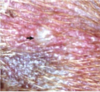
How would you describe this?

Tan-yellow firm plaque-like mass with an ulcerated surface.
Should point out that ulcers are typically depressed but this is elevated.


