Dermatology Flashcards
Multiple failed treatments for scabies

Infantile acropustulosis
intensely pruritic papulopustular lesions
tx: topical steroids and antihistamines
Staph Scalded Skin Syndrome vs. TEN/Steven Johnsons

SSSS: Plane of cleavage is higher, in the epidermis. Thin walled bullae
TEN/SJS: cleavage is at the basement membrane, thick walled bullae

Langerhans Cell Histocytosis
+/- systemic disease
non healing diaper dermatitis
hepatosplenomegaly,, FTT, lymphadenopathy, crhonically draining ears
Biosy: langerhans granules within cytoplasm of infiltrating cells

Scabies
tx: topical permethrin, x 2 courses (adult lice and 2nd for hatched eggs)

Serum sickness
urticarial lesions, periarticular swelling, extremity angioedema
nonpruritic
migratory stocking glove angioedema

Nevus sebaceus of Jadassohn
hairless, well circumscribed skin/yellow/orange waxy plaque

Infantile atomic dermatitis , birth-6months
ie eczema.
spartes diaper area

Scabies mites and eggs

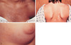
Urticaria pigmentosa
reddish-brown lesions, hyperpigmentation
cutaneous mastocytosis
Darrier sign: wheal and flare after stroking
Papular urticaria
severe, excoriated, papular reaction developed in response to recurrent flea bites
highly pruritic


Henoch Schonlein purpura

Pyogenic granuloma
benign vascular tumors, stem from grannulation tissue following foreing body/minor trauma
onset well after newborn period

Pityriasis rosea

Job syndrome
pruritic dermatitis (elevated IgE)

Auspitz sign
removal of psoriasis scale produces small points of bleeding from tortuous capilaries
KOH scraping

Candidal diaper dermatitis
pseudohyphae and spores

Guttate psoriasis
small droplike plaques with typical scales forllowing strep infection

Staphylococcal diaper dermatitis
thin walled pustules, erythamtous halos, collarette of scale

Granuloma annulare
NON pruritic, no scale

Psoriasis

Tinea versicolor
“spaghetti and meatballs” combination of hyphal and yeast forms

Ichthyosis vulgaris
Autosomal dominant
thick dry fish like scales

Lichen striatus
flat topped papules appearing in a linear or swirled districution along the lines of Blaschko
Self resolving, 1-3yrs

Erythema multiforme
commonly soles, palms, extensor surfaces

Lichen Planus
flat topped, pruritic polygonal violaceous papules and plaques
dorsal surfaces
tx: topical steroids

Mastocytoma
solitary reddish-brown plaque
postivie darier sign: after stroking, wheal and flare

Pityriasis alba
common in atopic dermatitis patients
poorly defined hypopigjmented, round scaly patches
not wood lamp enhancing

Juvenile Xanthogranuloma
infiltration and proliferation of histiocytes
if multiple lesions: refer to Ophtho, can be associated with hyphema

Nummular Eczema
annular/coin shaped
Tinea versicolor
caused by pityrosporum
Color change with Wood’s lamp
topical therapies (selenium sulfide, azoles)
KOH scrapings reveal the following

Tinea corporis
fungal hyphae seen as long septate branching rods at the margins and center of the scales

Tzanck preparation
multinucleated giant cell–> HSV or VZV

Tinea corporis, “ringworm”
Oral therapy (griseofulvin) when multiple lesions

X linked Ichthyosis
males
“dirty brown”, generalized scales
increased risk for undescended testes, testicular cancer

Dyshidrosis
chronic cracking, oozing,scaling after intiial tiny pruritic vesicles have been scratched
palms, soles, lateral fingers and toes
“tapioca” papules

Neonatal lupus erythematousis
annular rash and tissue infarction (right ear pinna)
thrombocytopenia, hypocomplementemia and elevated transaminases
ANA and AntiRo positive

Lanellar Ichthyosis
most severe
collodion membrane
flexural areas involved, eversion of eyelids and lips

Blistering distal dactylitis
tense blister filled with purulent fluid
Group A strep
larger size of initial vesicles compared to herpetic whitlow

Incontinentia pigmenti
linerarly distributed vesicles, warty papules, swirls and streaks
X linked dominant disorder, predominantly females (males lethal)
along lines of blaschko
CNS abnormalities,
