Derm Flashcards
What are the 3 most common skin and soft tissue infections? what is included in their differential dx?
Cellulitis Erysipelas Skin abscesses Gout DVT Venous stasis dermatitis (Bilateral)
Skin layers affected in Cellulitis, Erysipelas and Skin abscesses?
cell - deeper dermis and subcutaneous fat
erys - upper dermis and superficial lymphatics
skin ab - upper and deeper dermis
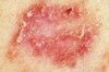
Name skin condition: unilateral presentation raised above level of surrounding skin with clear demarcations b/w involved and uninvolved skin. Non-purulent. acute onset of sx.
Erysipelas
b-hemolytic strep can present with butterfly rash on face**
pathogen responsible for: erysipelas cellulitis abscesses
ery - B-hemolytic strep
cell - b-hemolytic strep, staph aureus, MRSA
abscesses: sstaph aureus, MRSA
risk factors for Cellulitis, Erysipelas and Skin abscesses?
Skin barrier disruption
Preexisting skin conditions (eczema, impetigo, tinea)
Skin inflammation
Edema due to lymphatic drainage or venous insufficiency (venous stasis presents BILATERALLY)
Obesity
Immunosiuppression
Close contact w/ people w/ MRSA
complications of Cellulitis, Erysipelas and Skin abscesses?
NF
bacteremia and sepsis - blood cx
osteomyelitis - x-rays
septic joint - aspiration
Pasteurella multocida
cat bite
Capnocytophaga canimorsus
dog bite
Erysipelothrix rhusiopathiae
farm animals
Vibrio vulnificus
water borne = step on something at beach
Pseudomonas aeruginosa
must cover if pt is a diabetic
Sporothrix schenckii
rose gardener
Define impetigo
contagious superficial bacterial infection seen most commonly on the face seen in children age 2-5 more common in summer and fall
Primary vs secondary impetigo
primary - direct bacterial invasion of normal skin
secondary - infection at sites of skin trauma
what is the most common bacterial infection in children?
impetigo 3rd most common skin condition in children
Name the skin condition

non-bullous impetigo
Most common form
S auerus
Name skin condition?

bullous impetigo
vesicles enlarge to form flaccid bullae with clear fluid
becomes darker -> rupters leaving thin brown crust
fewer lesions - seen primarily in children
trunk more affected
S.aureus strain that produces a toxin that causes cleavage of superficial skin layer
Nsme skin condition?

Impetigo + Ecthyma
ulcrative form
lesions extend through epidermis to deep dermis
“punched out” ulcers covered in yellow crusts
Group A beta hemolytic strep pyrogenes
Treatment for Impetigo + ecthyma
ORAL
Dicloxacillin 250 mg QID
cephalexin 250 mg QID
erythromycin (penicillin allergy)
clindamycin (MRSA suspected)
Treatment for non-bullous and bullous impetigo?
TOPICALS
mupirocin (bactroban) TID
retapamulin (Altabax) BID
ORAL - if extensive
dicloxacillin
cephalexin
erythromycin (for penicillin allergy)
clindamycin (if mRSA suspected)
Complications of impetigo?
poststreptococcal glomerulonephritis
edema
HT
fever
hematuria
all seen 1-2 wks post infection
MRSA CA vs MRSA HA
CA -
- Sensitive to non-beta-lactam antibiotics
- Initially reported in IVDU
- Most frequent cause of SSTI presenting to US ERs and ambulatory clinics
HA
- Infection that occurs >48 hours following hospitalization
- Leading cause of surgical site infection
- Multidrug resistance
Treatment for MRSA
PO
trimethoprim-sulfamethoxazole
clindamycin
doxycycline
minocycline
IV
vancomycin
daptomycin
Define clinical presentation of Urticaria
intensley puritic raised erythematous plaques with central pallor
ANY area of body can be affected
waxing and waning (lesions appear and disapper w/in 24 hrs) - more severe at night
sometimes accompanied by angioedema (lips, extremeties, genitals)
Etiology and Pathophysiology of urticaria
infections
IgE mediated
direct mast cell activation (narcotics, muscle relaxants, radiocontrast agents, vanocmycin)
physical stimuli
mediated by mast cells in superficial dermis and basophils
release multiple mediators including histamine and vasodilatory mediators
Name medications that result in direct mast cell activation?
narcotics/opiods
muscle relaxants
radiocontrast agents
vancomycin
Management of urticaria
focus on short term relief of puritis and angioedema (2/3 will spontaneously resolve)
H1 antihistamines
- Diphenhydramine (Benadryl)
- Chlopheniramine (chlor-trimenton)
- Hydroxyzine (Vistaril)
- Certirizine (Zyrtec)
- Loratadine (Claritin)
- Fexofenadine (allegra)
H2 antihistamines
- Ranitidine (zantac)
- Nizatidine (Axid)
- Famotidine (Pepcid)
- Cimetidine (Tagamet)
glucocorticoids for sx lasting longer then 2-3 days (SEVERE)
prednisone 30-60 mg taper over 5-7 days
Name H1 antihistamines used to tx urticaria
- Diphenhydramine (Benadryl)
- Chlopheniramine (chlor-trimenton)
- Hydroxyzine (Vistaril)
- Certirizine (Zyrtec)
- Loratadine (Claritin)
- Fexofenadine (allegra)
Name H2 antihistamines used to tx urticaria
- Ranitidine (zantac)
- Nizatidine (Axid)
- Famotidine (Pepcid)
- Cimetidine (Tagamet)
Define Lipoma
most common benign soft tissue neoplasm (!% of population)
soft, painless, round or oval subcutaneous nodule
mature fat cells enclosed by a thin fibrous capsule - rarely inolve fascia or deeper muscles (superficial)
common on upper extremities and trunk
Lipoma and genetics
cause of lipomas are unknown - some have genetic predisposition
familial multiple lipomatosis - multiple lipomas in multiple family members
Gardner Syndrome - rare inherited condition characterized by familial adenomatous polyposis (multiple lipomas), common on face, scalp, neck and trunk
Tx for lipomas
none
if bothersome - surgical excision of fat cells and fibrous capsule (cosmetic or pain reasons)
What is the most common cutaneous cyst?
Epidermal inclusion cysts
firm, skin colored dermal nodules with visable central punctum
asymptomatic, can be red and inflammed
freely moveable
Pathophysiology of epidermal inclusion cysts
implantation and proliferation of epithelial elements into dermis (dead skin implants into hair follicle) from trauma or comedome
cyst wall consists of normal stratified squamous epithelium
Tx for epidermal inclusion cysts
asymptomatic - no tx, will resolve on own
Not inflammed - punch excision of cyst and drainage = must pull out all of “cheesy inside” along with capsule or cyst will reform
intralesional injections w/ triamcinolone - inject cyst w/ steroid to decrease size and inflammation
•Most common cutaneous disorder affecting adolescents and young adults (mostly males) that resolves in third decade of life
acne
Four main factors are involved in acne patho
- Follicular hyperkeratinization
- Increased sebum production - provides growth medium for c. acnes
- Cutibacterium ances (FKA Propionibacterium acnes) within the follicle - Microcomedones provides an anaerobic lipid-rich environment for bacteria
- Inflammation -Microcomedones provides an anaerobic lipid-rich environment for bacteria
cause of infantile acne?
androgens
Contributes to the development of acne through stimulating the growth and secretory function of the sebaceous gland
Labs ordered in pt w/ acne.
DHEA-S
total testosterone
free testosterone
4 types of Rosacea?
- Erythematotelangiectatic
- Papulopustular
- Phymatous
- Ocular rosacea
cause of Rosecea
Cause is unknown, possible factors include
- Immune dysfunction
- Inflammatory reactions to cutaneous microorganisms
- UV damage
- Vascular dysfunction
Dx? and Tx?

Erythematotelangiectatic or “classic rosecea”
FIRST LINE – avoid triggers, sun protection and decrease alc intake
SECOND LINE – laser and light-based therapy
Pharmacotherapy – alpha adrenergic agonists
- Topical brimonidine (Mirvaso)
- Topical oxymetazoline (Rhofade)
type of roseca more common in MEN
Phymatous
Pt presents w/ papules and pustules in central face. On examination you see reddness extending beyond the follicle.
Dx and Tx:
Papulopustular R
Topical metronidazole 0.75% (most common)
Azelaic acid 20% cream
Ivermectin 1% cream
Oral
- Tetracycline, doxy, minocycline
- Isotretinoin 0.3 mg/kg QD
Tx of phymatous Rosecea
Oral isotretinoin 0.3-1 mg/kg QD in early disease
Laser ablation and surgery in advanced disease
Side effect seen in more then 50% of pts w/ Rosecea
Ocular!!!!!
Lesions are characterized by well-demarcated erythematous plaques with silver scale. Dx??
Psoriasis
•When compared to normal epidermis, psoriasis epidermis shows:
- Increased number of epidermal stem cells
- Increased number of cells undergoing DNA synthesis
- A shortened cell cycle time of keratinocytes
- A decreased turnover time of epidermis
Most common type of psoriasis and where do the lesions present?
chronic plaque
symmetrically distributed on:
Scalp, extensor elbows, Knees and gluteal cleft most common sites
pt is a 10 y/o boy presnting to clinic w/ a rash on trunk and proximal extremties. you recognize the child as he came into clinic a few weeks ago to be tx for strep pharyngitis.
Dx and Tx????

guttate psoriasis
will resolve and not likely lead to chronic psoriasis
Pt presents w/ rash. you see no visable scaling. she was previously prescribed ketoconazole by another provider. after no relief of sx she presents to you.
dx??

Inverse psoriasis
most common clinical manifestation in pts w/ psoriatic arthritis?
nail psoriasis (nail pitting)
pts presents w/ fever, diarrhea, leukocytosis and hypocalcemia. on examination you see widespread erythema, scaling, and sheets of superficial pustules.
Dx and what could have caused this?
pustular psoriasis
pregnancy, infection or withdrawal of oral glucocorticoids
what is erythrodermic psoriasis
Generalized erythema and scaling from head to toe
inpatient management
what is auspitz sign
pinpoint bleeding after removal of plaque
dx of psoriasis
Tx choices for MILD psoriasis
less then 5% BSA
emollients
top corticosteroids - Hydrcortisone 1%, Triamcinolone 0.1%, Flucinonide 0.05%
Tar-T gel (neutrogena)
Vitamin D analogs
topical retinods (tazarotene)
Tx for mod-severe psoriasis ?
5-10% BSA
phototherapy (photochemotherapy)
- w/ either oral or bath psoralen followed by UVA radiation
- Increased cancer risk of non-melanoma skin cancer and melanoma
excrimer laser
methotrexate
cyclosporine
aprimelast
- •TNF- alpha inhibitors*
- •IL-17 inhibitors*
- •IL-23 and related cytokine inhibitor*
- •*specialist managed*
define hidradenitis suppurativa and the pathophys of the dz
Apocrine sweat gland dysfunction
- Follicular occlusion, follicular rupture and associated immune response
- Ductal keratinocyte proliferation -> ductal plugging -> expansion -> rupture and release of contents -> stimulating immune response and leading to sinus tracts in skin
Describe the 3 stages of the Hurley staging system for HS.
1: abscess formation
2: recurrent abscess formation w/ sinus tract formation and scarring
3: Diffuse involvement of multiple connected sinus tracts
Dx (stage) and tx

Avoidance of skin trauma
Smoking cessation
Weight management
Antiseptics – chlorhexidine 4% once/week
Emollients
Management of comorbidities
Dx (stage) and tx

Oral tetracyclines for several months
Clindamycin 300 mg BID and rifampin 600 mg QD
Oral retinoids
Antiadrenergic therapies – OCPS, spironolactone
Punch biopsy of fresh lesion (drain tracts
dx (stage) and Tx

TNF- alpha inhibitors
Adalimumab
Infliximumab
Systemic glucocorticoids – prednisone
Cyclosporin
Surgery
along w/ hair loss what is another common sx in pts w/ alopecia?
Onychorrhexis – longitudinal fissuring of nail plate
pathophys of alopecia
- Autoimmune disease in which hair follicles in the growth phase (anagen) prematurely transitions to the non-proliferative involution (catagen) and resting (telogen) phase
- This leads to sudden hair shedding and inhibition of hair regrowth
- T-cell mediated
- Inappropriate trigger of immune response against follicular antigens
Commonly presents
diagnostic finding of pts w/ alopecia
there are 2****
exclamation point at hair margins
•Skin biopsy -> Peribulbar lymphatic infiltrates surrounding follicles (swarm of bees)
first line tx for limited patchy hairloss
- Topical or intralesional corticosteroids
- Triamcinolone 2.5-5 mg/ml
- Betamethasone dipropionate 0.05%
or topical
Monoxidil
anthralin cream
PUVA
first line tx for extensive hair loss
- Topical immunotherapy
- Diphenylcyclopropenone (DPCP)
- Squaric acid dinutyl ester (SADBE)
Systemic therapies
Oral glucocorticoids
Sulfasalazine
Methotrexate
Cyclosporin
Biologics
what is eczema (dermatitis) closely related to?
asthma
allergic rhinitis
what type of hypersenitivity rxn is atopic derm
type I IgE mediated
•Intense itching produced by mast cells and basophils in dermis.
where does atopic dermatitis usually present
•Presents on flexor surfaces, neck eyelids, face, dorsum of hands and feet
** face and extensor/flexor in children
you dx your pt w/ atopic dermatitis but she DOES not want steroid creams and wants something she can use longer term?
What do you prescribe?
- Pimecrolimus (Elidel)
- Nonsteroidal
- Addition/alternative to topical steroids.
- Good for long term use.
OR
- Topical immunemodulators
- Tacrolimus (Protopic) -> Nonsteroidal (cytokine inhibitor)
- Used as an addition/alternative to topical steroids.
pt presents w/ coin shaped pruritic patches and plaques in clusters on her legs.
Name another condition this resembles and what you would do to rule it out.
Dx and TX

tinea corporis - usually clear in center
r/o w/ KOH swab
dx Nummular Eczema
tx
- Acute: Intermediate strength topcial steroid (triamcinalone cream 0.1%).
- Or if severe, high potency (Clobetasol ointment) +/- occlusion.
Long term: treatment with less potent topical steroids
pt presents w/ small vesicles appear on hands and feet complaning of pruritus
dx? and cause?

Dyshydrosis
•inflammation and foci of intercellular edema (spongiosis) which becomes loculated in the skin of the palm and soles.
tx dyshydrosis
- Mild cleansers (Cetaphil)
- Emollient barrier creams, protective gloves, avoidance of irritants.
- Burow’s solution. (Antibacterial Astringent)
- Topical corticosteroids are the mainstay.
- High: Clobetasol Ointment for acute flare
- Med: (triamcinolone 0.1% or Fluocinonide 0.05%) with or without occlusive dressing.
- Protopic and Elidel for long term management
Dx and Tx

stasis dermatitis - seen mostly in women
- Elastic compression stockings
- Burrow’s solution
- Moderate topical steroid: Desonide, Triamcinalone cream.
- Treat any secondary infection with po Abx. (Keflex)
woman presents in clinic w/ a rash showing clustered papulopustules on erythematous bases w/ scale around her mouth. she tells you she just finished using hydrocortisone cream.
dx and tx
- Topical antibiotics: Metronidazole or erythromycin.
- Severe cases may require oral minocylcin / doxycycline.
- *Avoid topical steroids* - will exacerbate sx
how do we tx Seborrheic Dermatitis on both scalp and face??
- Scalp: zinc shampoo, Ketoconazole shampoo
- Face, intertiriginous areas: Low potency topical steroids
(Desonide or Valisone Cream)
offending agent: p. ovale
pt presents w/ hardened area over wrist. It is a solitary pruritic eczematous eruption that appears Lichenified.
dx and tx
Lichen Simplex Chronicus
Intermediate strength topical steroid.
- Triamcinalone cream 0.1% prn
- Occlusion when able
- Oral antihistamines
- Protopic
- Elidel 1%
upon a yearly skin check of your 35 y/o pt. she complaing of a Purple, Polygonal, Pruritic, Papule on her scalp. w/ further examination of the mouth you see white lesions in the buccal mucosa.
Dx? Tx
Lichen Planus
Potent topical steroids with occlusion dressing, or intralesional steroid injections.
a 70 y/o pt comes in for a skin check. you see this. it appears to be stuck on and hace a warty appearance.
Dx tX?

Seborrheic Keratosis
none
•Vascular neoplasm brought on by genetic factors, hormonal factors, immunodeficiency or infection with Human Herpes Virus 8????
Kaposi sarcoma
Pt presents to clinic w/ this lesion on her face. on palpation you feel a sandpaper texture.
Dx and TX

actinic keratosis
Precursor for squamous cell carc.
tx of actinic keratosis w/ limited number of lesions
For extensive broad and numerous lesions???
•Cryotherapy
Imiquimod (less irritating then 5-Fu)
what is the most common cancer
basal cell
pathophys of basal cell
- BCC arise from immature pluripotential cells associated with the hair follicle. Mutations activate pathway that controls cell growth.
- Mutation also activates oncogenes and inactivates tumor suppressor genes, leading to tumor growth.
what is characteristic ft of all basal cell carc.
Bleeding w/out pain
Pt presents to clinic w/ waxy, pearly, semitranslucent nodules or papules with “rolled edge” forming around a central depression that is ulcerated, crusted and bleeding.
Dx ??

nodular basal cell
what form of skin cancer is most common in hispanics and asians??
Pigmented
on examination appears appears as a dry scaly lesion, superficial flat growths,
Dx?

Superficial BCC
pt arrives w/ complaints of a new plaque on side of nose. on physical exam you notice a white sclerotic plaque with telangiectasia.
Dx?
Morpheaform (sclerosing) BCC
gold standard tx for BCC
Mohs procedure
topical tx for BCC
- 5% imiquimod
- for the treatment of nonfacial superficial BCCs that are less than 2 cm in diameter. Applied 5 days per week, for a duration of 6-12 weeks. Has 80% cure rate.
- 5-FU
- approved for the treatment of superficial BCC, administered twice daily for 3-6 weeks.
where is SCC most frequently seen
areas of AK, sun exposed skin
face, backs of hands.
type of skin cancer w/ no preference for sun exposed areas
melanoma
over 50% of cases develop from preexisiting nevi
Define the ABCD of melanoma
A asymmetry
B Border (edges are irregular)
C Color is varied
Diameter >6mm
what type of melanoma?

superficial spreading
what type of melanoma?
most COMMON in sundamaged skin
starts as macular and becomes nodular
SLOW indidious growth

Lentigo maliga
you see this melanoma in sun exposed areas head, neck and trunk. it is usually Friable or ulcerated and bleeding

nodular
most common type of melanoma in darker skin types and most commonly seen on nails and feet.
acral-lentiginous
tx of melanoma
- Surgical excision
- Radiation
- Chemotherapy
Adjunct therapy
- Cytokines (IL-2)
- Vemurafenib
- Dabrafenib
- Trametinib
pt came into office last week and a questionable lesion was biopsied. results show the presence of keratin or “keratin pearls.” You read the pracitioners note where they add that there was Lymphadenopathy on palpation in adjacent lymph nodes.
Dx?
SCC
etiology of melanoma
- Damage to DNA of Melanocyte that promote oncogenes and inhibit tumor suppressor genes.
- Half have non inherited BRAF oncogene mutation
Familial inherited melanomas often have mutation in tumor suppressor genes like CDKN2A and CDK4
describe lesions seen in measles (rubeola)
start as macular or morbilliform rash on anterior scalp and behind ears then by day 2 or 3 down the trunk to extremities
Including palms/soles
what is pathogonomonic finding in rubeola (measles)
Koplick spots (white papules on buccal mucosa)
is there a prodrome w/ measles?
Yes
pathogen responsible for rubella?
toga virus
what is this called and a sign of what dz?

Forscheimer’s sign - rubella
is there a prodrome w/ rubella
NO
pt presents w/ pale pink morbilliform macules that began on face and spreads inferior, covering entire body in 24h. he also notes pain w/ upward lateral eye movement.
dx?
rubella
pathogen responsible for fifth dz (erythema infectiosum)
Parovirus
what are the 3 phases in fifth dz (erythema infectisum)
- 1st: abrupt asypmtomatic erythema of cheeks (slapped cheek) that is diffuse and macular.
- 2nd: by day 4 discrete erythematous macules and papules on proximal extremities and later the trunk evolving into lacey reticulate pattern by day 9.
- 3rd: recurring stage, eruption is reduced or invisible, only to reoccur with exposure to heat (bath) or sunlight.
what is a herald patch diagnostic of.
Pityriasis Rosea
what is the most common form of adverse drug rxn? where do the lesions begin and spread to?
morbilliform rash (type IV hypersen)
Erythema with macules and papules initially on trunk then generalizing within 2 days.
pt presents to clinic w/ strange mark on penis. he says it reoccurs and it sometimes itchy. The only changes he has made to his routine is taking ibprof after football practice to relieve muscle soreness.
Dx? and what causes this?

fixed drug rxn
NSAID, Sulfonamides, Barbiturates.
pt presents to clinic w fever and painful lesions. upon examination they have a target appearacne. currently they are localized to hands and feet. SHe is taking phenytoin and penicillin.
Dx? Tx?
Erythema Multiforme
if severe - systemic steroids
pt presents w/ blistering painful lesions on trunk. Dx?? and what are we concerned for?
SJS
progressing to TENS
Bullous pemphigoid patho?
- IgG Antibodies bind to basement membrane. à activates complement and inflammatory mediators à attracts inflammatory cells to the basement membrane which release proteases lead to blister formation.
- Blister formed by cleavage of the basal cells away from the basal lamina.
- Antibodies cause separation of epidermis from dermis.
tx for bollous Pemphigoid
potent topical steroids
occlusive dressing
Severe - prednisone
pt complains of burrowing lesions located on feet and web spaces b/w hands. lesions are itchy?
Dx? what does this dz spare?
scabies (delayed IV hypersen)
face
what type of bug manifestation would we only see immunocompromised pts
norwegian scabies
tx of scabies
topical
- Permethrin 5% cream. (Elimite)
- Lindane1 % lotion or cream (Kwell)
- More toxic (not for pregnant or kids
Oral
•Ivermectin (Stromectol)
how to differentiate b/w Pediculosis Corporis and scabies
Pediculosis Corporis SPARES hands and feet
scabies does NOT
pt dx w/ Pediculosis capitus along w/ intense itching of the scalp and lice within the hair follicles what other manifestation would we see?
posterior cervical lymphadenopathy,
most effective tx for head lice
Ovid Lotion – most effective for head lice NOT for children
Tx for lice
•OTC Nix cream rinse
Permethrin acts as neurotoxin resulting in paralysis of nerves in exoskeletal respiratory muscles of parasite à death
Ovid Lotion
bactrim
vaseline
along w/ the many systemic sx what sx is most severe in pts w/ a black widow spider bite
Abdominal pain
what spider bite leads to necrosis of the tissue
brown recluse
Dz and Tx?

black widow bite
ACLS
Antivenom administered in ER. (risk of allergic reaction).
Analgesics (Morphine)
Anithistamine (Benadryl)
Tetanus
pathogen responsible for tinea versicolor
Malassezia furfur (yeast).
pt arrives to clinic at the end of august complainig of well defined round macules with scaling on trunk and arms, or face. under woods light you see:
Dx/Tx

Tinea versicolor
Daily Selenium sulfide shampoo
Topical Ketoconazole cream daily x 3 weeks.
Oral Ketoconazole (careful of LFT’s).
diagnostic test when you suspect fungal infection:
KOH smear
a KOH smear was performed and results show hyphae and spores (Spaghetti and meatballs).
Dx?
tiinea versicolor
if you did not have access to a woods light. how do you differentiate b/w Vitilago and tinea versicolor?
ttinea vers:
Pt is asymptomatic and notices during the summer.
Well defined round macules with scaling on trunk and arms, or face.
vitilago
hypopigmentation macules may occur focally or generalized in pattern. NO SCALE
Hair in vitiliginous areas usually become white
pt arrives to clinic complaining of a lesion on her mouth. Physical exam shows tender grouped vesicles/blisters on an erythematous base.
Dx? and what test could you perform to comfirm your dx?
herpes
Tzanck smear (giant nucleated cells)
Tx for herpes
acyclovir
valcyclovir
pt comes to clinic complaining of
Rash, malaise, low grade temp
She says the rash stared as faint macules that develop into vesicular eruptions with “teardrop” vesicles on erythematous base. you notice the rash is only on the scalp and face.
Dx? and where shoudl you warn the pt that the rash will spread to?
varicella
trunk, then spreads to extremities. (May appear on palms/soles. )
pt comes to clinic complaining of lesions on the side and tip of the nose. she describes them as “burning”, “electrical”, “throbbing”. The lesion is unilateral.
what is this lesion called?
Dx?
How should you advise pt?
shingles (herpes zoster)
hutchinsons sign
Ophthalmic division of 5th cranial nerve must be seen by ophthalmologist due to complicating concerns of tetinal necrosis, glaucoma, optic neuritis.
which dz shows lesions usually unilaterally and along a dermatome?
herpes zoster
what is this?

hutchinsons sign - herpes zoster
what is this indicative of?

varicella
how do you differentiate herpes simplex from varicella based on Tzanck smear?
varicella
Tzank smear from vesicle show multinucleated giant cells
herpes
Tzanck smear -> Giant nucleated cells
what is Onychomycosis and how do we tx?
Lamisil
check LFTs
what dz is associated w/ break in the skin associated with trauma to the eponychium (cuticle) or nail fold and maceration of proximal nail fold. usually seen in people in the food industry/ always have hands in water?
what pathogens are responsible?
Chronic Paronychia
•pseudomonas aeruginosa or candida albicans
what dz is associated w/ aggressive manicure, nail biting?
pathogen responsible?
Acute Paronychia
Usually Gram + (Staphylococcus aureus)
tx for chronic paronchyia
Avoid inciting factors (moisture, manicuring)
Warms soaks
Topical steroid cream o
Antifungal : Spectazole
tx for acute paronchyia
Warm water soaks 3-4xday
PO Abx for Gr+ S.aureus (Augmentin 2gr x 5d)
Topical steroid cream
I&D if abscessed.
what is the most common wart that is associated w/ frequent exposure to water?
Veruca Vulgaris (common wart)
10 y/o comes into office and presents w/ 2-4mm flat topped flesh colored papules on face.
Dx?
Verruca Plana (flat wart)
what wart is found on the soles of feet/ pressure points?
Verruca Plantaris (plantar wart)
4 Ps of Lichn Planus
Purple
- Polygonal
- Pruritic
- Papule
tx of warts
IF has not resolved on own:
- Cryotherapy
- Salicylic acid / Cantharidin
- Occlusive dressing
- Intralesional injection of Bleomycin
Herpetic Whitlow:
occurring on the fingers or periungually, tenderness and erythema with deep seated blisters
Tzank smear from vesicle show multinucleated giant cells
varicella
Tzanck smear à Giant nucleated cells
herpes simplex
is erysipelas purulent?
no
most common bacterial infection in children
impetigo
Name the types of BCC
nodular
superficial
Morpheaform (Sclerosing):
pigmented
type of BCC seen in darker complextions
pigmented
type of BCC that may be misdiagnoses as eczema or psoriasis.
superficial
Name 4 types of melanoma
superficial spreading
lentigo maliga
nodular
acral-lentigous
most common melanoma in sundamaged skin
lentigo maliga
type of melanoma that is seen on sun exposed areas and is probable ulcerative or bleeding
Nodular
name most common melanoma in darker skin types
acral lentigous
where do acral lentigous melanoma commonly present
palms
soles
nailbeds
type of melanoma that starts macular then becomes nodular w/ a slow insidous growth rate
lentigo maliga
melanoma that is
Tendency to multicoloration including black, red, brown, blue and white.
Boarders tend to be more sharply defined.
superficial spreading melanoma
Lesions on sun exposed areas may be superficial papules, plaques or nodules, discrete and hard arising from an indurated, round elevated base.
Over months becomes larger and ulcerated, initially covered by crust.
SCC
hallmark of SCC on biopsy
•Biopsy: The histologic hallmark of SCC is the presence of keratin or “keratin pearls.” These are well-formed desmosome attachments and intracytoplasmic bundles of keratin tonofilaments.
what is the Most common epithelial precancerous lesion
actinic keratosis
•Spindle cells found in nodular lesions. Dx?
Kaptosi sarcoma
estimated that over 90% of adults over the age of 60 years have one or more
Seborrheic Keratosis
May be asymptomatic, but often pruritic. 2/3 of people will have lesions for < 1yr. May cause hair loss and damage nails. Variations can be ulcerative.
- Grouped together, flexor aspect of wrists, lumbar area, eyelids, shins, scalp.
- May have Reticulate white lesions on buccal mucosa.
- 4 P’s. Purple, Polygonal, Pruritic, Papule.
dx?
Lichens Planus
tx lichen plannus
Potent topical steroids with occlusion dressing, or intralesional steroid injections.
type of impetigo that forms thick adherent golden crusts
non-bullous (most common)
S. Aureus
enlarges vessicles w/ flaccid bullae w/ clear fluid become darker and ruptures leaving a thin brown crust.
Dx?
bullous impetigo
S. Aureus strain - that produce a toxin that cause clevage of superficial skin layer
Pathogen responsible for ecthyma
Group A beta hemolytic strep pyogenes
what skin condition is sometimes accompanies by angioedema
urticaria
pt presents w/ firm skin colored dermal nodules, visible central punctum. they are asymptomatic.
dx?
epidermal inclusion cysts
differentiate b/w wart and epidermal inclusion cysts
epidermal inclusion cysts have a central punctum
tx for mild acne
BPO + topical retinoid / abx
tx of moderate acne
BPO + topical retinoid + oral abx
tx for severe acne
everything
+oral isotretinoin
psoriasis seen in children/ young adults w/ no hx of psoriasis
guttate
name 4 types of rosacea
Erythematotelangiectati
Papulopustular
Phymatous
ocular
name 6 types of psoriasis
chronic plaque
guttate
pustular
Erythrodermic
inverse
nail
lower lip SCC
- Starts as actinic cheilitis
- Local thickening on keratosis then firm nodule that may grow outward as sizable tumor.
- Usually + hx of smoking
Periungual SCC
- Presents with signs of swelling, erythema and localized pain.
- Commonly in the nailfolds of hands resembling a wart
dry scaly lesion w/ threadlike border
Superficial BCC
misdx as psoriasis due to appearance of flat scaly lesion
melanoma w/ no apparent radial growth phase, Smooth, Dome-shaped and shows ulcerations/bleeding
nodular
primarily sun exposed areas of head, neck and trunk
melanoma w/ sharply defined borders
superficial spreading
appears pigmented- blue, black, white
melanoma that is Light brown, uniform pigmentation
acral lentigous
seen on darker skin types, nails ans palms of feet