Derm Flashcards

1
Q
A
2
Q

explain the location of atopic dermatitis

A
3
Q

atopic dermatitis

what age group is this found it? what are the three characteristics in a patient that make them higher risk for this? what are three things you might see on this patient? what type of hypersensitivity is this? where is this most commonly found (2) places and 4 others? what must you differentiate from? what is the diagnosis made from?

A

chronic relapsing skin disoder that starts in children and goes through adulthood

ALLERGY, ALLERGIC RHINNITIS, ASTHMA, PERIORAL PALOR, DENNIE MORGAN LINES, ALLERGIC SHINERS

type 1 IgE mediated hypersensitivity

-dry skin, puritis with “itch scratch cycle” flexor creases (antecubital and popliteal) most common

neck, eyelids, forehead, face, and dorsum of hands and feet, dermatographism characteristic

can be either atopic or contact (touching something you’re allergic to)

*make sure to culture for S. aureus and make sure not secondary infection and HSV in crusted lesions*

DX: IgE serum levels

4
Q

atopic dermatitis

what are 6 possible treatment options for this?

A

1. avoid irritants

  1. skin lubrication/emollients (moistureizers)

3. topical corticosteroids

4. antihistamines

  1. calcineurin inhibitors (tacrolimbus + pimecrolmus)
  2. UVB phototherapy is effective
5
Q

for atopic dermatitis….what can you do to figure out what is causing it?

A

PATCH TESTING!!!

6
Q

contact dermatitis: allergic

what cells initiati this? what are 5 common causes? what is the difference between the acute and chronic skin presentations?

A

t-cell mediated, occurs in those that have become sensitized

  • common causes: medication, jelwrey, rubber, disinfectants, cosmetics, plants*
    acute: erythema, vesicles, eroisions, crusting
    chronic: papules, plaques, crusts

**can do patch testing after dermatitis to figure out the cause**

7
Q

contact dermatitis: non allergic

what is this caused by? (3)

what do the acute and chronic skin presentations look like?

A

comes from contact with an irritating substance

direct toxin on the skin

common causes: abrasive, cleaning agents, caustic agents

Acute: erythema, vesicles, erosions, crusting

chronic: papules, plaques, crusts

8
Q

where is the rash for seborrheic dermatitis?

A
9
Q

seborrheic dermatitis

what does this occur in? what are the four most common places? what is the most common agent? what does this cause in infants and what does this cause in adults? what do the infected areas look like? what are the 7 treatment options for this?

A

subacute/chronic inflammation of the areas with increased SEBACEOUS GLANDS

body folds, face, scalp, genitalia

caustitive agent: pityrosporum ovale (yeast)

“cradle cap” in infants, “dandruff” in adults

scattered yellow gray scaly macules and papules with GREASY LOOK!!!

sticky crusts with fissures found behind the ears!!!

Tx:

  1. OTC dandruff shampoo
  2. cold tar shampoo
  3. Ketoconazole
  4. cradle cap: olive oil compresses, shampoo with sulfur, or ketoconazole shampoo
  5. UV radiation

6. shampoos with selenium sulfide

7. topical steroids hydrocorisone
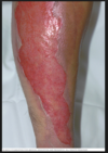
10
Q

stasis dermatitis

what causes this? what two populations of people is it worse in? the incompetency leads to what 5 things? what does the pt complain of? what 3 things do you see before the skin changes? what are two ways to make the DX and what do you see?

A

chronic rash on lower legs secondary to venous insufficiency

worse in pregnancy and women

see papules, scales and crusts

valvular incompetency leads to edema, dermatitis, brown stippled hyperpigmentation, fibrosis, ulceration (30%)

pt complain of aching in legs that is worse with standing, relieved by walking

typically see caricose veins, superficial phlebitis, and venous thrombus before skin changes

DX:

  1. doppler studies, sonography or venography will confirm chronic insufficiency
  2. biospy-show dilated tortuous veins, edema, and fibrin deposition
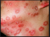
11
Q

dyshidrosis

what type of dermatitis is this and where does it occur? what age group? what is it commonly associated with? what is the funny food it is associated with looking like and what other characteristic is common? what are four triggers? what does it transform in to in late disease (5)? what two things are used to diagnose it?

A

acute/chronic puritic vesicular dermatitis on palms and soles

purititc, clusters of small vesicles in clusters “tapioca like appearence” on palms/soles

triggers: sweating, emotional stress, warm/humid weather, metals

Late disease: papules, scaling, lichenification, erosions ruptured from vesicles, painful fissures

DX: culture and KOH to rule out dermatophytosis

12
Q

dyshridrosis

what are the 5 treatment options?

A
  1. burrows solution and antihistamines (control itch)

2. topical high steroids

  1. systemic steroids if severe

4. intralesional triamcinolone

  1. fissures treated with: collodoin
13
Q

what are the treatment options for stasis dermatitis?

(6)

A

1. manage edema

2. topical corticosteroids

3. wet compress for oozing and crusting (caution with ulcers)

  1. compression stockings
  2. sclerosis of varicose veins to prevent dermatitis
  3. vascular bypass, endothelial thermal ablation or stenting (these are only mildy effective)
14
Q

explain the locations that are common for…:

  1. acne vulgaris
A
15
Q

acne vulgaris

what are the four causes?

A

1. increased sebum production seen with puberty and increased androgens

2. clogged sebaceous glands

3. propionibacterium acne overgrowth

  • anaerobic bacteria that is part of normal flora in the PS unit
  • digeste the sebum, but when large plug stimulates baterial for form lipase, this breaks down sebum in to fatty acids which spart inflammation process causing neutrophil attach to follcular wall

4. inflammatory response

16
Q

Acne Vulgaris

what are the three general types?

A
  1. comedomes

2. inflammatory pustules/papules

3. nodular or cystic acne

17
Q

acne vulgaris

comedomes

what are the two types? explain what causes each.

A
  1. open “blackheads”

  • infundibulum: hyperkeratosis, comecyte cohesiveness
  • androgen stimulations and sebum secretion
    2. closed “whiteheads”
  • accumulation of shed keratin and sebum
  • formation whorled lamellar concentrations
18
Q

what are the ratings for acne severity? (3)

what are the meds associated with each?

A

mild: comedones +/- small amounts of pustules retinoids, azelaic acids, salicyclic acid
moderate: comedones larger amounts of pustules and papules topical trentoin, erythromycin, clindamycin
severe: nodular or cystic oral minocycline, tetracycline, doxy, Isotrentoin

19
Q

erythmatotelangiectatic rosacea

what is it

A

facial flushing with telangiectasis, central face edema, SPARES PERIORBITAL AREAS

20
Q

papulopustular rosacea (PPR)

A

central face erythema with papules and pustules, less often stinging

SPARES THE PERIORBITAL AREA

21
Q

Phymatous rosacea

what is it and where does it occur?

A

follicular orficies thicken!!!! get nodularities that are disfiguring!

more common in men

get a rubery thickening of skin of the nose, chin, forehead, eyelids, or ears

22
Q

what are the treatment options for acne vulgaris? (6) options

A
  1. benzyl peroxide
  2. topical antibiotics (clindamycin, erythromycin, dapsone)
  3. azeleic acid-unique plant derived compound that has anti-bacterial and anti-comedogenic properties
  4. salycyclic and glycolic acids gels and washes
  5. topical retenoids-adapalenes (differin), tretoin, and tazarotene
  6. oral antibiotics minocycline, tetracycline, doxy! these are different than the topical abx!!
  7. isotretinoin-drastically attentuates acivitt of sebaceous glands and rate of keratinization in epi ONLY DERM CAN DO THIS HIGHLY TETRAGENIC!!!!

*****notice there is a difference between oral and topical antibiotics*******

23
Q

rosacea

what is this caused from? who is this most common in? what can be triggers for this? what are 4 unique presentations to this? what distinguishes this from acne? what are the 5 treatment options?

A

conrtoversy if cause is from inflammation or infectious

most common in fair skinned individuals

triggers: hot/cold,, HOT DRINKS, hot baths, spicy foods, ETOH, emotions

flushing and telangiectasis are key features of disease!!! causes phyma (enlargement of random area of the body), symmetrical presentation can have facial burning or stining, dry appearance, occular manifestifestations

***absense of comedones, distinguishes it from acne***

TX:

  1. topical metronidazole sodium, sulfacetamide, sulfur, erythromycin
  2. systemic abx: minocycline, doxy, metronidazole
  3. isotrentinoin
  4. ivermectin (anihelminthic)
  5. surgery for phyma
24
Q

cellulitis

where does this infection occur? what are the 3 organisms most likely to cause this in adults? what are the 3 organisms likely in kids? what are the 5 symptoms the patient will experience? what is the strange animal you can get this from? what do you do to DX this?

A

acute, spreading inflammation of the dermis and subcuteous tissues, occurs from breaks in the skin

adults: s. aureus, group A strep can get it from cat bite pasturella mulicida
children: hae. influenzae (periorbital), strep pneumoniae, s. aureus

expanding, red, swollen and tender, FEVER!! HEAD!! LYMPAHDENOPHATHY!! EDEMA!! NOT sharply demarkated…pt feels ILL

DX: culturing and drainage of discharge by needle aspiration

25
cellulitis ## Footnote what are the four treatment options? and what is really important to do when treating a cellulitis patient?
1. rest/heat 2. **begin treatment ASAP with abx that cover haemophilis influenzae, strep and staph!** 3. **dicloxacillin or cephalosporin**, if allergic to penicillin use **erythromycin** 4. if severe give **first generation cephalo IV!!** really important to mark the boarders so you can tell if it is improving or not!! may need surgery if necrotizing infection develops
26
erysipelas ## Footnote what aged people is this common in? what are the two places this commonly appeares? what is the #1 causing organism? what does the rash look like and what is it characterized yet? when does it disappeare? what is the DX and TX?
variant of cellulitis in ELDERLY on FACE AND EXTREMITIES **group A beta strep** painful macular rash with well defined margins, vessicles, FEVER, **rapid onsetand progression**"_fiery red face*"*_ * rash desquamates in 5-10 days* * DX:* bacterial culture TX: treat with antibiotics depending on organism!
27
impetigo contagiosa ## Footnote what two age groups is this common in? what two environmental things is it common with? where do you typically see this and is there a family hx? what **key** description of the lesions? what are the **3** organisms that commonly cause this? what are the three tx options?
common in INFANTS AND CHILDREN!! higher rates with poor hygiene and malnuitrition **HIGHLY CONTRAGIOUS!!! COMMONLY ON FACE!!! usually have family history of this!** **THICK CRUSTED "HONEY" colored macules and papules! starts as one, and spreads** **#1- staph aureus** **#2-GABHS** **#3-streptococcus pyoderma** **TX:** 1. _bactroban_ 2. _cephalexin_ or _dicloxacillin_ if severe and need systemic
28
bullous impetigo ## Footnote what organism causes this? who is it most common in? what three places do you see this rash most commonly present? what do you worry about as a SEVERE COMPLICATION?
Staph group II, **staphlococcus with endotoxin** **STAPH AUREUS** * most common in children/infants* overall not common * face, neck, and extremities* erythema that progresses to epidermal sloughing, worry about progression to scalded skin syndrome!!! TX: 1. bactroban 2. cephalexin or dicloxacillin if systemic needed
29
folliculitis ## Footnote what organism is most common for causing this? what is the second most common? what three areas does this most commonly effect? what are the two types of presentations you see? what are the two treatments?
**staph aureus infection of the hair follicles!** commonly seen on _butt, thighs and beard!_ "superficial hair" can be *pseudomonas* PAPULES AND PUSTULES Tx: antiseptic cleaners mupirocin
30
hidradentitis suppurativa
occurs in areas with **apocrine glands** like **axilla, angiogenital, and scalp leading to potential scarring and fibrosis** females: axilla more common men: angiogenital more common **TENDER INFLAMMATORY NODULES OR ABCESSES, open comedomes and _sinus tracts_ may drain _purlulent_ fluid** TX: 1. trimcinoloine 2. drainage of the abscess and excision of the sinus tracts 3. antibiotics until lesions resolve \*\*should consider psychological support for anogenital because it can be stressful\*\*\*
31
basal cell carcinoma ## Footnote how does this cancer most commonly present? fast or slow? why does the pt ususally seek help? where is it most common? what is the treatment?
MOST COMMON SKIN CANCER _pearly boarder (most common presentation), smooth nodule with telangectasis!!! KNOW THIS_ spreads **SLOW, easily treated _they can itch or bleed and this is why pt seeks help because it won't heal!!_** ear, face, neck most common!!! Can come in these 3 other presentations, but not as important: Flat scaly lesions Flesh colored or Brown Morpheaform-scar like Tx: complete eradication 1. excise 2. curettage 3. cryrotherapy 4. surgery 5. mohs
32
more pics of basal cell carcinoma
33
Melanoma ## Footnote how aggressive? what is it most commonly associated with? metastasis? what colors can it be? what abou borders? how does the pigment spread? what about lesion type? what is the treatment?
**VERRYYYYY AGRESSIVE!!!!** **80% ASSOCIATED WITH UV RADIATION!!!!** (can be in eye and anus) **HIGH METASTASIZEEEEEEE RATE!!!!!** **_BLACK OR DARK BROWN, SOMETIMES BLUE WITH MULTIPLE COLORS_** **_IRREGULAR BORDERS_** **_OUTWARD SPREADING PIGMENT_** **_CAN BE MACULAR TO NODULAR OR FLAT_** _Tx:_ _\*\*\*\*\*MUST EXCISE ALLL OF IT THROUGH MOHS OR FULL THICKNESS EXCISIONAL BIOPSY!!!\*\*\*\*\*_
34
what are the four types of melanoma and who/where would you find them?
**1. Superficial spreading malignant melanoma** \*\*\*\*MOST COMMON\*\*\* single melanoma upper back and legs mostly adults plaque irregular GREAT IRREGULARITY always growing **2. lentigo maligna melanoma** in eldery **3. nodular malignant melanoma** **4. acral lentiginous melanomas** **\*\*\***In palms, soles, nail beds\*\*\*
35
squamous cell carcinoma ## Footnote what happens in this? why do patients often seek help? metastasis? what are two things that commonly preced it? what are the treatments?
2nd most common type of skin cancer ## Footnote originates in the **keratinocytes of the mucosa and skin KERATINIZATION** _patients often seek help because they bleed or itch and they don't seem to heel (same as basal)_ red firm nodules, scaly with crust, sometimes bleed **METASTASIZES** often preceeded by **actinic keratosis or HPV** Tx: excision, curettage, cyrotherapy, radiation, mohs,
36
what is the most common melanoma type?
superficial spreading melanoma
37
where are the 5 places melanoma likes to metastasize?
1. lymph nodes 2. skin 3. liver 4. lungs 5. brain
38
NAIL AND GENITAL MELNOMA
39
LENTIGO MALIGNA
40
what determines the prognostic factor for melanoma mets? what are four regional places with worse prognosis?
the depth of the cancer=breslow's depth ## Footnote \*\*deeper=worse prognosis\*\* worse prognosis if on upper back, upper arm, neck, or scalp
41
what is key in melanoma?
early detection!!!
42
alopecia ## Footnote what percent have family history? what happens in this? what does the hair look like? and what about new hairs? what are the two categories of alopecia? what are the two treatment options
cause unknown can be assocaited with autoimmune like **SLE** 10-20% have family hx, usually toung disruption of normal hair cycle where hair follicles prematurely enter inactive phase exclaimation mark point hair pulls out easily from head, new hairs often gray or white **1. androgenic alopecia:** male pattern baldness, TX: minoxidil or finasteride **2. alopecia arata:** exclaimation pointhairs, *alopecial totalis or universalis* TX: **1. intralesional triamcinolone for small lesions** **2. systemia corticosteroids for large regions**
43
what are four RF for onchomycosis?
1. exzema 2. DM 3. immunosuppresion 4. occusive footwear
44
onychomycosis (tinea unguium) what fungus causes this? where is it most common? what does the look like? what MUST you do to dx this? what is the treatment for this? what percent are cured vs. clinical improvement?
_trchophyton rubrum_ most common in _urban areas_ infection of 1 or more fingernails or toe nails **opaque, thickened, discolored nails with subungual keratinzation, cracking nail** all dx must be confirmed with lab, NEVER DO on just clinical alone!! KOH This is because you must do _SYSTEMIC TREATMENT for 12 weeks **terbinafine**_ \*\*\*\*\*only cured in 30-50% but 75% improve clinically\*\*\*\*\*
45
paronychia ## Footnote what is this? what is the difference in the acute/chronic pathogen causes? what might this form that is a medical emergenct? what is the TX for mild, mod and severe?
acute infection of lateral or proximal nail fold, pain my extend into the proximal nail fold and eponychium **acute: staph aureus** **chronic: candidia** may form a felon _soft tissue infection of the pulp space, it can rupture and cause osteitis or osteomyelitis so MUST DRAIN THIS!!!!! THIS IS AN EMERGENCY!!!!!!_ TX: mild: local wound care, warm soaks mod: topical antibiotics like bacitracin +/- topical corticosteroids severe: oral antibiotics with or without excision/drainage with finger web block
46
alopecia totalis alopecia universalis what do these mean?
alopecia totalis=entire scalp alopecia universalis=entire body
47
actinic kerratosis "solar keratosis" what does this have the potential to develop into? what type of condition is this? what causes this? who is it more common in? what does it feel like and what cells are increasing? what are the 3 treatment options?
potential to develop ino _squamous cell carinoma_ ## Footnote **thickening of the horny layer of the epidermis, _premelignant condition_** caused by _sun exposure_ -more common in fair skinned individiuals. can also develop into cutaneous horn!!! **rough dry "sandpaper" appearence from _hyperkeratinization**_ and _**plaques_** TX: topical 5-fluorouricil, **cryrosurgery**, laser
48
seborrheic keratosis ## Footnote who is this most common in? is this cancerous? what causes this? what **are the 3 key buzz workds for this**? what are the 4 possible treatment options?
MC in fair skinned and ELDERY from _sun exposure_ **_BENIGN_** **_thickening of the keratin (keratinozation) of the horny layer of the epidermis_** "velvety wart apperance" "greasy" "stuck on appearence" benign plaque beige-brown-black TX: since begnin don't need to do anything but if bother the patient can... 1. cryrosurgery 2. electrodessication 3. 5-fluorocil 4. ecurettage
49
where do you see the rash for psoriasis?
50
epidermal turnover in psoriasis is....
28x normal
51
psoriasis ## Footnote what does this look like for lessions? where is it most common? what else is commonly assocaited with this? what sign might you see? what do you get an increase of? what can this lead to that you worry about?
papulopustular disease with scaly papules and plaques Raised annular plaque with thick silver/white scales on extensor surfaces elbows/knees, nail pitting, _auspitz sign!!_ **keratin hyperplasia from T cell activation** most common on _scalp and extensor surfaces like elbows and knees_, but can be anywhere extensive disease have _nail involvement with tiny pits, ridges, and seperated from nail bed (25%)_ **_CAN LEAD TO PSORATIC ARTHRITIS_**
52
psoriasis leading to psoratic arthritis...what are the characteristics of this?
occurs in 5-10% of patients Labs: elevated ESR, UA, decreased iron "sausage digits" with pencil in cup deformity, stiffness relieved after 30 mins activity \*\*LEADS TO HEART DISEASE\*\*\*
53
what pnenomenon do you see with psoriasis?
koebner phenomenon ## Footnote scratching makes it itch more, gets worse!
54
chronic stable plaque psorisis ## Footnote where are the three areas this is most common? what is it called when person get its it from repetitive trauma? what is a worry with this?
most common subtype of psoriasis ## Footnote _trunk, scalp, and extremities of extensor surfaces_ "**koebner pnenomenon"- occurs in areas with repetitive trauma, tile setter etc.** ITS REALLY UNPREDICABLE, CAN BE STABLE AND THEN ALL THE STUDDEN GET A LOST WORSE INTO 20-30% BSA WITHOUT REASON (life/stress) relatively fixed, and stable pink erythematous scaling plaque
55
psoriasis treatment options for: mild mod severe
**mild:** 1. topical corticosteroids 2. vitamin D **calcipotriene** 3. topical coal tar salycyclic acid prep and occulsive dressing to remove scales **mod:** 1. Retnoids- tazarotene gel **severe:** 1. UVB/PUVA 2. MTX (immune agent) 3. cyclosporine (immune agent)
56
palmoplantar pustular psoriasis (PPP) ## Footnote is this serious? what does it look like and where is it? what can cause it in the pt? what are the three treatment options and which one is the most important?
abrupt and _LIFE THREATENING_ condition characterized by _widespread pustules that coalasce to form lakes of pus, accompanied with fever and malaise_ **DEBILITATING chron eruption of PALMS AND SOLES** **painful and deeply seeded** \*\*can lead to depression in pt\*\* TX: 1. GO TO STRONGEST TOPICAL STEROID WITH OCCULSIVE DRESSING AND PLASTIC WRAP 2. retinoids and UV light
57
guttate psoriasis ## Footnote acute or chronic? what preceeds this and what do the lesions look like? what do you want to make sure you test a patient with this for?
**acute/ SUDDEN** papulosquamous eruption strep or viral URI, _"rain drop/red paint splatter"_ appearance _brightly_ erythematous round scaling disseminated papaules often persons first experience with their psoriasis, want to do **throat culture for strep and lymphatic and treat with empiric abx**
58
lipomas ## Footnote what are these a tumor of? what are there 4 characteristiscs? what are a subset of these you do worry about and what do you need to do about them? what is a treatment option? what can cause this from long term use?
**benign adipose subcutaneous tumors** neoplasm of _mature fat cells_ no harm to the patient SOFT, NONTENDER, LOBULATED, MOVEABLE MASSES under the skin, can be only one or a whole bunch of them and can be up to 6 cm! **angiolipomas: _have vascular component_,** should be excised, these are *tender in cold temp and with compression* Tx: lipomas are fine and only should be removed in irritating area or for cosmetic reasons. if soft and not connected to connective tissue can do LIPOSUCTION!! -angiolipomas with vacular component need to be removed chronic steroids can cause uneven and abnormal fat distributions= lipomas
59
_guttate psoriasis_ what is the difference in presentation between young and older individuals? what are treatment options?
**older:** pt has chronic stable plaque psoriasis gets URI and gets guttate flare **younger:** gets guttate psoriasis flare post URI which sets them up for the _progression to chronic plaque psoriasis_, so not ideal for younger pts because acute progess can set them up for long term issues TX: penicillin, erythromycin, azithromycin, topical steroids, UVB \*\*spontaneously resolve within 4-6 weeks, can develop into chronic plaque psoriasis\*\*
60
what is the most severe complication from psoriasis?
cardiovascular disease
61
should you ever use corticosteroids in psoriasis?
no because it can cause breakouts and precipitate pustular psoriasis during taper... ...corticosteroids are less effective....don't use them because of the risk
62
general dermaphyte infections "ringworm...but not a worm" what does the rash look like? where might you see other symptoms? what are the treatments? what is important to insure successful treatment? what do you need to monitor? and what can't you do?
**erythematous _ANNULAR_ patch with distinct boarders and _central clearing_, usually fine scale covers patch** itching, stining maceration or peeling fissures are common _between the digits_, _nail discoloration and onchyologys of the nail bed and plate_ Tx: keep area dry with powder, topical antifungals and then systemic if don't work _compliance and monitor is very important with these!! NEED TO MONITOR LIVER FUNCTION AND CANT DRINK WITH GRISEOFULVIN_
63
kerion
boggy inflammatory plaque studded with pustules can appear in any of the dermaphyte infections but most common in **tinea capitis**
64
tinea corporis (dermaphyte) ## Footnote what population of people do you worry about in this? what do you see on the hair? what does the rash look like? what do you treat with and for how long? how long should the treatment continue?
**_worry about this in wrestlers**_, _**broken hair shafts seen as black dots_** SEEN ON BODY!!! scaling, sharp, _**annular** ring shaped **plaques** with c**entral hair clearing with scales**_ Tx: topical antifungals, *usually responds within 4 weeks* \*\*\*\*continue treatment 1-2 weeks after clinical clearning\*\*\*
65
tinea capitits (dermatophyte) ## Footnote who is it most common in? what two things will you see? what is the treatment?
most common fungal infection in **children** causes **_ALOPECIA_**, can have presence of **_KERION_** TX: oral griseofulvin
66
Tinea pedis (dermatophyte) ## Footnote what is this called? where are the two most common presentations? what does the lesion look like? how long can it last? how can ou prevent it?
"**Athletes foot"**, most common 20-50s two most common presentations: 1. **in between the twos** **2. mocasin presentations with sole and heels** erythema, scaling, maceration or bullae formation with burning or stinging \*\*\*can last months to years to lifetime\*\*\* Prevention: shower shoes, keep it dry antifungal powder for tx
67
tinea versicolor aka pityriasis versicolor what organism causes this? what does it look like on the person? what does it look like under the microscope? what color does it fluoress? what type of yeast is this? when do people usually notice this? where is it most common? what are the 3 treatmetion options and how long does it last?
malassezia furfur "velvety" hyopigmented macules 4-5 mm _"spaghetti and meatballs"_ on KOH hyphae and spores, green/yellow flurescence on woods lamp, **_THIS IS A YEAST!!! lipophilic_** yeast that lives in te keratin of skin and hair follicles but can overgrow, NOT CONTAGIOUS **people usually get this in the summer when they go in the sun and get tan but get blotchy areas that don't tan** most common on _upper trunk and shoulders_ Tx: **ketoconazole shampoo, selenium sulfide, oral ketoconozole in extreme cases** \*\*dyspigmentation persists months after successful treatment\*\*
68
tinea cruris (dermaphyte) ## Footnote what is this? where is it common and what does it spare? what is it always associated with? what does lesion look like? what do you tx with?
"**JOCK ITCH'** dermaphyte infection in **upper thigh, groin and extend to butt** _scrotom and penis rarely involved!_ always associated with tinea pedis, weird!! large scaling with well demarked plaques with _central clearning_ Tx: antifungal
69
exanthematous/mobilform rash ## Footnote when can this happen? what does it show up as? what are the **5** most common drug causes? what do you treat with?
most commmon skin eruption following drug admin ## Footnote generalized **bright red macules and papuples that colasce to form plaques** _abx, NASAIDS, allopurinol, thaizide, diuretics_ TX: oral histamines
70
what is the most common dermaphyte?
Trichophyton rubrum
71
explain how tinea versicolor presents in different skin types?
untanned skin: light brown macules tanned skin: hypopigmented macules brown/black skin: dark brown macules \*\*since this is hypopigmentation, it dependswhat the backdrop looks like to determine the color\*\*
72
what are 4 risk factors for tinea versicolor?
1. sweating 2. tropical climate 3. aerobic exercise 4. cocoa butter application
73
where is the rash for pityriasis rosea?
74
pityriasis rosea ## Footnote what might this be caused by? what are two things that preceed the systemic rash? what does it look like and what pattern does the rash take? what is the treatment
unknown, thought **herpes virus 7**, fall/spring, teenagers and young adults, **URI prodrome first 1-2 weeks prior that leads to the herald patch** herald patch, salmon colored (solitary round salmon plaque with scales on trunk) which preceeds a _widespread symmetrical papular eruption_ by 1 week this new rash is _salmon colored and follows the the natural skin folds making a christmas tree distribution_ Tx: self limiting 6-8 weeks, UVB may be helpful if first week
75
varicella-zoster virus ## Footnote explain the differences seen between the primary and secondary eroptios of this virus? how are they descirbed? what sign do you watch out for? what is the order of the lesion developement? where does it begin and where does it spread to?
VARICELLA-ZOSTER virus **_varicella (chicken pox):_** 1st exsposure **vesicles on a erythematous base "DEW DROPS ON A ROSE PETAL"** describe the different stages macules-\>papules-\>vesicles "dew drops on a rose petal"-\>pustules-\>crusts \*\*appeare in crops!\*\* **BEGIN ON FACE AND TRUNK AND SPREADS TO EXTREMITIES** **_Herpes zoster (shingles)_**: VZV reactivation along a **Dermatone in THORACIC OR LUMBAR REGIONS**, reactivation from **ganglionic satelite cells****!** **-****Hutchinson's sign:**lesions on the nose mean lesions in the eye since**trigeminal nerve involvement CN #5**
76
how long can the post herpetic neuraligia with shingles last? what is a thing you worry about if eldery?
\>3 months...so give these people some pain meds ## Footnote occurence likelyhood is greater if over 60!
77
what are the two complications you worry about with herpes zoster virus reactivation (shingles)?
1. **eye involement herpes zoster opthalmicus:** look for _hutchinson's sign_ which is lesions at the end of the nose, if seen here likely it is already in the eye since it follows along the _trigeminal nerve or CN 5_ 2. **ear involvement herpes zoster oticus**: look for _ramsay hunt syndrome_ if lesions are seen on the ear, likely in the canal since it follows _facial nerve or CN 7_
78
verrucae ## Footnote what are the 6 treatment options??
1. spontaneous regression common 2. salicyclic acid plasters, cyrosurgery, electrocautery 3. imiquimod 4. intralesional interferon 5. surgical excision 6. ketolytic agent
79
verrucae ## Footnote what are they caused by? what is the most common? what are the 5 types of warts that can present? what are the three things that help you diagnose this?
caused by _100+ serotypes of HPV!_ _verrucae vulgaris mos common!_ *firm papules, skin colored, **_vegitation_**s, _red brow spots_* **1. skin warts:** flat/superficial **verrucae vulgaris** **2. plantar warts:** deep, "tiny heads of colliflower" **3. oral cavity warts or in pharynx:** can be life threatening if block the airway **4. angiogenital warts:** squamous epithelium of external genitalia CONDYLOMATA **5. cervical warts:** HPV 16 and 18 cause dysplasia Dx: microscope: **hyperplasia, hyperkeratosis,** **koiliocytic squamous cells present!** immunofluorescence-\>see HPV
80
what is the treatment options for varicella zoster virus? (4)
1. **acyclovir, valacyclovir** 2. **pain management for post therapeutic neuralgia** 3. **tricyclate antidepressants** 4. **corticosteroids**
81
erythema infectiosum
_human parvovirus B19_ fifth disease, red face"slapped cheek", arthropathy pink lacey rash with slapped face appearence, arthropathy is common in older children and adults infectious disease associated with arthropathy! spreads by respiratory droplets! \*\*\*CAUTION!!...this can cause an aplastic crisis in sickle cell patients\*\*\*
82
roseola infantum ## Footnote what virus causes this? what unique thing does the fever correlate with and how long does it last? what is interesting about the rash location and progession? who is this common in?
_herpes virus 6 or 7_ _fever for 4 days, this resolves before you get the pink macular rash!!\*\*_ during this time though the child still appeares well, doens't seem sick! children only childhood exanthem that starts on the trunk and migrates to the face!!
83
measles ## Footnote what virus causes this? what type of rash? what are the 3 things you should relate to this? what can you see in the mouth and what do they look like? where does the rash start? what is the treatment?
_paramyxovirus_=maculopapular rash **URI prodrome with 3 C's:** **COUGH, CORYZA, CONJUNCTIVITIS** FEVER, COUGH, ANOREXIA **_KOPLIK SPOTS IN THE MOUTH:_** small red spots in the buccal mucosa with _blue/white_ paler _center_ **_Brick red rash on skin begining at the hairline!!_** Tx: supportive and antiinflammatories!!
84
rubella (german measles) ## Footnote what virus is this caused by? how long does the rash last? what is the important thing to consider if the woman is pregnant and what 3 things can it cause? what do you see for lymphadenopathy? what is the buzz word rash?
_togavirus_ rash lasts _3 days!! pink maculopapular rash head to toe TERATOGENIC!_ can see lymphadenopathy **posterior cervical and posterior auricular** can see transient joint pain and photosensitivity in young women **TERATOGENIC IN 1ST SEMESTER:** congenital syndrome, sensineural deafness, **"BLUEBERRY MUFFIN RASH!"**
85
what can you do to prevent varicella-zoster virus? (2 options)
VACCINATION!! **child:** vaccinated 1-2 years old for varicella **adult:** Zostavax single dose \>60yrs...basically literally a booster of varicella, becuase it is the same virus, just marketed differently to apeal to elder adults! \*\*can't give if allergic to gellatin, neomycin, pregnant, or immunocomprimised!\*\*
86
peduculosis capitis ## Footnote how many legs does this have? who is it most common in? what do the eggs look like? can this cause infectious disease? what are the 4 treatment options and WHAT MUST YOU REMEMEBER TO DO??
"Head Lice" **6 legs 1-2 mm** common in overcrowding or poor hygiene, children! lice lay **white oval egges at base of hair follicle**, only survive few hours off scalp CANT CAUSE INFECTIOUS DISEASE THANK GOD TX: 1. permerthrin DOC 2. lindane/ivermectin (oral) DOC 3. special combes and petroleum jelly 4. wash all bedding and clothes, and put the in the dryer, or put them in a bag for 14 days \*\*\*\*\*\*\*KEY: MUST REAPPLY IN 7-10 DAYS TO KILL ANY NEWLY HATCHED EGGS AND LICE\*\*\*\*\*
87
pediculosis corporis ## Footnote where doe these live? can these transmit disease? what do you need to do? treatment?
**live in the seams of the clothing, NOT THE BODY!!** **_THEY DO TRANSMIT INFECTIOUS DISEASE_** dispose of infected clothing and bedding!! puritis!! Tx: **permethrin**
88
pediculosis pubis ## Footnote what is this? what does it look like? what is the treatment? who else must you treat?
"CRAB LICE/PUBIC LICE" _brownish gray specks_, with puritis for months TX: permethrin cream MUST TREAT SEXUAL PARTNER!!!
89
scabies ## Footnote what is the parasite that causes this? how many legs? where is this a major concern? where is the itching characteristic? what do you see on the skin? what is pathoneumonic if you see on the skin? what are the four treatment options and what must you do??????
sarcoptes scabei 8 legged mites, MAJOR COCERN IN LESS DEVELOPED COUNTRIES ## Footnote _webbed spaces between fingers and toes, wrists, around belt line or at edges of socks, **EXTREMELY PURITIC**_ burrows, papules or nodules on scrotom glands or penile shaft are indicative, brown dots visible microscopically...thats the poop TX: **1. 1% lidane, 5% permetherin lotion** 2. ivermectin (oral) **2. antihistamies for itching** 3. topical corticosteroids for itching \*\*\*\*\*\*MUST TREAT EVERYONE AROUND THEM REGARDLESS OF SYMPTOMS!!!\*\*\*\*\*\*
90
black widow spider bite ## Footnote what spider species is this? what does this cause? what does the lesion look like with which key thing? what are the four treatment options?
**_latrodectus​ mactans_** _neurological overstimulization_ (muscle aches, spasms, rigitity), OUTSIDE THE HOUSE, target lesions with _diaphoresis around effected site_ Tx: 1. muscles spasms: **diazapam** 2. pain: **opoids** 3. ridgity: **calcium gluconate** **4: antivenom** (requires horse serum senstivity testing first and reserved for elderly or infants!)
91
brown recluse spider bite ## Footnote what species of spider causes this? where are they most likely found? when does the pain start? what are 3 things/apparences the lesions takes? what symptoms does the patient get? WHAT DOES THIS LEAD TO? what are two treatment options?
_loxosceles reclusa_ ## Footnote most bites happen in the morning when somone puts on their clothes that have been lying on the ground MOSTLY INSIDE THE HOME **pain 3 horus after bite:** **1. infarct on skin with rapid blood coagulation** **2. "sinking" macule pale grey in color and eroded in the center with halo appearance (pance)** **3. _"red halo"_ hemmoragic bullae that undergoes eschar formation (pearls)** fever, headache, malasie and arthralgia leading to NECROSIS Tx: oral corticosteroids with consideration of early excision at bite site
92
what must you educate the pt about the tx of scabies?
puritis and dermatitis lasts several weeks after treatment because the feces is still irritating in the burrows of the skin... ## Footnote gross I'm so itchy now thats disgusting
93
what can you do when scraping the skin to get sample of scabies to facilitate yield?
drop of mineral oil helps facilitate yield, random!!!
94
spider bites are manages with....
local care and analgesics!
95
stevens johnson's syndrome ## Footnote what is this thought to be a sever form of? what percent of the BSA? what does it look like? what are the two most common drugs that cause this? what are the **3** complications you worry about? what does the patient **present with** and then what do they look like **4 days later?** what is the treatment? what is the _special sign_?
thought to be a severe form of *erythema multiforme but unknown cause* mucotaneous blistering reactions most often caused by _drug rxn sloughing **especially sulfanamides and anti convulsants**_ **worry bout complications!! infections, fluid loss, and electrolyte imbalance** pt presents with: fever, photophobia, sore throat with mucosal inflammation and cutaneous lesions on trunk 4 days later: **diffuse erythema, necrotic epidermis, wrinkled surfaces, sheet like loss of epidermis, raised flaccid blisters nikolsky sign** **tx: send them to the burn unit for care, fluid care and electrolyte imbalance!! treat it like a severe burn!**
96
what are the drugs that can cause steven johnson syndrome or toxic epidermal necrolysis?
sulfonamides, anticonvulsants, aninopenicillins, quinolones, cephalosporins, tetracyclines, phenobarbital, phenytoin, allopurinol, corticosteroids
97
Toxic epidermal necrolysis (TEN) what is this a severe form of? what is the difference between this and SJS? what are the presentations? what percent of sloughing? what are three things you worry about? how do you tx?
severe form of SJS, unknown cause but immune mediate \*\*\*Only difference, HIGHER TEMP AND MORE SLOUGHING\*\*\* life threatening **diffuse erythema, necrotic epidermis, wrinkled surfaces, sheet like loss of epidermis, raised flaccid blisters nikolsky sign** **sloughing \>30% BSA,** mucotaneous blistering reactions most often caused by drug rxn worry about infections, fluid loss, and electrolyte imbalance TX: send to burn unit and treat like severe burns!!!
98
what are two keys lab keys a patient with SJS or TEN will have?
anemia and lymphopenia
99
erythema multiforme ## Footnote what type of hypersensitivity is this? what two other things can it effect? what are the three main causes? 5 2 1 explain the characteristic rash appearance for this? what also accompanies this? what are the 3 treatments?
**type IV hypersensitivity rxn** (delayed cell mediated), \*\*\*can effect lungs and eyes\*\*\* causes: 1. drugs-**sulfonamides, phenytoin, barbituates, penicillin, allopurinol** 2. infections: _HERPES SIMPLEX (#1), mycoplasma_ 3. idiopathic Rash: macular-\>papular then vesicles and bullae form in the center of the papules get target or iris lesions "dusty violet" red, with mucosal lesions that erode and are painful TX: 1. avoid trigger 2. control herpes outbreak: acyclovir 3. systemic corticoids if severe
100
what are the two different types of erythema multiforme?
**erythema multiforme minor:** _target lesions_ distributed acrally, _no mucosal membrane lesions_ **erythema multiform major:** _target lesions_ that _involve \>1 mucous membrane, NO EPIDERMAL DETACHMENT!!_ (oral, genital, ocular mucosa)
101
urticaria and angiodema drug rxn what type of hypersenstivity is this? when does it develope? what four causes are this? what are the two treatment options?
**Type I IgE mediated** 2nd most common type, occurs within _miniutes to hours_ drugs: abx, NSAIDS, optiates, and radiocontrast Tx: systemic corticosteroids and anithistamines depending
102
what percent of pts with SJS have mucosal ulcers?
90% have mucosal ulcers anywhere from the mouth to the anus and are painful!!
103
1st degree burn ## Footnote what is damages and what does it look like?
minor damages to epidermis erythema, tenderness, _absence of blisters!!_
104
2nd degree burn ## Footnote what are the two types? what do they extend to? what characteristics do they have?
**1. superficial partial thickness burn** - goes to _papillary dermis_ - thinned walled _blisters, most blanche with pressure_, painful **2. deep superficial burns** - extend to _reticular dermis_ - _thicker_ walled _blisters_ which may *rupture*, mixture of erythema and pallor, painful with pressure application
105
3rd degree burn ## Footnote what does this destroy, what does the skin look like, what does this person lack
destroys the **epidermis and dermis** including **dermal appendages** skin appears **white and leathery or charred** **dry _without presence of sensation_**
106
4th degree burns ## Footnote what does this involve? pain?
destroy skin subcutaneous tissue, fascia, muscle or bone significant charring, with **_exposure of muscle fascia or bone_** **_EXTENSIVE DAMAGE TO NERVES_ resulting in little to NO PAIN!!**
107
5th degree burn
results in amputation of body part
108
what should you NEVER do for a burn! (2)
1. never immediately put water in it...could be a chemical burn and water could acitvate the chemicals and make it worse....NEED TO IDENTIFY CAUSE FIRST! 2. NEVER USE ICE!!!!!!!!!!! use only mild soap and water if indicated
109
vitiligo ## Footnote what type of condition is this and what is destroyed? what are two things this can be associated with? what does the condition present as? what are the 3 treatment options? what do you want to be aware of in these patients?
**_AUTOIMMUNE_** destruction of the **melanocytes** associations: *thyroid disease, pernicios anemia* 30% have family hx **macules of hypopigmentation that can occur focally, generalized pattern etc** tx: **1. SUNSCREEN** **2.** costmetic coverup 3. repigmentation by DERM \*\*caution can cause depression in patients ebcause it effects them socially\*\*
110
if chemical burn...what do you do for: non water activated chemical phosphorus hydrofluoric acid
1. **IF NON WATER ACTIVATED SUBSTANCE IDENTIFED:** irrigate profusely for _20 mins with running water_ 2. phosphorus use **copper sulfate** 3. hydrofluoric acid: copious lavage _30 mins!_
111
parkland formula used for burns ## Footnote what is this? and what is really really really important to measure in burn victums?
used to determine the amount of fluid needed for " aggressive intravenous resusitation" with _lactated ringers_ (preferred) \*\*need to monitor urine output as measure of circulation and hemodynic stability
112
what is the power of 9's used for burn victums?
allows you to quickly calculate the percent of a persons body is burned. _lund and browder chart_ head=9% each arm=9% front of leg=9% back of leg=9% front of torso=18% back of torso=18%
113
what are 4 labs you want to monitor with burn victums?
1. hematocrit 2. electrolytes 3. BUN 4. creatine
114
what is the treatment for burns?
sulfadiazine most common burn ointment ## Footnote unfortunately you just have to wait and watch....make sure they don't get infection...skin grafts are possible but it just takes time... :/
115
erythema infectiosum
_human parvovirus B19_ fifth disease, red face"slapped cheek", arthropathy pink lacey rash with slapped face appearence, arthropathy is common in older children and adults infectious disease associated with arthropathy! spreads by respiratory droplets! \*\*\*CAUTION!!...this can cause an aplastic crisis in sickle cell patients\*\*\*
116
roseola infantum ## Footnote what virus causes this? what unique thing does the fever correlate with and how long does it last? what is interesting about the rash location and progession? who is this common in?
_herpes virus 6 or 7_ _fever for 4 days, this resolves before you get the pink macular rash!!\*\*_ during this time though the child still appeares well, doens't seem sick! children only childhood exanthem that starts on the trunk and migrates to the face!!
117
pin worms "enterobiasis" NAME OF WORM? where? 4 sxs? _how to dx?_ 1 tx? _what must you do?_
ENTEROBIUS VERMICULARIS MC CHILDREN **lay eggs of perianal skin** **SXS:** **1. perianal itching worse at night** **2. insomnia** **3. irritabiltiy** **4. perianal excoriation** **DX: TAPE TEST 90% success over 3 nights** **TX: mebendazole AND TREAT ALL MEMBERS OF THE HOUSE** **\*\*HAND WASHING\*\***
118
Molluscum Contagiosum ## Footnote what cuases this? 4 descriptive words? tx?
POXVIRUS ## Footnote skin and mucous membranes in children groin and lower abdomen in adults 1. flesh colored waxy dome shaped **umbilicated** papules over face trunk and extremities 2. expell white material TX: self limited