Common Cutaneous Infections Flashcards
Major components of the normal skin flora
Aerobic cocci (Staphylococci and Streptococci)
Corynebacteria
Gram negative bacteria
Yeast (Malassezia)
Appearance of Staphylococcal lesions
- Usually appear as pustules, furuncles, or erosions with honeycolored crusts
- Bullae, widespread erythema and desquamation, or vegetating pyodermas may also indicate Staphylococci
Commensal Staphylococci
- Anterior nares of 20-40% of adults
- MAy also reside on hands and perineum
- Individuals usually infected via the nasopharyngeal route with their own Staphylococci
MRSA Risk Factors
- Exposure to children
- Prior antibiotic therapy
- Age >65
- Exposure to others with MRSA history
- Crowded housing
- Chronic skin disease
- Contact sports
- Pets
- Recent hospitalization for chronic illness
- HIV/AIDS
Treatment for Staphylococcal Infection
- First line treatment: Cephalexin (1st generation cephalosporin)
- For community-acquired infections withour MRSA risk factors, clindamycin, trimethoprim/sulfamethoxazole, doxycycline, or oral linezolid.
- MRSA Presumed without testing in patients with substantial risk factors
- IV Vancomycin or Linezolid for MRSA
- The underlined do not cover group A streptococci and should be perscribed with penicillin if coinfection is suspected. Clindamycin alone is sufficient to treat both.
Staphylococcal folliculitis
- May affect eyelashes, axilla, pubis, thighs
- Pubic follicultis may be sexually transmitted

Staphylococcal folliculitis of the pubis
Furuncle = boyle (acute, round, tender, circumscribed, perifollicular abcess that ends in central suppuration)
Carbuncle = two or more furuncles
Staphylococcal furunculosis
- Begins in hair follicles, continues by autoinoculation
- Often undergo necrosis and rupture, spilling out pussy necrotic discharge
- Usually begins with skin lesion, often from shaving
Impetigo contagiosa
- May be staphylococcal, streptococcal, or combined (70% staph aureus, rest GAS or combination, Group B streptococci common in newborn impetago)
- Characterized by discrete, thin-walled vesicles that rapidly become pustular and then rupture
- complication of pediculosis capitis
- Spread around the body via fingers or towels contaminated with discharge
- More common in hot humid weather
- May complicate other inflammatory skin conditions or skin infections
Diseases that impetigo may immitate
- Ringworm infection
- Toxicodendron dermatitis
Treatment of Impetago contagiosa
- Systemic antibiotics and topical treatment combination recommended
- Since most are staphylococcal, semisynthetic penicillins and 1st generation cephalosporines recommended
- Soak off crusts frequently to prevent autoinfection, and follow soaking with topical antibiotics
Bullous impetago
- Caused by bacteriophage 71- or 55-infected S. aureus
- Usually occurs in newborns (starts 4-10 days after birth)
- Neonatal type highly contagious and often affects family and nurses
- Often starts on hands and face, appear as large fragile bullae
- Rupture to leave impetigo circinata, which are circinate, weepy, crusted lesions
- Weakness and fever present as a late symptom
- Diarrhea or green stool, pneumonia, bacteremia, meningitis

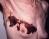
Bullous impetago
(impetago circinata also visible)
Erysipelas
- aka St. Anthony’s Fire
- Streptococcal infection (Usually GAS, sometimes goup C or G, often B in infants)
- Intense local redness, heat, swelling, with a raised, indurated border (may develop secondary features like bullae, vesiculation, sometimes with associated gangrene)
- Often, but not always, preceded by malaise and fever, headache, vomitting, joint pain
Complications of Erysipelas
Septicemia, deep cellulitis, necrotizing fasciitis, abcess development
Erysipelas is often confused with. . .
Contact dermatitis
Angioneurotic edema
The major distinguishing features are the absence of itchiness and the presence of fever
Treatment for erysipelas
- Systemic penicillin very effective
- General constitution improves within 24-48 hr, but skin may take longer to heal
- Treat w/ antibiotics for at least 10 days
- ice bags/cold compress locally
- Leg involvement and bullae may require hospitalization and monitoring with IV antibiotics
- Recurrence may occasionally occur, in which case long-term antibiotic prophylaxis is recommended
Cellulitis
- suppurative infammation involving the subcutaneous tissue
- Usually follows some discernable wound
- Mild local erythema and tenderness, malaise, fever, chills may be present at onset
- Spreads outward from central wound
- Pits upon pressure
- Central part may become vesicular or necrotic
- May be followed by gangrene, metastatic abscesses, and severe sepsis
Diagnosing cellulitis
- Usually made on clinical grounds
- uncommon for blood studies, including cultures, and skin biopsies or aspirates to be positive
- If, however, an open wound is present, there is a high probability of a culture being positive
Causes of cellulitis
- Streptococci ~75% of cases
- Remainder mostly staphylococci

Cellulitis
Intertrigo
- Superfcial infammatory dermatitis occurring where two skin surfaces are in apposition
- As a result of friction, heat, moisture, the affected fold becomes erythematous, macerated, and secondarily infected
- may be erosions, fssures, and exudation, with symptoms of burning and itching
*
Risk factors for intertrigo
- Heat, humidity
- Obesity
- Extremes of age