CNS Flashcards
Meninges
Layers of membranes covering brain and spinal cord
Cerebrum (locate)
A

Epithalamus (locate)
B
Thalamus (locate)
C

Hypothalamus (locate)
Diencephalon (locate)
B, C, D

Cerebellum (locate)
G

Brain stem (locate)
E, F, H

Medulla oblongata (locate)
H

Midbrain (locate)
E

Pons (locate)
F

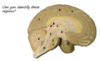
What is this image?

Cerebrum
What is the “tree” in the middle and what is it made of?

Arbor vitae
made of white matter
What is the outer rim and what is it made of?
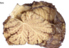
Cerebellar cortex
gray matter
Label the meninges

Left - Arachnoid mater
Middle - Subarachnoid space
Right - Dura mater
Label the Image

A - Vertebrae
B - Adipose tissue in epidural space
C - Dura mater
D - Arachnoid mater
E - subarachnoid space
F - Pia mater
G - Spinal cord
H - Cenral canal
Label the numbered parts of the spinal cord

1 - Gray matter
2 - white matter
3 - soma of neurons
4 - myelinated axons
What is A?

A - Posterior white column
What is C?

C - Lateral white column
What is E?

E - Anterior White column
What is F?

F - Subarachnoid space
What is D?

D - Anterior Gray Horn
What is L?

L - Dura mater
What is K?

K - Anterior median fissure
What is J?

J - Central canal
What is I?
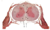
I - Gray commissure (bridge across central canal)
What is H?

H - Posterior median sulcus
What is G?

G - Pia mater
What is C?

C - Lateral white column
Where is the cervical enlargement and why does it exist?
C3/C4-T1
To allow room for nerves entering and exiting arms
Where is the lumbar enlargement and why does it exist?
T9-T12
To allow room for nerves entering and exiting legs
What information travels on the posterior side of the spinal cord?
Sensory
What information travels on the anterior side of the spinal column?
motor
What are the colored regions and their functions?


