Chapter 9.1 Flashcards
The Human Gas Exchange System
Gas exchange takes place in the human thorax. This is a collection of organs and tissues in the chest cavity
Trachea
The airways that leads from the mouth and nose to the bronchi.
- The trachea is lined with mucus-secreting Goblet cells and cilia.
- The cilia sweep microorganisms and dust away from the lungs
Lungs
Humans have two longs, both which are a central part of the respiratory system and where gas exchange takes place
Bronchi
‘Bronchi” is the plural of ‘Bronchus’. The left and right bronchi are at the bottom of the trachea and are similar in structure, but narrower. The bronchi lead to bronchioles
Bronchioles
These are narrow tubes(less than 1mm) which carry air from the bronchi to the alveoli.
As they are so narrow, they have no supporting cartilage and so can collapse
Alveoli
The main site of gas exchange in the lungs. These are tiny sacs with many structural adaptations to enable efficient gas exchange, such as their thin walls and large surface area to volume ratio
Capillary network
An extensive network of capillaries surrounds the alveoli and are an exchange surface between the lungs and the blood.
=During gas exchange, oxygen diffuses from the alveoli and into the capillaries, while carbon dioxide diffuses the other way and is exhaled
Cartilage is a strong and flexible tissue found in various places around the body. What makes it so strong ?
- One place is in rings along the trachea, called Tracheal rings
- These rings help to support the trachea and ensure it stays open, while allowing it to move and flex while we breathe
Goblet cells can be found scattered throughout the ciliated epithelium in the trachea. what are and function
- They are mucus-producing cells that secrete viscous mucus which traps dust, bacteria and other microorganisms and prevents them from reaching the lungs
- The mucus is then swept along by the cilia of the ciliated epithelium upwards and is swallowed
- The mucus and any microorganisms will then be destroyed by the acid in the stomach
The alveoli have a lining of thin squamous epithelium, that allows for gas exchange why
The squamous epithelium forms the structure of the alveolar wall and so is very thin and permeable for the easy diffusion of gases
Smooth muscle can be found throughout the walls of the bronchi and bronchioles why
It helps to regulate the flow of air into the lungs by dilating when more air is needed and constricting when less air is needed
Each alveolus is surrounded by an extensive network of capillaries why
Carbon dioxide diffuses out of the capillaries and into the alveoli to be exhaled, while oxygen diffuses the other way from alveoli and into the capillaries to be carried around the body
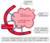
what ensures that there is sufficient time and opportunity for gas exchange to occur
capillaries which have a diameter of around 3-4µm, which is only wide enough for one red blood cell to travel through at any one time
Ciliated epithelium is a specialised
tissue found along the trachea down to the bronchi. Each cell has small projections of cilia which sweep mucus, dust and bacteria upwards and away from the lungs and the epithelium itself
Explain how the lining of the trachea, bronchus and bronchioles provide protection against pathogens
Cilia beat to move mucus away from the lungs and up towards the mouth.
- Mucus is produced by goblet cells and acts as a barrier to pathogens from entering the ciliated epithelium.
- The mucus also traps bacteria and microorganisms, while blood vessels bring macrophages to engulf any pathogens in phagocytosis.
Cartilage
-The cartilage in the trachea has a ‘glassy’ appearance due to its translucent protoplasm -It contains no nerves or blood vessels
Ciliated epithelium
- Ciliated epithelial cells are distinctive due to their narrow cell bodies and hair-like cilia located along the apical layer
- The cilia are tiny projections which greatly increase the surface area of the cell
Goblet cells
- Goblet cells can be found scattered among the ciliated epithelium of the trachea and bronchi
- They are distinctive in the epithelium due to their lack of cilia (although they still have some microvilli projecting outwards)
- The nucleus is found towards the basal (bottom) layer of the cell, with a large Golgi apparatus and mucus granules found towards the apical layer
Squamous epithelium
Squamous epithelium is made of thin, flat squamous cells
Smooth muscle
- The cells of smooth muscle are tightly packed and are found beneath the ciliated epithelium
- Unlike skeletal muscles, they are not striated and so don’t show any cross stripes under a microscope
Capillaries
-Capillaries are distinctive from other blood cells due to their tiny diameter (~4 μm) -Their walls consist of a single layer of epithelial cells
Trachea
- A tracheal cross-section shows the large lumen which air has to travel through
- The innermost cells of the trachea are the ciliated epithelia with projections called cilia -The cells of the ciliated epithelium are shown here
– the cells are tightly packed and interspersed with goblet cells,
- The cilia are essential for sweeping bacteria and dust-filled mucus away from the lungs and up the trachea into the back of the mouth
- This mucus is then swallowed, with any pathogens hopefully destroyed by the acidic conditions in the stomach
Bronchi
- Bronchi are distinctive from the trachea because their lumen is narrower; 8.7mm instead of 18mm
- However, like the trachea, they are lined by ciliated epithelium
Bronchioles
- Bronchioles are approximately 1mm or less in diameter
- Smooth muscle and cuboidal epithelium are found in their walls