Chapter 17. Pupillary and Eyelid Abnormalities Flashcards
Questions 17-1: Which of the following are features of light-near dissociation?
A. Pupils constrict to light but dilate with near stimuli
B. Pupils have limited constriction to light, but constrict better to near stimuli
C. Pupils have dilation to light but constriction to near stimuli
D. Pupils dilate in response to light and dilate to near stimuli
Answer 17-1: B. Light-near dissociation is a disorder of pupil response, where pupilary constriction in response to light is blunted, but the response to near stimuli is better. Syphilis is commonly considered first in the differential diagnosis of light-near dissociation, but other lesions may produce this as well, especially including diabetes.
Question 17-2: Which of the following are potential causes for irregular pupils?
A. Syphilis
B. Degenerative disease of the iris
C. Midbrain lesions
D. Posterior synechiae
E. All of the above
Answer 17-2: E. All of these are potential causes for irregular pupils. Midbrain lesions produce oval or eccentric pupils. Posterior synechiae is adhesion of the iris to the lens. Other potential causes Include trauma to the iris ocular ischemia, and Holmes-Adie syndrome
Question 17-3: Hippus is oscillation in pupillary diameter with steady iIIumination of the eye. Which of the following statements concerning interpretation is true?
A. Hippus indicates a defect in retinal transduction, with variable stimulation of midbrain pathways
B. Hippus is normal variation in pupillary diameter and does not indicate pathology
C. Hippus is due to defective iris contraction resulting from partial denervation
D. Hippus indicates a defect in optic nerve transmission to the lateral geniculate
Answer 17-3: B. Hippus is the normal variation in pupillary diameter With steady illumination. The cause may be a cyclic constriction of the pupil due to light, resultant reduction in light impacting on the retina, then reflexive pupil dilation because of the reduced illumination. This causes increased retinal illumination which begins the cycle again
Question 17-4: A patient presents with marked anisocoria with the left eye dilated and unresponsive to light. The dilated pupil does not constrict to convergence, either. There are no other ocular motor deficits, and the right eye dilates and constricts normally to stimuli. The dilated pupil does not constrict to pilocarpine at 0.05% or I %. What is the most likely diagnosis?
A. Third nerve palsy
B. Demyelinating disease
C. Horner’s syndrome
D. Pharmacologic dilation of the pupil
E. Simple anisocoria
Answer 17-4: D. Pharmacologic pupillary dilation from agents such as atropine produces dilation of the affected pupil which does not constrict in response to any stimulus, including light, convergence. low- or high-dose pilocarpine. Third nerve palsy would usually be associated with other ocular motor problems. and even if isolated should respond to pilocarpine. Demyelinating disease typically produces ocular motor defects without pupillary abnormalities. Homer’s syndrome produces pupillary constriction on the side of the lesion, and both pupils (affected and unaffected) respond to light
Question 17-5: A patient presents with anisocoria with the right pupil smaller than the left. There appears to be mild ptosis on the right. There are no other ocular motor abnormalities and no other deficits. Which is the most likely diagnosis and what might be done to confirm the diagnosis ?
A. The patient has Horner” s syndrome which can be confirmed by failure of response to cocaine administration
B. The patient has Horner’s syndrome which can be confirmed by constriction to 0.05% pilocarpine administration
C. The patient has tonic pupil which can be confirmed by constriction to 0.05% pilocarpine
D. The patient has tonic pupil which can be confirmed by constriction to 1 % pilocarpine
Answer 17-5: A. The patient appears to have Horner’s syndrome which can be confmned by ad:ninistration of cocaine to the eye with the smaller pupil. Pilocarpine at low and high doses is used for examination of the dilated pupil. and the fonner can differentiate tonic pupil from other causes, the latter can differentiate pharmacologically dilated pupil
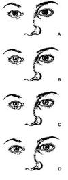
Question 17-6: Figure 17-1 shows a series of pupillary responses to light, as labeled. What type of response is demonstrated here?
A. Tonic pupil on the left side
B. Simple anisocoria
C. Horner’s syndrome on the right side
D. Partial third nerve palsy on the left side

Answer 17-6: A. The patient has a tonic pupil on the left side. The anisocoria is minimal in the dark state. The normal right side constricts in response to light and near stimuli and dilates upon return to distant gaze. The abnormal left side has an impaired response to light, a good constriction to near stimulation, and slowed dilation in response to the relum to distant gaze
Question 17-7: Which of the following statements regarding tertiary syphilis is true?
A. The Venereal Disease Research Laboratory test is preferable to the fluorescent treponemal antibody absorption test for diagnosis of tertiary syphilis
B. Patients with bilateral tonic pupils should be screened for syphilis
C. Horner’s syndrome is the most common ocular manifestation of syphilis
D. All are true
Answer 17-7: B. Patient with bilateral tonic pupils should be screened for syphilis. Note that bilateral tonic pupils are harder to detect clinically than unilateral tonic pupil. Homer’s syndrome is not a common complication of syphilis. The FTA-ADS and microhemagglutination assay for Treponema pallidum are preferable to VDRL for tertiary syphilis because of the high frequency of false-negative tests
Question 17-8: Which is the most likely cause of lid retraction?
A. Diabetes
B. Orbital tumors
C. Hypothyroidism
D. Hyperthyroidism
Answer 17-8: D. Hyperthyroidism is the most common cause of lid retraction. The upper and lower lids are often affected. The lid retraction is noticed most when the patient looks straight ahead or down. Lid lag can additionally be demonstrated by having the patient follow 3 finger with their gaze and observing the delay in lid depression with downward gaze. Diabetes and orbital tumors can both produce ocular motor abnormalities and include ptosis as a component, but these are uncommon causes of lid retraction.
Question 17-9: Which of the following are causes of excessive lid closure?
A. Blepharospasm
B. Apraxia of lid opening
C. Hemifacial spasm
D. Myokymia
E. All of these
Answer 17-9: E. All of these are potential causes of excessive lid closure. Blepharospasm is uncontrolled contraction of the orbicularis oculi causing lid closure. and may be associated with dystonic posturing of the face. Apraxia oflid opening is inappropriate inhibition of the levator palpebrae. Hemifacial spasm is episodic contractions of all muscles innervated by the facial nerve on one side. Myokymia is involuntary twitches of the orbicularis muscle. Myotonia is another major cause not listed here.
Question 17-10: Which of the following are potential causes of insufficient lid closure?
- Proptosis
- Parkinsonism
- Facial nerve palsy
- Myotonic dystrophy
Select: A = 1,2,3. B = 1,3. C = 2. 4. D = 4 only. E = All
Answer 17-10: EAll of these are potential causes of insufficient lid closure. Proptosis results in difficulty of the lids covering the globe. Parkinsonism is associated with decreased blinking. a form of insufficient lid closure. Facial nerve palsy causes impaired function of the orbicularis oculi with incomplete closure of the eye; there is significant risk of damage to the cornea. Myotonic dystrophy is associated with weakness of the orbicularis oculi


