Chapter 14: Anemias Flashcards

1
Q

In general, what are macrocytic vs. microcytic anemias each caused by?

A
  • Macrocytic = abnormalities that impair maturation of erythroid precursors in the bone marrow
  • Microcytic = disorders of hemoglobin synthesis (most often iron deficiency)
2
Q

Which mean cell volume (fL) is indicative of micro- vs. macrocytic anemia?

A
  • MCV <80 = microcytic
  • MCV >100 = macrocytic
3
Q

What does an elevated Red Cell Distribution Width (RDW) tell us?

A
  • That marrow is pumping out reticulocytes (larger cells)
  • Elevated RDW is a reactive phenomenon observed in states of anemia with a functioning marrow
4
Q

Which changes are seen in red cells, white cells, and platelets with acute blood loss?

A
  • Leukocytosis
  • Reticulocytosis
  • Thrombocytosis
5
Q

A male or post-menopausal female presenting with iron-deficiency anemia is indicative of what until proven otherwise?

A

GI tract lesion

6
Q

Hemolytic anemias share what 3 features?

A
  • A shortened red cell life span below typical 120 days
  • EPO levels and compensatory ↑ in erythropoiesis
  • Accumulation of hemoglobin degradation products as part of red cell hemolysis
7
Q

What ar the 3 principal clinical features of extravascular hemolysis?

A
  • Anemia
  • Splenomegaly
  • Jaundice
8
Q

Patients with extravascular hemolysis often benefit from what?

A

Splenectomy due to much of the pathologic destruction of RBC’s occurring in the spleen

9
Q

What are the 5 main manifestations of intravascular hemolysis?

A
  • Anemia
  • Hemoglobin-emia
  • Hemoglobin-uria
  • Hemosiderin-uria
  • Jaundice
10
Q

In all types of uncomplicated hemolytic anemias, the excess serum bilirubin is of what type?

A

Unconjugated

11
Q

What happens to the levels of serum haptoglobin with extra- and intravascular hemolysis?

A
  • Extravascular = variably decreased as some Hgb escapes phagocytes
  • Intravascular = becomes markedly reduced–> freeHgboxidizes tomethemoglobin
12
Q

Regardless of the cause of type of hemolytic anemia what morphological changes are seen in the peripheral blood and BM?

A
  • ↑ numbers of erythroid precursors (normoblasts) in the marrow
  • Prominent reticulocytosis in the peripheral blood
13
Q

With chronic hemolysis, elevated biliary excretion of bilirubin promotes what?

A

Formation of pigment gallstones (cholelithiasis)

14
Q

Hereditary spherocytosis is an inherited disorder caused by intrinsic defects in what?

A

Red cell membrane skeleton –> cells are spheroid, less deformable, and vulnerable to splenic sequestration

15
Q

What is the inheritance pattern of hereditary spherocytosis; prevalence is highest where?

A

Autosomal Dominant (75% of cases); highest in Northern Europe

16
Q

What is the characteristic morphology of the red cells seen on smears in pt with hereditary spherocytosis?

A

Small, dark-staining (hyperchromic) red cell lacking central zone of pallor

17
Q

Characteristc morphology of the spleen in hereditary spherocytosis?

A

Moderate splenomegaly (500-1000 gm)

18
Q

Red cells of hereditary spherocytosis are abnormally sensitive to what; what is the MCHC finding?

A
  • Osmotic lysis when incubated w/ hypotonic salt solution
  • HS red cells also have ↑ MCHC, due to dehydration caused by loss of K+ and H2O
19
Q

Characteristic clinical findings of hereditary spherocytosis are what; some may be asymptomatic but most will have what type of anemia?

A
  • Splenomegaly + jaundice + anemia +/- gallstones
  • CHRONIC hemolytic anemia of mild to moderate severity
20
Q

Aplastic crises occurring in the setting of hereditary spherocytosis is often triggered by what?

A

Parvovirus B19 infection (ssDNA virus)

21
Q

How is the anemia and its complications in hereditary spherocytosis treated?

A

Splenectomy

22
Q

What are the most common triggers responsible for the episodic hemolysis seen with G6PD deficiency?

A
  • Infections: viral hepatitis, pneumonia, and typhoid fever
  • Drugs: ASA, diphenhydramine, antimalarials, sulfonamides, and nitrofurantoins
  • Foods: including simple CHO’s, tonic water, sulfites (wine and dried fruit), and fava beans** (endemic in Mediterranean, ME, and Africa)
23
Q

The acute intravascular hemolysis is greater in pt’s with which variant of G6PD deficiency?

A

Mediterranean variant

24
Q

What is seen 2-3 days following exposure of G6PD-deficienct pt’s to oxidants?

A

Acute intravascular hemolysis marked by anemia, hemoglobinemia, andhemoglobinuria

25
Q

Sickle cell disease is due to a point mutation at which codon of β-globin; causes the replacement of which AA’s?

A

6th codon of β-globin causing replacement of glutamate —> valine

26
Q

Why do patients with hereditary persistence of HbF experience less severe sickle cell disease?

A

HbF inhibits polymerization of HbS even more than HbA

27
Q

Why do individuals who are homozygous for HbS but also have co-existent α-thalassemia have decreased severity of disease?

A
  • α-thalassemia reduces Hgb synthesis which ↓ MCHC
  • ↑ MCHC levels facilitate sickling
28
Q

What factor is responsible for the vascular occlusion seen most prominently in the spleen, bone marrow, and inflammed tissues in a pt with sickle cell disease?

A
  • BM/Spleen = sluggish blood flow through microvascular beds
  • Inflammed = slowed blood due to adhesion of leukocytes to activated endothelial cells and transudation of fluid thru leaky vessels
29
Q

Which AA’s are swapped in the HbC variant?

A

Glutamic acid —> lysine (“lyCine”) in 6th AA of β-globin

30
Q

How does the release of free hemoglobin from lysed sickle red cells contribute to vasoconstriction, ↑ platelet aggregation, stasis, sickling, and thrombosis?

A

Free Hbg can bind and inactivate NO, which is a potent vasodilator and inhibitor of platelet aggregation

31
Q

Which crises due to vaso-occlusion is extremely common in children w/ sickle cell disease; how does it manifest?

A
  • Painful bone crises and often difficult to distinguish from osteomyelitis
  • Most frequently manifest as hand-foot syndrome or dactylitis of the bones of the hands or feet, or both
32
Q

Up to 45% of males with sickle cell disease are affected by what after puberty?

A

Priapism –> may lead to hypoxic damage and ED

33
Q

How does acute chest syndrome as a complication of sickle cell disease present?

A

Fever + cough + chest pain + pulmonary infiltrates

34
Q

Sequestration crises occur in which pt’s with sickle cell disease and how does it manifest?

A
  • Children w/ intact spleens
  • Massive entrapment of sickle red cells leads to rapid splenic enlargement + hypovolemia, and sometimes shock
  • May be fatal
35
Q

A sudden worsening in the anemia of a patient with sickle cell disease should prompt you to consider what underlying cause?

A

Aplastic crises due to Parvovirus B19

36
Q

Chronic hypoxia in children with sickle cell disease is responsible for impairment in what?

A

Growth and development, as well as organ damage affecting spleen, heart, kidney, and lungs

37
Q

Sickling provoked by hypertonicity in the renal medulla causes damage and eventually leads to what?

A

Hyposthenuria (inability to concentrate urine) –> ↑ propensity for dehydration

38
Q

What is the most common cause of death in adults vs. children w/ sickle cell disease?

A
  • Adults = Acute Chest Syndrome
  • Children = Haemophilus influenzae
39
Q

How does the morphology of the spleen change from childhood to early adulthood in pt with sickle cell disease?

A
  • Children have splenomegaly due to red pulp congestion caused by trapping of sickled cells in the cords and sinus
  • Early adulthood will have autosplenectomy as chronic erythrostasis leads to splenic infarction, fibrosis, and progressive shrinkage
40
Q

Leg ulcers are a common finding in which patients with sickle cell disease?

A

Adults

41
Q

Which is more common and serious, Hgb C or Hgb SC disease?

A

Hgb SC is more common and severe

42
Q

What is the distinctive morphology of Hgb C which may be seen on routine peripheral blood smear?

A

Crystalline form

43
Q

The defects in globin synthesis that underlie thalassemia disorders cause anemia through what 2 mechanism?

A
  • ↓ red cell production
  • ↓ red cell life-span

*NOT primarily hemolysis*

44
Q

How is β-globin synthesis affected in β0 mutations vs. β+ mutations; what is the most common underlying mutation for each?

A
  • β0 mutation: absent β-globin synthesis; most commonly due to chain terminator mutation
  • β+ mutation: reduced (but detectable) β-globin synthesis; most commonly due to splicing mutations
45
Q

Impaired β-globin synthesis in β-thalassemia causes a deficit in HbA synthesis producing what type of red cells?

A

Underhemoglobinized” hypochromic, microcytic red cells

46
Q

What occurs to the unpaired α-chains seen in β-thalassemia; what is the proximal cause of most red cell pathology?

A
  • Precipitate within red cell precursors, forming insoluble inclusions
  • Prox. cause of red cell pathology is membrane damage caused by these inclusions; also undergo apoptosis
47
Q

What are some of the complications due to ineffective erythropoiesis seen in severe β-thalassemia?

A
  • Massive erythroid hyperplasia in marrow and extensive extramedullary hematopoiesis —> erosion of bony cortex + hepatosplenomegaly
  • Metabolically active erythroid progenitors steal nutrients from already hypoxic tissues causing severe cachexia in untreated pt’s
  • Suppresses hepcidin = ↑ iron absorption on top of repeat transfusions can lead to seconary hemochromatosis
48
Q

β-thalassemia major is most common where?

A

Mediterranean countries, parts of Africa, and SE Asia

49
Q

How soon after birth does the anemia of β-thalassemia major present?

A

6-9 months after birth as Hgb synthesis switches from HbF –> HbA

50
Q

What are the major manifestations of bone seen in pt with untreated β-thalassemia major?

A
  • Hypoxia –> ↑ EPO –> Expansion of marrow erodes existing cortical bone and induces new bone formation of the skull and face
  • Giving rise to “crewcut” appearance on X-ray
51
Q

Blood transfusions may improve the anemia and suppress complications related to excessive erythropoiesis in β-thalassemia, but may lead to what complications?

A

Cardiac disease and 2’ hemochromatosis from excessive iron overload = important cause of death

52
Q

What is survival like in pt’s with β-thalassemia major who receive transfusions and iron chelation; what is the only therapy offering a cure?

A
  • Transfusions + chelators = survival into the 3rd decade
  • Hematopoietic stem cell transplantation = only cure
53
Q

What are the levels of HbF and HbA2 like in β-thalassemia minor?

A
  • ↑ levels of HbA2
  • HbF levels are normal or sometimes slightly ↑ (will be in major)
54
Q

Recognition of β-thalassemia minor (trait) is important for what 2 reasons?

A
  • Superficially resembles the hypochromic microcytic anemia of iron deficiency
  • Has implications for genetic counseling
55
Q

Which value is diagnostically useful in women of childbearing age who are at risk for both β-thalassemia trait and iron deficiency?

A

HbA2 –> ↑ in β-thalassemia minor (trait)

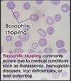
56
Q

Basophilic stippling seen on smears of β-thalassemia major are indicative of what?

A

Toxic injury to RBC’s

57
Q

α-thalassemias are caused by what?

A

Inherited DELETIONS that result in reduced or asbent synthesis of α-globins (compared to β-thalassemias which are due to mutations)

58
Q

How do the tetramers formed in infants with α-thalassemia differ from that of older children/adults?

A
  • Infants form γ4 tetramers known as hemoglobin barts
  • Older children/adults form β4 tetramers known as HbH
59
Q

α-thalassemia trait is caused by deletion of how many α-globin genes and what 2 ways can this occur; which populations more affected?

A
  • Deletion of 2 α-globin genes
  • From single chromosome = α/α -/- = Asians
  • From two chromosomes = α/- α/- = African, Asian
60
Q

Clinically significant α-thalassemia is more common in children born to a parent with what type of halotype?

A

At least one parent with -/- halotype; more often seen in Asians

61
Q

What is the clinical picture like in someone with α-thalassemia trait?

A
  • Small red cells (microcytosis) + minimal or no anemia
  • No abnormal physical signs
  • HbA2 levels are normal or low

*Resembles β-thalassemia minor*

62
Q

Hemoglobin H Disease (HbH) is caused by deletion of how many α-globin genes and is most common in which population?

A
  • Deletion of 3 α-globin genes
  • Most common in Asian populations
63
Q

Why is there tissue hypoxia disproportionate to the level of hemoglobin in patients with HbH disease?

A

HbH has extremely ↑ affinity for O2

64
Q

What is the anemia like in HbH disease and it resembles that of which β-thalassemia?

A
  • Moderately severe anemia
  • Resembles that of β-thalassemia intermedia
65
Q

What is the most severe form of α-thalassemia called and is due to what?

A
  • Hydrops fetalis
  • Due to deletion of all 4 α-globin genes
  • Excess γ-globin chains form hemoglobin barts that have such a high affinity for O2 that they deliver little to tissues
66
Q

When does hydrops fetalis become evident and what must be done for survival of the infant?

A
  • Fetal distress becomes evident during 3rd trimester
  • Require intrauterine transfusions
  • There will be lifelong dependence on blood transfusions for survival w/ the assoc. risk of iron overload
67
Q

What is the only hemolytic anemia caused by an acquired genetic defect?

A

Paroxysmal Nocturnal Hemoglobinuria (PNH)

68
Q

What is the inheritance pattern and dysfunctional enzyme in Paroxysmal Nocturnal Hemoglobinuria (PNH)?

A
  • X-linked and subject to lysonization (random inactivationof one X chromosome in cells of females)
  • Mutation of PIGA which encodes enzyme essential for GPI-linked proteins that regulate complement activity
69
Q

In Paroxysmal Nocturnal Hemoglobinuria (PNH) blood cells are deficient in which 3 GPI-linked proteins that regulate complement; which is most important?

A
  • Decay-accelerating factor (CD55)
  • Membrane inhibitor of reactive lysis (CD59) = most important; potent inhibitor of C3 convertase, prevents spontaneous activation of alternative complement path
  • C8 binding protein
70
Q

What is the leading cause of death in pt’s with Paroxysmal Nocturnal Hemoglobinuria (PNH)?

A

Venous Thrombosis, often involving hepatic, portal, or cerebral veins

71
Q

5-10% of patients w/ Paroxysmal Nocturnal Hemoglobinuria (PNH) are at risk for developing which hematopoietic disorders?

A

AML or a myelodysplastic syndrome

72
Q

Which monoclonal antibody can be used in PNH to prevent conversion of C5 to C5a and reduce risk of venous thrombosis by 90%?

A

Eculizumab

73
Q

How is the diagnosis of Paroxysmal Nocturnal Hemoglobinuria (PNH) made?

A

Flow cytometry to detect red cells deficient in GPI-linked proteins such as CD59

74
Q

What are the secondary causes of the warm antibody type of immunohemolytic anemia?

A
  • Autoimmune disorders
  • Drugs
  • Lymphoid neoplasms
75
Q

Most of the causative antibodies of the warm antibody type of immunohemolytic anemia are of what class?

A

IgG

76
Q

What occurs in the pathophysiology of warm antibody types of immunohemolytic anemias?

A
  • IgG-coated red cells bind Fc receptors on phagocytes, which remove the red cell membrane via “partial” phagocytosis
  • Loss of membrane creates spherocytes which are sequestered and destroyed in the spleen —> moderate splenomegaly
77
Q

Explain how antigenic drugs can cause warm antibody type of immunohemoltyic anemias and what are 2 drugs this is commonly seen with?

A
  • 1-2 weeks after initiating therapy w/ large IV doses of drugs such as penicillin or cephalosporin
  • Drugs bind red cell membrane and antibodies can act as opsonins that promote extravascular hemolysis
78
Q

What is the prototypical tolerance breaking drug responsible for warm antibody type of immunohemoltyic anemias; how does this occur?

A
  • α-methyldopa
  • Induces production of antibodies againt red cells antigens, particularly the Rh antigen
79
Q

What are some conditions associated with microangiopathic hemolytic anemia?

A
  • DIC = most common
  • Thrombocytopenic purpura (TTP)
  • Hemolytic-uremic syndrome (HUS)
  • Malignant HTN
  • SLE
  • Disseminated cancer
80
Q

What is the morphology of both red cells and neutrophils seen on a blood smear in megaloblastic anemia?

A
  • Red cells = macrocytic and oval (macro-ovalocytes) w/ variation in size (anisocytosis) and shape (poikilocytosis)
  • Neutrophils = larger than normal (macropolymorphonuclear) and show nuclear hypersegmentation, having 5+ nuclear lobes
81
Q

Why is there pancytopenia in megaloblastic anemia if there are increased hematopoietic precursors being produced in the marrow?

A

Abnormal DNA synthesis causes most precursors to undergo apoptosis in the marrow

82
Q

Pernicious anemia is a specific form of megaloblastic anemia caused by what?

A

Autoimmune gastritis impairiring production of IF needed for Vit B12 uptake

83
Q

What is the proximate cause of anemia in vitamin B12 deficiency?

A

Lack of folate

84
Q

What is thought to initiate the gastric mucosal injury underlying autoimmune gastritis and pernicious anemia?

A

Autoreactive T-cell response initiates the injury and triggers formation of autoantibodies

85
Q

What are the characteristic morphological changes seen in the stomach of pt w/ autoimmune gastritis (pernicious anemia)?

A
  • Fundic gland atrophy
  • Intestinalization –> glandular epithelium undergoes metaplasia to mucus-secreting goblet cells that resemble the lining of the large intestine
86
Q

Long-standing pernicious anemia causes what finding of the tongue?

A

Atrophic glossitis = beefy red tongue

87
Q

Which sx’s of B12 deficiency respond to folic acid administration and which do not?

A
  • Hematologic sx’s (anemia) may improve
  • Neuro sx’s will NOT and may even worsen
88
Q

Diagnosis of pernicious anemia is based on what 4 findings?

A
  • Moderate to severe megaloblastic anemia
  • Leukopenia w/ hypersegmented granulocytes
  • Low serum B12
  • ↑ levels of homocysteine and methylmalonic acid
89
Q

Diagnosis of pernicious is confirmed how?

A

Parenteral B12 causes an outpouring of reticulocytes and a rise in hematocrit levels around 5 days after beginning administration

90
Q

Elevated homocysteine levels as seen in vitamin B12 deficiency are a risk factor for what?

A

Atherosclerosis and thrombosis

91
Q

Porphyria accumulation in the liver is associated with what conditions?

A
  • Liver damage w/ hepatic insufficiency

and

  • ↑ risk for hepatocellular carcinoma
92
Q

What are the clinical signs/sx’s associated with chronic porphyrias?

A
  • Skin = very sensitive to sunlight –> blistering, abnormal hair growth
  • Teeth = staining
93
Q

What are the clinical signs/sx’s associated with acute porphyrias?

A
  • Referred pain from the thorax and abdomen
  • Seizures, hallucinations, and general psychosis
94
Q

Which lab values and clinical signs/sx’s distinguish a true folate deficiency from that of Vit B12 as a cause of megaloblastic anemia?

A
  • Serum homocysteine levels are ↑, but [methylmalonate] = normal
  • Neurological changes do NOT occur w/ pure folate deficiency
95
Q

What is the effect of diseases with ineffective erythropoiesis leading to 2’ hemochromatosis (i.e., β-thalassemia) on hepcidin levels?

A

Suppresses hepatic hepcidin production, even when iron levels are high

96
Q

What are the HCT, Hgb, serum iron, ferritin, TIBC, and hepcidin levels like with iron deficiency anemia?

A
  • HCT, Hgb =
  • Serum iron =
  • Serum ferritin =
  • TIBC = ↑
  • Serum hepcidin = ↓
97
Q

What is a diagnostically significant finding in the bone marrow with iron deficiency anemia?

A

Dissapearance of stainable iron from macrophages; best assessed with Prussian blue stain on smear of aspirated marrow

98
Q

What is seen morphologically on peripheral blood smear of iron deficiency anemia?

A

Red cells are hypochromic microcytic w/ modest poikilocytosis

99
Q

The chronic illnesses associated w/ anemia of chronic disease can be grouped into what 3 categories?

A
  • Chronic microbial infections, such as osteomyelitis, bacterial endocarditis, and lung abscess
  • Chronic immune disorders, such as RA and regional enteritis
  • Neoplasms, such as carcinomas of the lung and breast, and Hodgkin lymphoma
100
Q

Which inflammatory mediator associated with anemia of chronic disease is responsible for the increased production of hepcidin?

A

IL-6

101
Q

Which 2 lab values distinguish anemia of chronic disease from iron deficiency anemia?

A

High ferritin and low TIBC

102
Q

Aplastic anemia is a syndrome characterized by what?

A

Chronic primary hematopoietic failure + attendant pancytopenia (anemia, neutropenia, and thrombocytopenia)

103
Q

What is the inheritance of Fanconi anemia and the dominant clinical features?

A
  • AR caused by defects in multiprotein complex required for DNA repair
  • Marrow HYPOfunction early in life, often other congenital anomalies, such as hypoplasia of kidney and spleen
  • Bone anomalies, most commonly of thumbs or radii
104
Q

List 6 physical agents/infections which may cause aplastic anemia?

A
  • Whole-body irradiation
  • Viral infections
  • Hepatitis (non-A,B,C, or G type)
  • CMV
  • EBV
  • Herpes zoster (varicella zoster)
105
Q

What are 2 types of mutations which are associated with adult-onset aplastic anemia?

A
  • Inherited defects in telomerase
  • Abnormally short telomeres
106
Q

What are the 2 major etiologies which have been proposed for the pathogenesis of aplastic anemia?

A
  • Extrinsic, immune-mediate suppression of amrrow progenitors thru activated TH1 cells producing IFN-y and TNF
  • Intrinsic, abnormality of stem cells; may be antigenically altered via exposure to drugs, infectious agents, etc.
107
Q

How is the diagnosis of aplastic anemia made and what are you looking for?

A
  • Examination of the bone marrow via biopsy
  • Markedly HYPOcellular BM largely devoid of hematopoietic cells; only fat cells, fibrous stroma, and scattered lymphocytes + plasma cells
108
Q

What are the presenting signs/sx’s that may be seen with aplastic anemia?

A
  • Anemia —> weakness, pallor, and dyspnea
  • Thrombocytopenia –> petechiae and ecchymoses
  • Neutropenia –> frequent/persistent minor infections or sudden onset chills, fever, and prostration
109
Q

What is the morphology of the red cells in aplastic anemia; what is the rule?

A
  • Slightly macrocytic and normochromic
  • Reticulocytopenia is the rule
110
Q

What is the treatment of choice for aplastic anemia; prognosis?

A

Bone marrow transplant; 5-year survival of 75%

111
Q

Pure red cell aplasia may occur in association with what conditions?

A
  • Thymoma
  • Large, granular lymphocytic leukemia
  • Drug exposures
  • Autoimmune disorders
  • Parvovirus B19
112
Q

With the exception of parvovirus, most pure red cell aplasias have what type of basis?

A

Autoimmune

113
Q

How does the pure red cell aplasia associated with Parvovirus differ in a healthy pt vs. pt with moderate/severe hemolytic anemia?

A
  • Normal = the aplasia is transient and infection cleared 1-2 weeks
  • Pt w/ moderate to severe hemolytic anemia, even a brief cessation of erythropoiesis results in rapid worsening of the anemia –> aplastic crisis
114
Q

What is Myelophthisic anemia and the most common cause?

A
  • Marrow failure due to space-occupying lesions replacing normal marrow elements
  • Most commonly due to metastatic cancer, often carcinoma of the breast, lung, and prostate
115
Q

Myelophthisic anemia is also a feature of which phase of myeloproliferative disorders?

A

Spent phase

116
Q

What is the seen in both the marrow and peripheral blood with Myelophthisic anemia?

A
  • Marrow distortion and fibrosis
  • Abnormal release of erythroid precursors and immature granulocytic forms (leukoerythroblastosis)
  • Appearance of tear-drop shaped red cells
117
Q

How does chronic renal failure lead to anemia and what is the severity of the anemia proportional to?

A
  • Due to diminished synthesis of EPO by the kidney
  • Anemia tends to be proportional to the severity of the anemia