CH. 2 Neurophysiology- The Generation, Transmission, and Integration of Neural Signals Flashcards
(19 cards)
Neurophysiology
Neurophysiology – Study of the specialized life processes that allow neurons to use chemical and electrical signals to process and transmit information.
- Brain function is an alternating series of electrical signals within neurons and of chemical signals between neurons.
- We need to understand how electrical signaling works in the brain.
CLASSIC PATTERN OF NEURAL FUNCTION – Information flows within a neuron via electrical signals (action potentials) and passes between neurons through chemical signals (neurotransmitters).
Electrical Signals of the Nervous System
Electrical Signals of the Nervous System – Like all living cells, neurons are more negative on the inside than on the outside, so we say they are polarized, meaning there is a difference in electrical charge between the inside and outside of the cell.
The inside of the neuron is more negative than the fluid around it.
RESTING POTENTIAL – An electrical difference across the membrane.
- Of about –50 to –80 thousandths of a volt, or millivolts (mV)
- Specifically, a neuron at rest exhibits a characteristic
- To understand how this negative membrane potential comes about, we have to consider some special properties of the cell membrane, as well as two forces that drive ions across it.
- One important type of membrane-spanning protein is the ion channel.
ION CHANNEL – Tube-like pore that allows ions of a specific type to pass through the membrane.
- The cell membrane of a neuron contains many such channels that selectively allow potassium ions (K+ ) to cross the membrane, but not sodium ions (Na+).
- Because it is studded with these K +channels, we say that the cell membrane of a neuron exhibits selective permeability.
SELECTIVE PERMEABILITY – Allowing some things to pass through, but not others.
The large, negatively charged protein molecules stay inside the neuron.
CONCENTRATION GRADIENT** – Particles move from areas of high concentration to areas of low concentration. That is, they move down their **CONCENTRATION GRADIENT.
- The resting potential of the neuron reflects a balancing act between two opposing processes that drive K + ions in and out of the neuron.
- The first of these is diffusion.
DIFFUSION – Which is the tendency for molecules of a substance to spread from regions of high concentration to regions of low concentration.
- Particles that can pass through the membrane, such as K+ , will diffuse across until they are equally concentrated on both sides. Other ions, unable to cross the membrane, will remain concentrated on one side.
- The second force at work is electrostatic pressure.
ELECTROSTATIC PRESSURE – The property of a membrane that allows some substances to pass through, but not others.
- Charged particles exert electrical force on one another: like charges repel, and opposite charges attract.
- Positively charged cations like K+ are thus attracted to the negatively charged interior of the cell; conversely, anions are repelled by the cell interior and so tend to exit the extracellular fluid.
- Much of the energy consumed by a neuron goes into operating specialized membrane proteins called sodium-potassium pumps.
SODIUM-POTASSIUM PUMPS – The energetically expensive mechanism that pushes sodium ions out of a cell, and potassium ions in.
- This action results in a buildup of K +ions inside the cell (and reduces Na + inside the cell).
- But as we explained earlier, the membrane is selectively permeable to K +ions (but not Na +ions).
- Therefore, K +ions can leave the interior, moving down their concentration gradient and causing a net buildup of negative charges inside the cell.
- As negative charge builds up inside the cell, it begins to exert electrostatic pressure to pull positively charged K +ions back inside.
- Eventually the opposing forces exerted by the K +concentration gradient and by electrostatic pressure reach the equilibrium potential.
EQUILIBRIUM POTENTIAL – The electrical charge that exactly balances the concentration gradient: any further movement of K + ions into the cell (drawn by electrostatic attraction) is matched by the flow of K + ions out of the cell (moving down their concentration gradient).
- The resting potential of a neuron provides a baseline level of polarization found in all cells.
- But unlike most other cells, neurons routinely undergo a brief but radical change in polarization, sending an electrical signal from one end of the neuron to the other, as we’ll discuss next.

The Sodium Pump Diagram (attached)
EQUILIBRIUM POTENTIAL – The electrical charge that exactly balances the concentration gradient: any further movement of K + ions into the cell (drawn by electrostatic attraction) is matched by the flow of K + ions out of the cell (moving down their concentration gradient).
(A) The sodium-potassium pump:
- Cells contain many large, negatively charged molecules.
- The sodium-potassium (Na + -K + ) pump continually pushes Na + ions out and pulls K + ions in.
(B) Membrane permeability to ions:
- The membrane is permeable to K + ions, which pass back out again through channels down their concentration gradient.
- The departure of K + ions leaves the inside of the cell more negative than the outside.
- Na + ions cannot pass back inside.
(C) Equilibrium potential:
- CATIONS – like Na + push against the membrane’s exterior, attracted to the negative interior. Likewise, anions coat the interior of the cell membrane, attracted to cations on the other side.
- ANIONS –coat the interior of the cell membrane, attracted to cations on the other side.
- When enough K + ions have departed to bring the MEMBRANE POTENZTIAL to –65 mV or so.
- The electrical attraction pulling K + in is exactly balanced by the concentration gradient pushing K + out.
- This is the K + equilibrium potential, approximately the cell’s resting potential.

A Threshold Amount of Depolarization Triggers an Action Potential
A Threshold Amount of Depolarization Triggers an Action Potential:
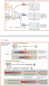
ACTION POTENTIAL – Are very brief but large changes in neuronal polarization that arise in the initial segment of the axon. And then move rapidly down the axon.
- The information that a neuron sends to other cells is encoded in patterns of these action potentials, so we need to understand their properties.
Two concepts are central to understanding how action potentials are triggered.
HYPERPOLARIZATION – An increase in membrane potential (i.e., the neuron becomes even more negative on the inside, relative to the outside).
HYPERPOLARIZATION –An increase in membrane potential (the interior of the neuron becomes even more negative).
- So if the neuron already has a resting potential of, say, –65 mV, hyperpolarization makes it even farther from zero, maybe –70 mV.
DEPOLARIZATION – A decrease in membrane potential (the interior of the neuron becomes less negative). Depolarization is the reverse, referring to a decrease in membrane potential. The depolarization of a neuron from a resting potential of –65 mV to, say, –50 mV makes the inside of the neuron more like the outside.
- In other words, depolarization of a neuron brings its membrane potential closer to zero.
- Applying a hyperpolarizing stimulus to the membrane produces an immediate response that passively mirrors the stimulus pulse.
GRADED RESPONSES – The greater the stimulus, the greater the response, so these changes in the neuron’s potential are graded responses.
LOCAL POTENTIALS – If we measured the membrane response at locations farther and farther away from the stimulus location, we would see. Across the membrane get smaller as they spread away from the point of stimulation.
- Up to a point, the application of depolarizing pulses to the membrane follows the same pattern as for hyperpolarizing stimuli, producing local, graded responses.
- However, the situation changes suddenly if the stimulus depolarizes the axon to –40 mV or so.
THRESHOLD – The stimulus intensity that is just adequate to trigger an action potential in an axion.
ACTION POTENTIAL – Sometimes referred to as a spike because of its shape—is provoked. An action potential is a rapid reversal of the membrane potential that momentarily makes the inside of the neuron positive with respect to the outside.
- Unlike the passive graded potentials that we have been discussing, the action potential is actively reproduced (or propagated) down the axon,
- larger depolarizations do not produce larger action potentials. In other words, the size (or amplitude) of the action potential is independent of stimulus size.
ALL-OR-NONE PROPERTY – Either it fires at its full amplitude, or it doesn’t fire at all.
- It turns out that neurons encode information by changes in the number of action potentials rather than in their amplitude. With stronger stimuli, more action potentials are produced, but the size of each action potential remains the same.
AFTERPOTENTIAL – The form of the action potential shows that the return to baseline membrane potential is not simple. Many axons exhibit small potential changes immediately following the spike; these changes are called afterpotentials

Ionic Mechanisms Underlie the Action Potential
Ionic Mechanisms Underlie the Action Potential:
- The action potential is created by the sudden movement of Na +ions into the axon.
- At its peak, the action potential reaches about +40 mV, approaching the equilibrium potential for Na+ , when the concentration gradient pushing Na + ions into the cell would be exactly balanced by the positive charge pushing them out.
- The action potential thus involves a rapid shift in membrane properties, switching suddenly from the potassium-dependent resting state to a primarily sodium-dependent active state and then swiftly returning to the resting state.
- This shift is accomplished through the actions of VOLTAGE-GATED Na+ CHANNEL.
VOLTAGE-GATED Na+ CHANNEL – Its central Na+-selective pore is gated. The gate is ordinarily closed. But if we electrically stimulate the neuron. Then the axon may be depolarized. If the axon is depolarized enough to reach threshold levels, the channel’s shape changes, opening the “gate” to allow Na + ions through for a short while.
- It monitors the axon’s membrane potential, and at threshold the channel changes its shape to open the pore, shutting down again just a millisecond later.
- The channel then “remembers” that it was recently open and refuses to open again for a short time.
- These properties of the voltage-gated Na + channel are responsible for the characteristics of the action potential.
- As long as the depolarization is below threshold, Na + channels remain closed. But when the depolarization reaches threshold, a few Na + channels open at first, allowing a few ions to start entering the neuron. The positive charges of those ions depolarize the membrane even further, opening still more Na + channels. Thus, the process accelerates until the barriers are removed and Na +ions rush in.
- The voltage-gated Na + channels stay open for a little less than a millisecond, and then they automatically close again. By this time, the membrane potential has shot up to about +40 mV. Positive charges inside the nerve cell start to push K + ions out, aided by the opening of additional voltage-gated K + channels that let lots of K + ions rush out quickly, restoring the resting potential.
- As we bombard the axon with ever-stronger stimuli, an upper limit to the frequency of action potentials becomes apparent.
Similarly, applying pairs of stimuli that are spaced closer and closer together reveals a related phenomenon: beyond a certain point, only the first stimulus is able to elicit an action potential. The axonal membrane is said to be REFRACTORY(unresponsive) to the second stimulus.
REFRACTORY – Temporary unresponsive or inactivated.
Refractoriness has two phases:
- ABSOLUTE REFRACTORY PHASE – A brief period immediately following the production of an action potential, no amount of stimulation can induce another action potential, because the voltage-gated Na + channels can’t respond. Brief period of complete insensitivity to stimulus.
-
RELATIVE REFRACTORY PHASE – During which only strong stimulation can depolarize the axon to threshold to produce another action potential. Period of reduced sensitivity during which only strong stimulation produces an action potential.
- The neuron is relatively refractory because K +ions are still flowing out, so the cell is temporarily hyperpolarized after firing an action potential.
- The overall duration of the refractory phase is what determines a neuron’s maximal rate of firing.
- In general, the transmission of action potentials is limited to axons.
- Cell bodies and dendrites usually have few voltage-gated Na + channels, so they do not conduct action potentials.
- The ion channels on the cell body and dendrites are stimulated chemically at synapses.
Because the axon has many such channels, an action potential that occurs at the origin of the axon regenerates itself down the length of the axon. Continued on next card…

Action Potentials are Actively Propagated Along the Axon
Action Potentials are Actively Propagated Along the Axon:
Because the axon has many such channels, an action potential that occurs at the origin of the axon regenerates itself down the length of the axon.
VOLTAGE-GATED CHANNELS
ACTION POTENTIALS
- Another function for which voltage-gated channels are crucial.
HOW ACTION POTENTIALS SPREAD DOWN THE AXON:
How does the action potential travel?
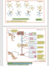
- The action potential is regenerated along the length of the axon. Remember, the action potential is a spike of depolarizing electrical activity (with a peak of about +40 mV), so it strongly depolarizes the next adjacent axon segment.
- Because this adjacent axon segment is similarly covered with voltage-gated Na + channels, the depolarization immediately creates a new action potential, which in turn depolarizes the next patch of membrane, which generates yet another action potential, and so on all down the length of the axon.
- An analogy is the spread of fire along a row of closely spaced match heads in a matchbook. When one match is lit, its heat is enough to ignite the next match, and so on along the row.
- Voltage-gated Na + channels open when the axon is depolarized to threshold.
- In turn, the influx of Na + ions—the movement of positive charges into the axon—depolarizes the adjacent segment of axonal membrane and therefore opens new gates for the movement of Na + ions.
- The axon normally conducts action potentials in only one direction—from the axon hillock toward the axon terminals—because, as it progresses along the axon, the action potential leaves in its wake a stretch of refractory membrane.
- The action potential does not spread back over the axon hillock and the cell body and dendrites, because the membranes there have too few voltage-gated Na + channels to be able to produce an action potential.
CONDUCTION VELOCITY – The speed at which an action potential is propagated along the length of an axon.
- Varies with the diameter of the axon.
- Larger axons allow the depolarization to spread faster through the interior.
- Conduction velocity in large fibers may be as fast as r 300 miles per hour.
MYELIN – Sheathing also greatly speeds conduction.
- The myelin sheath is provided by glial cells. This sheath surrounding the axon is interrupted by NODES OF RANVIER.
NODES OF RANVIER – Small gaps spaced about every millimeter along the axon (see Figure 1.5A). Because the myelin insulation resists the flow of ions across the membrane, the action potential “jumps” from node to node. This process is called SALTATORY CONDUCTION.
MULTIPLE SCLEROSIS (MS) – Is a disease in which myelin is compromised.
- Most cases of multiple sclerosis (MS) are due to the body’s immune system generating antibodies that attack one or more molecules in myelin. If the myelin is damaged enough, then saltatory conduction of axon potentials is disrupted, throwing off the brain’s timing in coordinating behavior and interpreting sensory input
HOW IS an AXON LIKE a TOILET?:
- A toilet flush is similar to an action potential.
- All-or-none property Pushing the toilet lever harder does not produce a bigger flush. Pushing a neuron past threshold does not increase the size of the action potential.
- Refractory phase Until the tank is full, the toilet will not flush again. Until the Na + channels recover, the neuron cannot produce another action potential.
- Direction Like water in a properly operating toilet, an action potential always travels in one direction only.

SYNAPSES CAUSE LOCAL CHANGES in the POSTSYNAPTIC MEMBRANE POTENTIAL
SYNAPSES CAUSE LOCAL CHANGES in the POSTSYNAPTIC MEMBRANE POTENTIAL:
When the action potential reaches the end of an axon, it causes the axon to release a chemical, called a NEUROTRANSMITTER into the synapse.
NEUROTRANSMITTER – The chemical released from the presynaptic axon terminal that serves as the basis of communication between neurons.
- When an axon releases neurotransmitter molecules into a synapse, they briefly alter the membrane potential of the other cell.
- Because information is moving from the axon to the target cell on the other side of the synapse, we say the axon is from the PRESYNAPTIC CELL, and the target neuron on the other side of the synapse is the POSTSYNAPTIC cell.
PRESYNAPTIC CELL – Located on the “transmitting” side of a synapse.
POSTSYNAPTIC – Referring to the region of a synapse that receives and responds to neurotransmitter.
- The vast majority of synapses use neurotransmitters to produce postsynaptic potentials.
When an EXCITATORY PRESYNAPTIC NEURON (dashed) fires, it shows a normal action potential and causes depolarization (EPSP) in thePOST SYNAPTIC NEURON(solid). See attached image
- Remember that EXCITATORY and iINHIBITORY neurons get their names from their actions on postsynaptic neurons, not from their effects on behavior.
- After a brief delay, the postsynaptic cell (yellow) displays a small local depolarization, as Na +channels open to let the positive ions in.
- This postsynaptic membrane depolarization is known as an excitatory postsynaptic potential (EPSP) because it pushes the postsynaptic cell a little closer to the threshold for an action potential.
EXCITATORY POSTSYNAPTIC POTENTIAL (EPSP) – A depolarizing potential in a neuron that is normally caused by synaptic excitation. EPSPs increase the probability that the postsynaptic neuron will fire an action potential.
- The action potential of the inhibitory presynaptic neuron (blue in Figure 2.10) looks exactly like that of the excitatory presynaptic neuron; all neurons use the same kind of action potential.
- But the effect on the postsynaptic side is quite different. When the inhibitory presynaptic neuron is activated, the postsynaptic membrane potential becomes even more negative, or hyperpolarized. This hyperpolarization moves the cell membrane potential away from threshold—
- it decreases the probability that the neuron will fire an action potential—so it is called an inhibitory postsynaptic potential (IPSP).
INHIBITORY POSTSYNAPTIC POTENTIAL (IPSP) – A hyperpolarizing potential in a neuron. IPSPs decrease the probability that the postsynaptic neuron will fire an action potential.
- Usually IPSPs result from the opening of channels that permit chloride ions (Cl– ) to enter the ceii.
What determines whether a synapse excites or inhibits the postsynaptic cell? :
- One factor is the particular neurotransmitter released by the presynaptic cell.
- Some transmitters typically generate an EPSP in the postsynaptic cells; others typically generate an IPSP.
- Whether a neuron fires an action potential at any given moment is decided by the balance between the number of excitatory and the number of inhibitory signals that it is receiving, and it receives many signals of both types at all times.

SYNAPTIC INPUT
SYNAPTIC INPUT:
- Complex behavior requires more than the simple arrival of signals across synapses. Neurons must also be able to integrate the messages they receive. In other words, they perform information processing.
- We’ve seen that postsynaptic potentials are caused by transmitter chemicals that can be either depolarizing (excitatory) or hyperpolarizing (inhibitory). From their points of origin on the dendrites and cell body, these graded EPSPs and IPSPs spread passively over the postsynaptic neuron, decreasing in strength over time and distance.
- Whether the postsynaptic neuron will fire depends on whether a depolarization exceeding threshold reaches the axon hillock, triggering an action potential. If many
- EPSPs are received, the axon may reach threshold and fire. But if both EPSPs and IPSPs arrive at the axon hillock, they partially cancel each other. Thus, the net effect is the difference between the two:
- Summed EPSPs and IPSPs do tend to cancel each other out. But because postsynaptic potentials spread passively and dissipate as they cross the cell membrane, the resulting sum is also influenced by distance. For example, EPSPs from synapses close to the axon hillock will produce a larger effect there than will EPSPs from farther away.
SPATIAL SUMMATION – The summation of postsynaptic potentials that reach the axon hillock from different locations across the cell body. If this summation reaches threshold, an action potential is triggered.
- The summation of potentials originating from different physical locations across the cell body is called SPATIAL SUMMATION.
TEMPORAL SUMMATION – The summation of postsynaptic potentials that reach the axon hillock at different times. The closer in time the potentials occur, the greater the summation.
- Postsynaptic effects that are not absolutely simultaneous can also be summed, because the postsynaptic potentials last a few milliseconds before fading away. The closer they are in time, the greater is the overlap and the more complete is the summation, which in this case is called TEMPORAL SUMMATION.
- Imagine a neuron with only one input. If EPSPs arrive one right after the other, they sum, and the postsynaptic cell eventually reaches threshold and produces an action potential (FIGURE 2.11B). But these graded potentials fade quickly, so if too much time passes between successive EPSPs, they will never sum and no action potentials will be triggered.

Spatial Versus Temporal Summation
Spatial Versus Temporal Summation:
SPATIAL SUMMATION – The summation of postsynaptic potentials that reach the axon hillock from different locations across the cell body. If this summation reaches threshold, an action potential is triggered.
The summation of potentials originating from different physical locations across the cell body is called SPATIAL SUMMATION.
TEMPORAL SUMMATION – The summation of postsynaptic potentials that reach the axon hillock at different times. The closer in time the potentials occur, the greater the summation.
- Postsynaptic effects that are not absolutely simultaneous can also be summed, because the postsynaptic potentials last a few milliseconds before fading away. The closer they are in time, the greater is the overlap and the more complete is the summation, which in this case is called TEMPORAL SUMMATION.
- Imagine a neuron with only one input. If EPSPs arrive one right after the other, they sum, and the postsynaptic cell eventually reaches threshold and produces an action potential (FIGURE 2.11B). But these graded potentials fade quickly, so if too much time passes between successive EPSPs, they will never sum and no action potentials will be triggered.
- Although action potentials are all-or-none phenomena, the postsynaptic effect they produce is graded in size and determined by the processing of many inputs occurring close together in time.
AXON HOLLOCK – The membrane potential at the axon hillock thus reflects the moment-to-moment integration of all the neuron’s inputs, which the AXON HOLLOCK encodes into action potentials.

Synaptic Transmission Requires a Sequence of Events
Synaptic Transmission Requires a Sequence of Events:
- The action potential arrives at the presynaptic axon terminal.
- Voltage-gated calcium channels in the membrane of the axon terminal open, allowing calcium ions (Ca2+ ) to enter.
- Ca 2+Watson/Breedlove causes synaptic vesicles filled with neurotransmitter to fuse with the presynaptic membrane and rupture, releasing the transmitter molecules into the synaptic cleft.
- Transmitter molecules bind to special receptor molecules in the postsynaptic membrane, leading—directly or indirectly—to the opening of ion channels in the postsynaptic membrane. The resulting flow of ions creates a local EPSP or IPSP in the postsynaptic neuron. – (4a) – SYNAPTIC DELAY – Is the time needed for Ca 2+to enter the terminal, for the vesicles to fuse with the membrane, for the transmitter to diffuse across the synaptic cleft, and for transmitter molecules to interact with their receptors before the postsynaptic cell responds.
- The IPSPs and EPSPs in the postsynaptic cell spread toward the axon hillock.(If the sum of all the EPSPs and IPSPs ultimately depolarizes the axon hillock enough to reach threshold, an action potential will arise.)
- Synaptic transmission is rapidly stopped, so the message is brief and accurately reflects the activity of the presynaptic cell.
- Synaptic transmitter may also activate presynaptic receptors, resulting in a decrease in transmitter release.
Let’s look at these seven steps in a little more detail:
Action Potentials Cause the Release of Transmitter Molecules into the Synaptic Cleft: (See next card)

Action Potentials Cause the Release of Transmitter Molecules into the Synaptic Cleft:
Let’s look at these seven steps in a little more detail:
Action Potentials Cause the Release of Transmitter Molecules into the Synaptic Cleft:
SYNAPTIC VESICLE – A small, spherical structure that contains molecules of neurotransmitter.
SYNAPTIC CLEFT – The space between the presynaptic and postsynaptic cells at a synapse. This gap measures about 20–40 nanometers.
CALCIUM ION (Ca2+) – A calcium atom that carries a double positive charge.
RECEPTOR MOLECULES RECOGNIZE TRANSMITTERS:
- The action of a key in a lock is a good analogy for the action of a transmitter on a receptor protein. Just as a particular key can open a door, a molecule of the correct shape, called a LIGAND.
LIGAND – A substance that binds to receptor molecules, such as a neurotransmitter or drug that binds to postsynaptic receptors.
- Can fit into a receptor protein and activate or block it.
- So, for example, at synapses where the transmitter is ACETYLCHOLINE (ACh) the ACh fits into areas called ligand-binding sites in NEUROTRANSMITTER RECEPTOR molecules located in the postsynaptic membrane.
ACETYLCHOLINE (ACh) – A neurotransmitter that is produced and released by parasympathetic postganglionic neurons, by motor neurons, and by many neurons in the brain.
NEUROTRANSMITTER RECEPTOR – Also called simply receptor. A specialized protein, embedded in the cell membrane, that selectively senses and reacts to molecules of a corresponding neurotransmitter.
- The nature of the postsynaptic receptors at a synapse determines the action of the transmitter.
- For example, ACh can function as either an inhibitory or an excitatory neurotransmitter, at different synapses. At excitatory synapses, binding of ACh to one type of receptor opens channels for Na + and K + ions. At inhibitory synapses, ACh may act on another type of receptor to open channels that allow Cl – ions to enter, thereby hyperpolarizing the membrane (i.e., making it more negative and so less likely to create an action potential).
- The chemical nicotine, found in tobacco products, mimics the action of ACh at some synapses, increasing alertness and heart rate.
AGONISTS – A substance that mimics or boosts the actions of a transmitter or other signaling molecule. Molecules such as nicotine that act like transmitters at a receptor are called agonists.
ANTAGONISTS – A substance that blocks or reduces the actions of a transmitter or other signaling molecule. molecules that interfere with or prevent the action of a transmitter, like curare, are called antagonists.
- Just as there are master keys that fit many different locks, there are submaster keys that fit a certain group of locks, as well as keys that each fit only a single lock. Similarly, each chemical transmitter binds to several different receptor molecules.
CHOLINERGIC – Referring to cells that use acetylcholine as their synaptic transmitter.
- Nicotinic cholinergic receptors—yes, the subtype on which nicotine exerts its effects—are found at synapses on muscles and in autonomic ganglia; it is the blockade of these receptors that causes paralysis brought on by curare and bungarotoxin.
- Most nicotinic sites are excitatory, but there are also inhibitory nicotinic synapses. The many “flavors” of receptors for each transmitter have evolved to enable a variety of actions in the nervous system.
- The nicotinic ACh receptor resembles a lopsided dumbbell with a tube running down its central axis (see Figure 2.13). The handle of the dumbbell spans the cell membrane, with two sites on the outside that fit ACh molecules (Karlin, 2002). For the channel to open, both of the ACh-binding sites must be occupied. – (TJM)** – But these receptors could be filed by **EITHER ACH or NICOTINE.
- Different receptor systems become active at different times in fetal life. The number of any given type of receptor remains plastic in adulthood: not only are there seasonal variations, but many kinds of receptors show a regular daily variation of 50% or more in number, affecting the sensitivity of cells to that particular transmitter.

Neural Circuits Underlie Reflexes
Neural Circuits Underlie Reflexes:
For simplicity, so far we have focused on the classic:
AXO-DENDRITIC SYNAPSE – From axon to dendrite.
AXO-SOMATIC SYNAPSES – From axon to cell body, or soma.
- But many nonclassic forms of chemical synapses exist in the nervous system.
AXO-AXONIC SYNAPSES – form on axons, often near the axon terminal, allowing the presynaptic neuron to strongly facilitate or inhibit the activity of the postsynaptic axon terminal.
DENDRO-DENDRITIC SYNAPSE – A synapse at which a synaptic connection forms between the dendrites of two neurons.
- Allowing coordination of their activities.
KNEE-JERK REFLEX – A variant of the stretch reflex in which stretching of the tendon beneath the knee leads to an upward kick of the leg.
- This reflex is extremely fast: only about 40 milliseconds elapse between the hammer tap and the start of the kick. Several factors account for this speed:
- (1) both the sensory and the motor axons involved are myelinated and of large diameter, so they conduct action potentials rapidly;
- (2) the sensory cells synapse directly on the motor neurons; and
- (3) both the central synapse and the neuromuscular junction are fast synapses.

EEGs Measure Gross Electrical Activity of the Human Brain
EEGs Measure Gross Electrical Activity of the Human Brain:
ELECTROENCEPHALOGRAMS (EEGs) – The electrical activity of millions of cells working together combines to produce electrical potentials large enough that we can detect them with electrodes applied to the surface of the scalp. Recordings of these spontaneous brain potentials (or brain waves), called electroencephalograms (EEGs)
EVENT-RELATED POTENTIALS (ERPs) – Are EEG responses to a single stimulus.
- Such as a flash of light or a loud sound.
- ERPs have very distinctive characteristics of wave shape and time delay (or latency) that reflect the type of stimulus, the state of the participant, and the site of recording.
Electrical Storms in the Brain Can Cause Seizures
Electrical Storms in the Brain Can Cause Seizures:
EPILEPSY – A disorder in which SEIZURES lasting for a few seconds or minutes may produce dramatic behavioral changes such as alterations or loss of consciousness and rhythmic convulsions of the body. A disorder of electrical potentials in the brain.
- In the normal, active brain, electrical activity tends to be desynchronized; that is, different brain regions carry on their functions more or less independently.
- In contrast, during a seizure there is widespread synchronization of electrical activity: broad stretches of the brain start firing in simultaneous waves, which are evident in the EEGs as an abnormal “spike-and-wave” pattern of brain activity.
Major Categories of Seizure Disorders:
TONIC-CLONIC SEIZURE – Most severe, with loss of consciousness and rhythmic convulsions. Accompanied by abnormal EEG activity all over the brain.
SIMPLE PARTIAL SEIZURE – More subtle. The characteristic spike-and-wave EEG activity is evident for 5–15 seconds at a time. Behaviorally, people experiencing simple partial seizures show no unusual muscle activity; they just stop what they’re doing and seem to stare into space.
COMPLEX PARTIAL SEIZURES – A type of seizure that doesn’t involve the entire brain and therefore can cause a wide variety of symptoms.
- Seemingly random set of behavioral symptoms actually reflected the functions of the particular brain regions activated by the seizure.
- Others experiencing seizures would produce a completely different set of behaviors. In some individuals, complex partial seizures may be provoked by stimuli like loud noises or flashing lights.
Surgical Probing of the Brain Revealed a Map of the Body
Surgical Probing of the Brain Revealed a Map of the Body:
- In the twentieth century, neurosurgeons began taking drastic measures to help people with severe epilepsy that did not respond to medication: surgical removal of the part of the brain where the seizures begin. The trick, of course, is to remove the part of the brain where the seizures begin.
- One strategy for people whose epileptic seizures were preceded by an aura was to try to find the point where stimulation recreated the aura.
- Using this refined technique, Penfield was able to cure about half of his patients, and seizures were reduced in another 25%.


