Ch 1 Anatomy Flashcards
Vermis of cerebellum is responsible for control of? hemispheres?
Axial = vermin
appendicular = hemispheres
AX the VERMIN!
Parts of cerebellum
Anterior Hemi
Posterior Hemi
Vermis
Flocculonodulus

Anterior Lobe gets input from?
Dorsal and ventral Spinothalamic Tract
Cuneocerebellar Tract
APe’S
Posterior Cerebellum is responsible for what function?
Precise movement
Flocculonodular Lobe is responsible for?
Equilibrium and Eye Movements
Way to remember old names of cerebellar lobes?
APeS FAVorite PreNuP
Ant = paleo or SPINAL cerebellum (ataxia)
Floc = Archi or VESTIBULOcerebellum (eyes and equilibrium)
Post = neo or PONTOcerebellum (precise mvmt)
Superior Middle and Inferior Cerebellar peduncles connect to?
Midbrain
Pons
Medulla
(makes sense)

Cerebellar Nuclei from lateral to medial?
Dentists Emulate Great Friends
Dentate
Emboliform
Globose
Fastigial

The dentate nucleus contributes to? What tract is it part of?
Dentatorubrothalamic tract which contributes to dexterity and synergy of movement.
What is in the dentatorubrothalamic pathway?
Purkinje cells -> dentate nucleus -> contralateral red nucleus -> ventral lateral thalamus -> motor cortex area 4,6

How does the dentate signals in the dentatorubrothalamic tract connect to the red nucleus?
Superior cerebellar peduncle
Fastigial nucleus contributes to ?
stance and walking
“stance and walking fast”
emboliform and globose nuclei are known as?
They contribute to?
Interposed nuclei
stability and speed in initiation of movement.
Lesions in the interposed nuclei cause?
TARDy
Titubation (makes sense cant keep the head steady)
Action tremor (cant move smoothly)
Rapid alternating movements off (cerebellum allows RAM)
DYsmetria (also makes sense)
Layers of the cerebellar cortex?
MPG
molecular
purkinje
granule cell layers
What cells are in molecular layer of the cerebellum?
Molecular
Basket cells
Stellate cells
Purkinje cell dendrites
Parallel fibers of granule cells
Golgi cell dendrites
Moleculer = miles of cells
What cells are in the Purkinje layer of the cerebellum?
just purkinje
Purkinje = Per
What cells are in the granular layer of the cerebellum?
Granule
Golgi
Glomeruli
Miles per GaLLon
Golgi cells make? role? acts upon what cells?
GABA
INHIB
GRANULE cells
Granule cells make? role? acts upon what cells?
Glutamate
Excitatory +++
Basket, Golgi, Stellate
ONLY GRANULE CELLS ARE EXCITATORY
Pukinje cells make? role? acts upon what cells?
GABA
INHIB
deep cerebellar nuclei and vestibular nuclei
Stellate cells make? role? acts upon what cells?
Taurine
inhib
Purkinje
Purkinje cells are inhibited by ? (2)
Basket cells (via GABA)
Stellate cells (via taurine)
Summary of Cerebellar cortex?
Only granule cells are excitatory
Stellate cells use taurine,
Basket, golgi and purkinje use GABA

Two main sources of input into the cerebellar cortex?
mossy and climbing fibers
moseying and climbing into the cerebellar cortex
Mossy fibers use? to snapse on?
aspartate
to synapse on granule cells, which they excite
Axons from the granule cells form? What do those fibers do?
Parallel fibers in the molecular layer, which excites purkinje cells
1 mossy fiber can stimulate >1000 purkinje cells.
1 mossy -> multiple granule cells -> 1000 purkinje cells
mossy are bossy

Climbing fibers provide input to the cerebellum by (explain tract)?
mossy fibers start in contralateral olivary nucleus
through the inferior cerebellar peduncle
to the molecular layer of the cerebellum where they excite 1000s of Purkinje cells

Output from the cerebellar cortex is handled by what cells?
Purkinje
The cortex provides input to the cerebellum via what pathways (3)?
Corticoponocerebellar
Cerebro-olivocerebellar
Cerebroreticulocerebellar
Info from the corticoponto cerebellar pathways enter through the ____ peduncle
middle cerebellar
Pons is in the middle
Info from the cerebro-olivocerebellar tract enter the cerebellum as?
climbing fibers (excitatory to Purkinje cells)
“climbing an olive tree”
The spinal cord sends info to the cerebellum via what pathways (4)?
Cuneocerebellar
Dorsal (posterior) spinocerebellar
Rostral spinocerebellar
Ventral spinocerebellar
Cuneocerebellar tract carries what info?
movement of the IPSIlateral upper extremity and rostral body to the cerebellum
Describe cuneocerebellar tract?
ipsi UE muscle -> up the tract to synapse in the accessory cuneate nucleus -> inferior cerebellar peduncle into the cerebellum.
Dorsal spinocerebellar tract carries what info?
afferent info about IPSIlateral movement of the LOWER extremity limbs and trunk to the cerebellum.
Fibers in this tract give rise to mossy fibers that into the inferior cerebellar peduncle to the cerebellum
Ventral spinocerebellar tract carries what info?
unconscious proprioceptive movement from the trunk and LE to the cerebellum
What is unique in how the ventral spinocerebellar tract travels? compared to other spino-cerebellar tracts?
Ventral goes through the SUPERIOR cerebellar peduncle
Rostral spinocerebellar tract carries what info?
proprioceptive info about the UE and rostrum (similar to cuneo)
Summary of spino cerebellar tracts function - cuneo vs rostral vs dorsal spino vs ventral spino?
UE and rostrum
Movement = cuneo
Proprio = rostral
LE and TRUNK
movement = dorsal
proprio = ventral
Info from the vestibular nerve enters the cerebellum through?
Inferior cerebellar peduncle
Efferent pathways from the cerebellum are?
Globo-emboliform-rubral pathway
Dentatothalamic/Dentatorubral pathway
Fastigial Vestibular Pathway
Fastigial reticular pathway
Describe the pathway of the globose-emboliform-rubral pathway?
leaves globose and emboliform nucleus and leaves via SUPERIOR cerebellar peduncle where it decussates into the contralateral red nucleus

Describe the pathway of the dentatothalamic tract?
leaves dentate and goes to the CONTRALATERAL ventral lateral (VL) nucleus of the thalamus
DENTists are eViL
Describe the dentatorubral pathway?
dentate to CONTRAlateral red nucleus
DENTists also cause a lot of RED blood
Main function of dentato/rubrothalamic pathway? Descibe the whole pathway starting from Purkinje in the cerebellum?
Synergy of movement
Purkinje - Dentate - contralateral RED - VL nucleus of thalamus - motor cortex (areas 4,6)

Fastigial Vestibular pathway travels from the fastigial nucleus to the vestibular nucleus via?
INFERIOR cerebellar peduncle

Fastigial reticular pathway travels from the fastigial nucleus to the reticular nucleus via
INFERIOR cerebellar peduncle

AFFERENT pathways through the
INFERIOR PEDUNCLE?
Arcuatocerebellar
Cuneo
Dorsal spino
olivo
reticulo
trigemino
vestibulo
Theres a lot of INput into the INferior cerebellar peduncle
Try Real VODCA
AFFERENT pathways throught the MIDDLE cerebellar peduncle?
Pontocerebellar
AFFERENT pathways through the superior cerebellar peduncle?
cerulocerebellar tract
tectocerebellar tract
trigeminocerebellar tract
Ventral spinocerebellar
EFFERENT pathways through the inferior cerebellar peduncle?
Fastigial vestibular
fastigial reticular
Efferent pathways through middle cerebellar peduncle?
none
EFFERENT pathways through the superior cerebellar peduncle?
Dentatorubral tract
Dentatothalamic tract
Globose-emboliform-rubral pathway
Uncinate bundle of Russell
“Following your GED, you can get superior education, like your Uncle Russell
Which cerebellar peduncle is primarily responsible for outputs? inputs?
input = inferior
outputs = superior
(middle only has afferent through the pontocerebellar)
What makes up the Triangle of Mollaret?
A triangle of signals between Red Nucleus, IO, and dentate
Neural fibers travel from the Red Nucleus to ipsilateral inferior olive via central tegmental tract
Climbing fibers from the inferior olive travel through the ICP to the contralateral dentate via climbing fibers
Dentate fibers travel via SCP to Red Nucleus

A lesion in the pathways of the Triangle of Mollaret causes?
palatal myoclonus
Layers of the Cerebral Cortex
Superficial to Deepest
Molecular
External Granular
External Pyramidal
Internal Granular
Internal Pyramidal aka ganglionic layer
Multiform Layer
Which layer of the cerebral cortex is responsible for cortical-cortical connections?
2 and 3
External granular and pyramidal
Which layer of the cerebral cortex gets info from the thalamus?
4
Internal granular layer
What is the brodmans area of Primary visual cortex? Wernicke? Broca?
17
22 and 44 (they are double)
“At 17, you take your primary view at an R rated movie”

Cranial nerves in order?
Which ones are sensory/motor/both?
1 - olfactory S
2 - optic s
3 - oculomotor m
4 - trochlear m
5 - trigeminal b
6 - abducens m
7 - facial b
8 - vestibular s
9 - glossopharyngeal b
10 vagus b
11 spinal accessory m
12 hypoglossal m
Some say marry money, but my brother says big brains matter most!
Function of Trigeminal Nerve?
BOTH
facial sensation and motor (mastication), mylohyoid muscle, Anterior belly of digastric, tensor muscles
If you try to masticate a mile of food, your A+ belly becomes more tense.

Function of facial nerve?
both sensory and motor
facial movement, taste in ant 2/3 of tongue, salivation, lacrimation.
Braches of Facial nerve?
Temporal
Zygomatic
Buccal
Mandibular
Cervical
To Zanzibar By Motor Car

Functions of glossopharyngeal nerve?
both motor and sensation
stylopharyngeus muscle
taste in post 1/3 of tongue
sensation to middle and external ear
pharynx
parotid gland
carotid body and sinus
Function of vagus nerve?
Motor and sensory
Motor to lift palate (pharynx, larynx, viscera)
carotid sinus reflexes
sensory - taste in the pharynx
Which CN are involved in parasympathetic system?
3, 7, 9, 10
Through which foramen does each CN exit skull?
1 - Cribiform Plate
2- Optic canal
3,4, 5(1) - Superior Orbital Fissure
5 (2) - Foramen rotundum
5 (3) - Ovale
6 - Superior orbital fissure
7 - Internal auditory meatus (then stylomastoid foramen)
8 - Internal auditory meatus
9, 10, 11 - Jugular foramen
12 - hypoglossal foramen
“Cleaners Only Smelling Salty Scents Right Onto Smelly Iguanas Is Justified, Justified, Justified, However!”
What foramen does the ICA travel through?
Foramen Lacerum
RUM on ICe.
What foramen does the middle meningeal artery travel through?
foramen spinosum
Explain the tract relay for olfaction?
olfactory neurons - olfactory bulbs to synapse where the olfactory tracts carry information to the olfactory cortex

What makes up the olfactory cortex (2)?
piriform cortex
periamygdaloyoid cortex
Once in the olfactory cortex, where do the fibers project?
orbitofrontal cortex
entorhinal cortex (Secondary Olfactory cortex)
hippocampus
mediodorsal nucleus of the thalamus

Which CN does NOT relay through the thalamus
CN 1
CN 1 is smelly and the other CNs dont want it there
What syndrome occurs when there is a lesion or meningioma in the olfactory groove?
Foster Kennedy Syndrome
What makes up Foster Kennedy Syndrome?
- ipsilateral optic atrophy
- contralateral papilledema
- ipsilateral anosmia
- Think of JFK if he couldnt see or smell, he’d still be groooovin man.*
Lesions of the uncus cause what sx?
olfactory hallucinations
Think of Uncle Olaf (olfactory) in Lemony Snicket
Functions of SO eye muscle innervated by CN 4?
Depression and Intorsion of the eye
People that are SO depressed become introverted.
What is unique about CN4?
Longest and smallest cranial nerve
only CN that crosses midline
only CN that exits from the dorsal brainstem
Trochlear nerve is small, depressed, and SO introverted that it exits out the back to avoid everyone.

branches of CN V and where they exit the skull?
v1 - ophthalmic (superior orbital fissure)
V2 - Maxillary (foramen rotundum)
V3 - mandibular (Ovale)
You move your mandible to drink ovaltine

How does V3 differ from V1 and V2?
V3 is motor and sensory (1 and 2 are just sensory)
does not travel through cavernous sinus like V1 and 2

Sensory functions of CN V
- face, mouth, sinuses, meninges AND
- SENSATION (not taste) to anterior 2/3 tongue

Where is the trigeminal ganglion located?
Meckel’s cave

V3 motor functions (4)?
Mastication muscles (mandibular)
mylohyoid muscle
anterior belly of digastric
Tensors (tympani and tensor veli palatini)
If you try to MASTICATE a MYLe of food, you BELLY becomes TENSE
WHat nerve supplies the Anterior and posterior belly of digastric muscle?
Ant = V3 and Post = CN 7
Explain the corneal reflex (afferent and efferent)?
Afferent V1
Efferent CN 7 via orbicularis oculi muscle which causes you to blink when your cornea is touched
Explain the jaw jerk reflex (afferent and efferent)?
mandibular both aff and eff
if jaw jerks with reflex hammer = UMN
Explain the tearing reflex (afferent and efferent)?
afferent V1, efferent CN 7
reflex to start tearing if something is sensed to be lodged in eye
Explain the oculocardiac reflex (afferent and efferent)?
afferent V1
efferent CN 10
pressing on the eye causes bradycardia
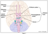
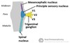
Main 4 nuclei of CN5? Their function?
Main sensory nucleus - fine touch sensation
Mesencephalic nucleus - position sense
Motor nucleus - all V3 motor shit (tense belly masticating)
Spinal nucleus - pain and temp sense (think spinothalamic tract)
WHat is unique about the mesencephalic nucleus?
It is the only primary sensory neuron located in the CNS rather than peripherally
Me’s in the cephalic!
All the trigeminal sensory functions converge where?
Ventral posteriormedial nucleus of the thalamus
which then projects to the postcentral gyrus (main sensory cortex)
VP Meeting area
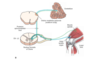
What tract carries PAIN AND TEMP info from face and mouth to trigeminal ganglion?
Explain entire tract all the way to sensory cortex
Ventral trigeminothalamic tract
1 - information carried to trigeminal ganglion via above
2 - then carried to spinal trigeminal nucleus via spinal trigeminal tract
3 - then to contralateral VPM nucleus of the thalamus where it radiates via posterior limb of internal capsule to the somatosensory cortex responsible for sensation to the face
People vent when they are in pain

What tract carries TOUCH AND PRESSURE info from face and mouth to trigeminal ganglion?
Explain entire tract all the way to sensory cortex
Dorsal trigeminothalamic tract
1 - to trigeminal ganglion via above
2 - to principal sensory nucleus of CN V
3 - to ipsilateral VPM of the thalamus to radiate via posterior limb of internal capsule to the ipsilateral somatosensory cortex
(purple dotted line in picture)

explain the route of CN 7 out of the brainstem and exiting the skull
exits brainstem at the cerebellopontine angle, then travels through the internal auditory meatus and the facial canal
exits the skull through the stylomastoid foramen
Roles of CN 7? (Long - divide into motor, parasympathetic and sensory)
MOTOR: - facial muscles - besides mastication
MOTOR: - innervates stapedius, post. belly of digastric, stylohyoid, ant and sup. auricular muscles
PARASYMP: - innervates glands: lacrimal, parotid, submandibular, sublingual
SENS: - taste to ant. 2/3 of tongue
SENS: - sensation of the external ear
Motor branches of CN7?
temporal
zygomatic
buccal
mandibular
cervical

buccal branch of the facial nerve innervates?
buccinator
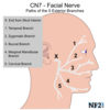
cervical branch of the facial nerve innervates?
platysma

mandibular branch of the facial nerve innervates?
orbicularis oris

temporal branch of the facial nerve innervates?
frontalis

zygomatic branch of the facial nerve innervates?
orbicularis oculi

Difference in upper vs lower facial muscle innervation in CN 7?
Upper facial muscles recieve innervations from both cerebral hemispheres
Lower only receieves contralateral cortical input
UMN lesions cause lower paralysis
LMN lesions cause ipsi upper and lower facial paralysis
First order sensory neurons of CN7 are located in?
geniculate ganglion

Which nerve branch of CN 7 is responsible for sensation of taste in ant 2/3 tongue?
chorda tympani
Explain the route for taste starting with ant 2/3 tongue
chorda tympani -> geniculate ganglion -> nucleus tractus solitarius -(via central tegmental tract)-> VPM of thalamus -> cortical taste area

Taste involves 3 sets of CTs: Chorda Tympani, Central Tegmental, cortical taste center
Preganglionic parasympathetic fibers for CN 7 are located in the ?
superior salivatory nucleus

Which CNs are involved in afferent and efferent of corneal reflex?
afferent V1, efferent CN 7
Which CNs are involved in afferent and efferent of corneal reflex?
afferent V1, efferent CN 7
Ramsay Hunt Syndrome?
Herpes zoster infection of the geniculate ganglion
- patients have unilateral facial palsy, pain and vesicles around the ear, as well as loss of taste on ant 2/3 of tongue
spreading can lead to hearing loss
Review the entire routes of CN 7

Where is the vestibular ganglion located?
internal auditory meatus

What detects linear vs angular acceleration?
Linear - utricle and saccule
Angular - semicircular canals

Whiuch subnuclei form the vestibular nuclear complex?
- Lateral vestibular nucleus
- inferior vestibular nucleus
- medial nucleus
- superior nucleus
Lateral vestibular nucleus gives rise to:
lateral vestibulospinal tract
fibers from the inferior and medial vestibular nuclei travel to:
flocculonodular lobe of the cerebellum via the inferior cerebellar peduncle.
The medial nucleus is the main source of fibers for the:
medial vestibulospinal tract
All 4 vestibular subnuclei contribute to:
ascending MLF, which helps with visual fixation while the head is moving.
Describe the transmission of sound:
- Sound waves cause the tympanic membrane to vibrate.
- Sound is amplified by the middle ear ossicles (malleus, incus, stapes)
- Sound waves then reach the oval window, which connects with the vestibule of the inner ear.
- then sound travels to the scala vestibuli, which contains perilymph
- perilymph movements are transmitted to the cochlear duct. This results in movement of the basilar membrane in reference to the tectorial membrane.
- This activates mechanoreceptor cilia on hair cells.
- At the base of the hair cells, synapses activate the dendritic processes of bipolar cells of cochlear divison of CN8.
Where are the cell bodies of the cochlear division of CN8 located?
in the spiral ganglion in the temporal bone.

The hair cells of the cochlea and their supporting structures form the:
Organ of Corti
The hair cells of the organ of Corti are the:
auditory receptor cells
Describe the organization of the organ of corti
“tonotopic organization”
The hair cells at the base of the cochlea(near the oval window) are activated by higher frequency sounds.
Apex is activated by lower freq.

Brainstem auditory evoked potentials causes 7 waves, where are these waves from?
I - Nerve (CN8)
II - Cochlear nuclei (medulla)
III - Superior olivary complex (pons)
IV - Lateral lemniscus (pons)
V - Inferior Colliculus (midbrain)
VI - Medial Geniculate (thalamus)
VII - Auditory radiations (thalamocortical)
Above the level of the ______, unilateral lesions to the auditory tract do not cause deafness, why?
cochlear nuclei
bilateral connections
Weber test - conductive vs sensorineural hearing loss?
Conductive - vibration is better heard in the affected ear
Sensorineural - better in the normal ear
Sensorineural makes sense.
Rinne test - conductive vs sensorineural
Conductive - cannot hear it vibration in affected ear (bone>air)
Sensorineural - vibration can be heard
CN supplies motor to?
One muscle, stylopharyngeus
Style magazine has glossy photos
The main motor nucleus of CN9 is formed by ?
nucleus ambiguus
CN9 supplies sensation to?
- middle and external ear
- pharynx
- posterior 1/3 of tongue
sensory nucleus of CN9 is part of ?
nucleus of the tractus solitarius
Parasympathetic fxs of CN9?
innervates the parotid gland
Parasympathetic nucleus of CN9 is the?
inferior salivatory nucleus
What reflexes involve CN9?
carotid sinus reflex - dec HR and dilates vessels
afferent limb of the gag reflex
Lesions of CN9?
Glossopharyngeal neuralgia - brief sharp pains on the tongue and radiating to the ear. Usually assoc with swallowing/talking.
CN 10 innervates which muscles?
muscles of the palate
pharynX, larynX
palatoglossus
(except stylopharyngeus - CN9 and Tensor Veli Palatini CNV)
Your X-PALS, pharynX, larynX, wont watch Style TV
CN10 is responsible for taste sensation of the
pharynX
Parasympathetic functions of CN10?
parasymp nucleus of CN10 is the dorsal motor nucleus of the vagus. efferent fibers travel down to the distal 1/3 of the transverse colon.
R SCM muscle moves head in which direction?
Left

CN 10 innervates which muscles?
SCM and trap
Causes of medial vs lateral winging of the scapula?
Serratus Anterior - innervated by long thoracic nerve
when injured = medial winging
Trapezius - CN 11
When injured = lateral winging
Eat a SALTy wing meal, and you wont get caught in a late, 11th hour accessory trap!
Hypoglossal Nerve CN12 innervates the muscles of?
tongue, except palatoglossus (CN9)
Injury of CN12 causes?
tongue deviation towards the side of the lesion (ONLY IF LMN)
UMN = fibers project to the contralateral side so point away from the injured side.
CN 12 LMN injury Licks the Lesion
Symptoms of cavernous sinus thrombosis?
Papilledema
Proptosis
ophthalmoplegia
“PAP, PROP and painful OP”
What structures are in the cavernous sinus?
CN 2, 3, 4, V1, V2, 6
postganglionic sympathetic fibers
ICA
What nerve is closest to the ICA in the cavernous sinus?
CN 6
Abducens abuts the ICA
What is Tolosa Hunt Syndrome?
Tx?
Granulomatous inflammation in the cavernou sinus that results in painful ophthalmoplegia
Tx with prednisone
Function of posterior hypothalamic nucleus?
heat conservation
A&P grocery store is usually too hot or too cold
Function of anterior hypothalamic nucleus?
detects elevated body temperature and triggers cooling mechanism
stimulates the parasymp nervous system
A&P grocery store is usually too hot or too cold
Function of posteror lateral hypothalamic nucleus?
role in transition between sleep and wake
Function of lateral hypothalamic nucleus?
controls appetite
Function of ventromedial hypothalamic nucleus?
inhibits appetite
Function of paraventricular hypothalamic nucleus?
synthesizes ADH and oxytocin
responsible for neuroendocrine and autonomic responses to stress
provides excitatory input for preganglionic sympathetic neurons
Function of supraoptic hypothalamic nucleus?
synthesizes ADH and oxytocin
Function of arcuate hypothalamic nucleus?
produces dopamine
Function of medial preoptic hypothalamic nucleus?
controls release of gonadotropic hormones from the pituitary
Function of posterior tuberomammillary hypothalamic nucleus?
histaminergic innervation to cortex
Function of suprachiasmatic hypothalamic nucleus?
circadian rhythm
Which hypothalamic nuclei regulate appetite?
Ventromedial and Lateral
Very Much Late for dinner!
Components of the limbic system
amygdala
basal forebrain
cingulate gyrus
habenula
hippocampus
hypothalamus
mamillary body
olfactory cortex
septal nuclei
thalamus
ventral striatum
The main limbic system pathway is known as ? Components
Papez Circuit
Hippocampus > Fornix > Mammilary Body > Anterior Nucleus of Thalamus > Cingulate nucleus > Entorhinal cortex
Hippocampus named PEPE with a spraycan of MACE
Function of stria terminalis
connects the amygdala to septal nuclei/ anterior hypothalamus
Function of stria medullaris
connects septal nuclei/Ant hypothalamus to habenula
Median forebrain bundle connects?
Midbrain
Orbitofrontal
Septal area
hypothalamus
MOSH
Klüver Bucy Syndrome?
bilateral anterior temporal lobe damage, which causes patients to be
- hyperoral
- hypersexual
- placid
2 dots over the u represent the bilateral ant temporal lobes.
Korsakoff Syndrome?
amnestic disorder usually assoc with alcoholism/malnutrition in which patients tend to confabulate.
Wernicke Disease?
due to thiamine deficiency
- Confusion
- Ataxia
- Nystagmus
Basic breakdown of spinal cord components
Each spinal nerve has an anterior and posterior rootlet.
dorsal rootlets are primarily sensory, and ventral are primarily motor.
Spinal nerves divide into rami after leaving the intervertebral foramen.
The anterior rami are responsible for supplying the muscles and skin of the anterolateral body
The posterior rami are responsible for supplying the skin and muscles of the back.

What type of sensation is communicated through free nerve endings?
Pain and temperature
Parking and Transportation is free at the end of the day.
What type of sensation is communicated through Paccinian receptors?
Touch, pressure, vibration
To play Puccini, you must apply perfect touch, pressure, and vibration in your instrument.
What type of sensation is communicated through Merkel receptors?
Light touch
Angela Merkel is the first female Chancellor of Germany and so she has a light touch.
What type of sensation is communicated through Meissner receptors?
Two point discrimination
A meiser is someone who is cheap and discriminates their two cents for everything!
Dermatomal correlate for shoulder?
C2
Dermatomal correlate for thumb?
C6
Dermatomal correlate for middle finger?
C7
Dermatomal correlate for little finger?
C8
Dermatomal correlate for axilla?
T2
Dermatomal correlate for nipple?
T4
Dermatomal correlate for umbilicus?
Dermatomal correlate for anterior thigh?
L2
Dermatomal correlate for knee?
L3
Dermatomal correlate for great toe?
L5
Dermatomal correlate for small toe?
S1
Dermatomal correlate for posterior thigh?
S2
Dermatomal correlate for perianal area, genitals?
S3-5
Region of the cord most vulnerabel to ischemia?
mid thoracic cord
Cord segment where lumbar cistern ends?
S2

Cord segment where conus medullaris terminates in adults?
L1/L2
Cord segment where conus medullaris terminates in newborns?
L3
Area of the cord AND cord levels where dorsal spinocerebellar tract arises?
Clarke’s column
C8-L3

Area of the cord AND cord levels where sympathetic innervation of the body occurs?
Intermediolateral cell column
T1-L2

Area of the cord AND cord levels where sacral outflow of the parasympathetic nervous system occurs?
Intermediolateral cell column
S2-S4
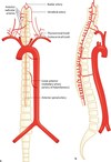
Anterior 2/3 of the spinal cord is supplied by what artery? Where does it originate?
Anterior spinal artery
comes off the verts
Anterior spinal artery supplies what tracts?
Anterior 2/3 of cord
- lateral corticospinal tract
- lateral spinothalamic tract
- anterior horns

Anterior spinal artery infarction causes what sx?
paralysis
loss of pain and temp
urinary and fecal incontinence
(DCML system is preserved)
What artery supplies most of the lumbar and sacral spinal cord
Radicular artery of adamkiewicz
Artery of Adamkiewicz arises from?
Descending aorta usually comes off around T9-T12
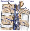
Venous return of the cord enters epidural veins called? What is unique about these veins?
Batson’s Plexus
Lack valves - allow spread of infection or cancer to spread easily.
Cuneocerebellar tract carries what info and brings it where?
Carries afferent information about movement of the ipsilateral upper extremity and rostral body to the cerebellum.
DCML carries?
Vibration, proprioception and light touch
DCML - nucleus gracilis vs cuneatus
gracilis is from lower body and cuneatus is more rostral
Describe DCML tract
information travels up the DC, synapses in gracilis/cuneatus, decussates in the medulla in the medial lemniscus via arcuate fibers and goes to VPL, synapses again and then goes to prim somatosen cortex.
Dorsal spinocerebellar tract carries?
unconscious proprioceptive information from the lower limbs and trunk to the cerebellum
(cuneocerebellar tract but for the lower body)
Rostral Spinocerebellar tract carries?
unconscious proprioceptive information from the upper limbs and rostral body to the cerebellum, similar to the cuneocerebellar tract.
What types of fibers mediate pain and temp via spinothalamic tract?
small myelinated (fast) A-delta fibers
and unmyelinated (slow) C fibers
Course of spinothalamic tract?
Enter the spinal cord and synapse in lamina II, then cross the anterior commisure and ascend in the spinothalamic tract to the VPL of the thalamus, synapses again and goes to primary somatosensory cortex.
Ventral spinocerebellar tract carries?
similar to dorsal spinocerebellar, carries proprioceptive info from the lower limbs and trunk, however it enters the cerebellum via the superior cerebellar peduncle and some fibers cross.
Dorsal enters via the inferior peduncle.
Corticospinal tract is responsible for
voluntary motor activity
Describe the lateral corticospinal tract
Starts in lamina V of cerebral cortex (primary motor cortex)
fibers travel through the corona radiate, to the posterior limb of the internal capsule and VENTRAL brainstem.
In the caudal medulla, 90% of fibers cross and descend in the lateral corticospinal tract.
The other 10% travel in the ventral corticospinal tract (ipsilateral), these fibers cross in the ventral white commisure and terminate in the cervical and upper thoracic regions.

Intermediolateral columns are located in what levels of the spinal cord? What kind of fibers come from this region?
T1-L2
Preganglionic sympathetic autonomic fibers

Function of medial reticulospinal tract?
facilitates antigravity muscles
Function of lateral reticulospinal tract?
inhibits antigravity muscles and facilitates the antagonizing muscles.
Rubrospinal tract is involved in?
similar to lateral reticulospinal tract and ventral corticospinal tract, rubrospinal tract inhibits antigravity muscles and facilitates antagonizing muscles.
Medial vestibulospinal tract is responsible for?
changes in head and trunk position in reponse to information from semicircular canals.
Medial vestibulospinal tract arises from what nucleus and descends to?
arises from medial vestibular nucleus and descends to anterior horn cells.

Lateral vestubulospinal tract is responsible for?
Arises from what nucleus
facilitates antigravity muscles (like medial reticulospinal tract)
lateral vestibular nucleus
Which descending tracts faciltate anti-gravity muscles?
lateral vestibulospinal tract
medial reticulospinal tract
Which descending tracts antagonize ant-gravity muscles?
corticospinal tract
lateral reticulospinal tract
rubrospinal tract
Site of lesion and tracts involved in the following lesion: Brown Sequard
spinal cord hemisection
affects the lateral corticospinal tract, lateral spinothalamic, anterior horn, dorsal column
Site of lesion and tracts involved in the following lesion: B12 deficiency
dorsal columns, lateral corticospinal tracts, spinocerebellar
Tracts involved in the following lesion:
Friedrich’s
dorsal columns
lateral corticospinal tract
spinocerebellar tract
Site of lesion and tracts involved in the following lesion: Polio
Anterior horns
Site of lesion and tracts involved in the following lesion:
Syphilis ( tabes dorsalis)
dorsal columns
Site of lesion and tracts involved in the following lesion: Syringomyelia
cavitation of the cervical cord.
affects decussating lateral spinothalamic axons and anterior horns
Site of lesion and tracts involved in the following lesion:
Ventral spinal artery occlusion
anterior 2/3 of the cord.
affects lateral corticospinal tracts, lateral spinothalamic tracts, anterior horns
Site of lesion and tracts involved in the following lesion: spinal muscular atrophy
anterior horns
Site of lesion and tracts involved in the following lesion: ALS
anterior horns
corticospinal tracts
What is the only CN with no relay in the thalamus
CN1
Anterior nucleus of the thalamus is involved in? Part of what circuit? Recieves info from?
limbic function
part of papez circuit
mamillary bodies
Anterior nucleus of the thalamus sends info from mamillary bodies to?
cingulate gyrus
intralaminal nuclear thalamic group sends info to ?
the striatum
mediodorsal/dorsomedial thalamic nucleus is involved in relay from? to?
amygdala, temporal lobe, and substantia nigra to the prefrontal and frontal association cortex
affects motivation, memory
Largest thalamic nucleus?
Pulvinar
Pulvinar thalamic nucleus is involved in?
visual attention
Largest thalamic nucleus attracts the most visual attention,
Sensory info from the limbs meets where in the thalamus?
VPL of the thalamus
VPLimbs
Sensory info from the face meets where in the thalamus?
VPM
What else relays at the VPM?
sensation of the face via CN5
taste info from nucleus tractus solitarius
VPMeeting
VPmmm or VPyum (taste info)
How is taste transmitted to from the tongue/epiglottis to the cortex?
Ant 2/3 tongue => CN 7 => geniculate nuc
Posterior 1/3 tongue = CN 9 => inferior nucleus 9
epiglottis/esophagus = CN 10 => inferior nucleus 10
these all carry to the NTS, which relays to the VPM and then to the cortex (brodman area 43)
LGN of the thalamus receives what info?
visual info from the optic tract.
MGN of the thalamus receieves what info?
auditory info from the inferior colliculi
and then projects it to the primary auditory cortex.
VA nucleus of the thalamus - function?
receives info from? and sends it to?
important role in movement regulation via GABA
input from GPi and SNr to the VA nucleus -> sends GABA signals to the cortex.
VL nucleus of the thalamus - function?
receives info from? and sends it to?
part of the dentatorubrothalamic pathway, which is how the cerebellum influences synergy of movement.
Purkinje cells > dentate nucleus > contralateral red nucleus > VL thalamus > areas 4,6 motor nucleus
Explain the full CSF flow?
choroid plexus => lateral ventricles => IV foramina of monroe => 3rd vent => aqueduct of sylvius => 4th vent => foramen of Luschka and Magendie => subarachnoid space => arachnoid granulations

How many cc of CSF do adults have?
~150
How many cc of CSF does the choroid plexus make?
20 cc/hour or 500 cc/day
if you work in the choir (choroid), youll get 20$ an hour, or 500$ a day!
normal CSF pressure in the lateral recumbant position?
10-15 cm H2O
If you are lying down on the job, youll only make 10-15$ an hour
Circumventricular organs are unique why? which organs are in this?
these organs have an interrupted Blood brain barrier.
- Neurohypophysis
- Organum vasculosum
- Subcommisural organ
- Pineal gland
- Area postrema
- Median eminence
- sub-fornical organ
NO SPAM about FORNication
area postrema function? what makes it unique?
on the surface of the medulla adjacent to the 4th ventricle, connects to the NTS (only paired circumventricular organ)
detects toxins and causes VOMITING
Eating pastrami makes you wanna vomit.
function of the median eminence?
Where the hypothalamus releases hormones that are carried to the anterior pituitary
Function of the neurohypophysis?
synonymous with the posterior pituitary. Secretes oxytocin and vasopressin which are synthesized from the thalamus.
Only paired circumventricular organ?
Area postrema (not sure what they mean by paired organ, maybe with the NTS? look into it)
only CN that does not relay to the thalamus?
CN 1 olfactory
