Cell Injury Flashcards
List four possible causes of cell injury and death.
Hypoxia
Toxins
Physical agents - direct trauma, extremes of temperature, changes in pressure, electric currents.
Radiation
Micro-organisms
Immune mechanisms
Dietary insufficiency and deficiencies, dietary excess.
What is hypoxia?
Hypoxia is when the body or some tissue within the body is deprived of oxygen.
What are the causes of hypoxia?
Hypoxaemic hypoxia - arterial content of oxygen is low.
Anaemic hypoxia - decreased ability of haemoglobin to carry oxygen.
Ischaemic hypoxia - interruption to blood supply.
Histotoxic hypoxia - inability to utilise oxygen in cells due to disabled oxidative phosphorylation enzymes.
List four toxins.
Glucose and salt in hypertonic solutions
High concentration of oxygen
Poisons
Pollutants
Insecticides
Herbicides
Asbestos
Alcohol
Narcotic drugs
Medicines
Where are the principal structural targets for cell damage?
Cell membranes - plasma membrane, organelle membranes
Nucleus - DNA
Proteins - structural, (enzymes)
Mitochondria - oxidative phosphorylation
List three free radicals that are of particular biological significance in cells.
OH. (Hydroxyl) - the most dangerous
O2- (superoxide)
H2O2 (hydrogen peroxide)
How are free radicals removed from our bodies?
Spontaneous decay
Enzymes - superoxide dismutase (SOD) - produces H2O2 which is less toxic to cells.
Also, catalases and peroxidases complete process of removal: H2O2 to O2 and H2O.
Free radical scavengers - vit E, A and C
What happens to the morphology of cells when they are damaged/die?
Detachment and loss of ribosomes and accumulation of denatured proteins. Nuclear changes - clumped chromatin - very subtle (reversible) followed by various combinations of pyknosis (shrinkage), karryohexis (fragmentation) and karryolysis (dissolution) (irreversible).
What is pyknosis?
Shrinkage
What is karryohexis?
Fragmentation
What is karryolysis?
Dissolution
What are abnormal accumulations which respect to cell injury and cell death?
Abnormal accumulations in reversibly injured cells reflect damaged proteins and accumulation of abnormal metabolites.
E.g. Mallory’s hyaline, seen in alcoholic liver disease - accumulation of altered keratin filaments.
Define oncosis.
Spectrum of changes in injured cells prior to death. - process of death.
Define necrosis.
Morphological changes that follow cell death in living tissue, largely due to progressive degradative action of enzymes on lethally injured cell.
Define apoptosis.
Cell death induced by regulated intracellular program - cells activate enzymes that degrade cells’ own nuclear DNA and proteins. Cell suicide.
List the different types of necrosis.
Two main types: coagulative and liquefactive.
Two other special types: caseous (TB) and fat necrosis.
NB. Gangrene and infarct are clinical terms - NOT types of necrosis.
Where are you most likely to see liquefactive necrosis?
In the brain because it is a fragile tissue without support from a robust collagenous matrix.
Where is fat necrosis most typically seen?
Pancreas as a consequence of pancreatitis.
Pancreatitis causes release of lipases from injured pancreatic acinar cells. Lipases act on fatty tissue of pancreas and fat elsewhere in the abdominal cavity causing fat necrosis.
What is gangrene?
Gangrene is a clinical term for necrosis that is visible to the naked eye.
What are the different types of gangrene?
Dry- coagulative necrosis
Wet - liquefactive necrosis typically due to infection.
NB. Everyone has had gangrene….remnant of the umbilical cord in a newborn.
What type of necrosis is typically associated with tuberculosis?
Caseous
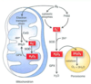
Briefly describe the two pathways of apoptosis initiation.
Intrinsic - (cell stress pathways) p53 activation, cytochrome C release from mitochondria. Combines with APAF1 and caspase 9. Forms an apoptosome. This activates downstream caspases.
Extrinsic - (death receptor pathway) death ligands TRAIL & FAS bind receptors, activating procaspase 8, which activates downstream caspases.
What is the pathway of paracetamol metabolism? What happens when glutathione runs out?

What happens in a paracetamol overdose?
After an overdose, glutathione is depleted and NAPQI binds with sulphydryl groups on liver cell membranes, eventually causes hepatocyte necrosis and liver failure.
Some people have lower reserves of glutathione and paracetamol OD is more dangerous.
