Causes of Chest Pain Flashcards

1
Q
A

C - left anterior descending artery

2
Q
A

A - aspirin and clopidogrel

this is a typical presentation of a silent heart attack in a diabetic

3
Q

What is the definition of ischaemic heart disease?

A
  • decreased blood supply to the myocardium which results in chest pain (angina pectoris)
  • it is primarily due to atherosclerosis of the coronary arteries
  • this results in a mismatch between the oxygen supply to the heart and oxygen demand
4
Q

What are the different types of ischaemic heart disease?

A
  • IHD is an umbrella term
  • it can be divided into atherosclerotic and vasospastic causes
  • atherosclerotic causes can be further divided into stable angina and ACS
  • ACS is an umbrella term that covers:
    • unstable angina
    • non ST-elevation MI
    • ST-elevation MI
5
Q

What is Prinzmetal angina?

A
  • a condition characterised by angina-type chest pain at rest
  • it occurs due to abnormal coronary artery spasm
6
Q

What are the risk factors for ischaemic heart disease?

A
  • hypertension
  • diabetes
  • smoking
  • family history of IHD
  • hyperlipidaemia and hypercholesterolaemia
    • this can be due to diet or familial hypercholesterolaemia
7
Q

What are some signs of hyperlipidaemia that may be picked up on examination?

A
  • xanthelasma
    • this describes yellow coloured deposits of cholesterol around the eyes
  • xanthomata
    • this describes yellow cholesterol-rich deposits that can appear anywhere in the body
  • corneal arcus
    • a cholesterol-rich deposit that forms a ring within the cornea
8
Q

What is the definition of stable angina?

A
  • chest pain brought on during exertion and relieved by rest
  • the pain occurs due to myocardial ischaemia, most commonly due to atherosclerotic plaques reducing blood flow to the heart
9
Q

What signs might be seen on examination of someone with stable angina?

What investigations should be performed?

A
  • examination is normal in chronic stable angina
    • unless there is an underlying heart condition (e.g. HF)
  • bloods should be performed:
    • ​FBC
    • lipids - to check for hyperlipidaemia
    • glucose - to check for diabetes
  • ECG should be performed to make sure more sinister causes aren’t missed (e.g. silent MI)
10
Q

What is involved in the conservative managment of stable angina?

A
  • management aims to reduce risk factors
  • weight loss
  • improving diet
  • smoking cessation
  • if conservative measures are ineffective, then medical management is considered
11
Q

What is involved in the medical management of stable angina?

A
  • ACE inhibitors
  • antiplatelets - usually aspirin
  • statins
  • anti-anginals
    • beta-blocker or calcium channel blocker
  • GTN spray
    • ​this should be used when anginal pain is felt to relieve it
    • if the pain is not relieved by GTN, patient needs to go to hospital as they could be having an MI
12
Q

Why does anginal pain actually occur in stable angina?

A
  • there is increased oxygen demand by the body (and heart) during exercise
  • vessels containing stable atherosclerotic plaques are unable to dilate enough to allow adequate blood flow to meet the myocardial demand
  • this leads to myocardial ischaemia (lack of oxygen), which causes the pain
13
Q

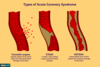
What is the definition of acute coronary syndrome?

A
  • a range of conditions due to a sudden reduction in blood flow to the heart
  • this is usually due to atherosclerotic plaques within the coronary arteries
  • these plaques may cause complete or substantial occlusion of the artery
14
Q

What investigations can be performed to identify which type of ACS is present?

A
  • first an ECG is performed to look for any visible changes
    • if there is ST elevation - it is a STEMI
  • if there are no ECG changes then look at troponin
    • troponin is released when myocardial cells infarct
  • in an NSTEMI there is cell infarction so troponin is raised
    • ​it would also be raised in STEMI
  • there is no infarction in unstable angina, so troponin is not raised
15
Q

What is unstable angina?

A
  • chest pain at rest due to ischaemia but without cardiac injury
  • it occurs when an atherosclerotic plaque ruptures and a thrombus forms around the ruptured plaque
  • this causes partial occlusion of the coronary artery, which leads to angina-type pain
16
Q

What are the signs and symptoms associated with ACS?

Which groups do special attention need to be paid to?

A
  • acute central chest pain that is gripping / heavy
    • pain may radiate to the neck, arm and/or jaw
  • sweating
  • pallor
    • ​reduced CO might lead to patient becoming hypotensive
  • +/- shortness of breath
  • can be silent in the elderly and diabetics
    • ​often there is no central chest pain
    • they may be nauseous or have a different type of pain
17
Q

What investigations are performed in ACS?

A
  • ECG
  • troponin
    • elevated troponin suggests myocardial injury - STEMI or NSTEMI
18
Q

What ECG changes can be seen in ACS?

A
  • ST segment elevation
  • ST segment depression
  • T wave inversion
  • (look at the difference between the baseline and where the ST segment starts to see if there is elevation / depression)*
19
Q
A
20
Q

What ECG changes are seen in a STEMI?

A
  • hyperacute T waves
  • ST elevation
  • new LBBB
  • there may also be T wave inversion
21
Q

What is shown in these images?

A

they all show ST elevation

there are many different forms of ST elevation

the ST segment must just be raised from the baseline

22
Q

What ECG changes are present in an NSTEMI / unstable angina?

A
  • ST depression
  • T wave inversion
23
Q

What ECG change is associated with an old infarct?

A

pathological Q waves

these indicate that the patient has had some form of myocardial ischaemia in the past

the Q wave appears deeper

24
Q

How much of the artery is occluded in a STEMI?

What are the different treatments available depending on the time since symptom onset?

A
  • in a STEMI, there is complete occlusion of a coronary artery
  • if < 12 hr since symptom onset AND PCI available in 120 mins then PCI is performed
    • this involves placing a stent or balloon into the artery to open it up
  • if < 12 hr since symptom onset and PCI NOT available in 120 mins then thrombolysis is performed
    • this aims to dissolve the clots and open up the arteries
  • if > 12 hr since symptom onset, there are too many risks with invasive procedures

angiography is performed, then possibly followed by PCI

  • this involves inserting a needle in the femoral / radial artery and a catheter into the heart to visualise the perfusion of the heart
25
What medications are given immediately to someone presenting with a STEMI?
remember **MONAC** - these drugs are all given **_immediately_**: * M - **_morphine_** * O - **oxygen** (if sats \< 90%) * N - **nitrates** * A - **_aspirin_** * C - **_clopidogrel_** * (aspirin & clopidogrel oral tablets - **300mg** of each* * then obtain IV access to give **2.5mg diamorphine*** * nitrates are not always given - they help with the pain and open up the arteries to prevent further damage)*
26
What medications are given for the long-term management of someone who has had a STEMI?
* **_beta-blocker_** * do not give in acute heart failure * **_ACE-inhibitor_** * this will help with BP and should be given within 24 hours of presentation * **_statin_** * this will reduce the cholesterol
27
What type of occlusion is present in NSTEMI? How is it managed differently to STEMI?
* in an NSTEMI there is a **_near complete occlusion_** of a coronary artery * management is the **SAME** as the STEMI drugs but with **_LMWH_** * this is usually in the form of **_fondaparinux_** or **_enoxaparin_**
28
What score is used to determine the risk of someone who has just had an NSTEMI having another CVS event?
the **_GRACE score_** is used * **low risk** patients have **_outpatient_ angiography** * **moderate / high risk** patients have **_inpatient_ angiography** with **possible PCI** * also given GpIIb/IIa inhibitors (abciximab)
29
What mnemonic is used to remember the management of NSTEMI and STEMI?
MONAC BAS * M - morphine * O - oxygen * N - nitrates * A - aspirin * C - clopidogrel * B - beta blocker * A - ACE inhibitor * S - statin * MONAC are given immediately at presentation * BAS are part of long term management
30
What mnemonic can be used to remember the complications of STEMI / NSTEMI?
**DARTH VADER** * D - death * A - arrhythmia * R - rupture * T - tamponade * H - heart failure * V - valve disease * A - aneurysm * D - Dressler's syndrome * E - embolism * R - reinfarction
31
D -Dressler's syndrome * this is a presentation of **pericarditis** that is occurring **2 - 10 weeks following an MI**
32
D - aspirin + colchicine + NSAIDs * this is the management for anyone with pericarditis * aspirin is given as she has a history of MI / heart failure
33
What is the definition of pericarditis? What can cause this?
* **inflammation of the pericardium** ***_Causes:_*** * idiopathic * viral - **Coxsackie A9**, **mumps**, **EBV** * bacterial - **pneumococcus**, **staph**, **strep** * connective tissue disorders - **sarcoidosis** * system autoimmune disorders - **SLE**, **rheumatoid arthritis** * **_Dressler syndrome_** (2 - 10 weeks post-MI) * **malignancy**
34
What is Dressler's syndrome?
* this is a form of **pericarditis** that occurs **_2 to 10 weeks following an MI_** * it is thought to be **antibody-related** after damage to the pericardium
35
What are the risk factors for pericarditis?
* male gender * 20 - 50 years old * transmural MI * cardiac surgery * neoplasm * uraemia / dialysis
36
What type of pericarditis can occur in heart failure? What is the result of this on cardiac output?
* **_constrictive pericarditis_** can occur * the pericardium becomes **thicker** as part of a **chronic process** * a thicker pericardium does not allow for as much movement of the heart, so both **_ventricular filling_** and **_CO are reduced_**
37
What are the signs and symptoms associated with pericarditis?
* **sharp, stabbing _pleuritic chest pain_** that is **_relieved on sitting forwards_** * this may radiate to the trapezius ridge * **corysal symptoms** if it is of viral origin * ​e.g. runny nose, viral / flu-like symptoms * **_pericardial friction rub_** on examination * **tamponade** * ​Beck's triad is present on examination if this is present
38
What is meant by pleuritic chest pain?
* pain that **varies with breathing movements** and is **_worse on inspiration_** * it is described as **stabbing, knife-like** or **catching**
39
Where is pericardial friction rub best heard? What does this sound like?
* heard best on the **_lower left sternal edge_** * it is a **scratching sound** that sounds like 2 pieces of paper rubbing together
40
What is tamponade?
* this occurs when the **pericardial sac is _filled with blood_** * this is **life-threatening** as the blood filling the pericardium **compresses the heart,** meaning it cannot beat properly * there is not enough blood being ejected to the body / brain
41
What triad is present on examination if tamponade is present?
**_Beck's triad_** * low arterial blood pressure * muffled heart sounds * distended neck veins
42
What investigations are performed in pericarditis?
* bloods * to ensure all electrolytes are balanced * CXR to check for **pericardial effusion** * ECG
43
What is a pericardial effusion?
this occurs when there is **_fluid in the pericardium_** there is not as much fluid present in the pericardium as there would be in tamponade
44
What ECG changes are associated with pericarditis?
***widespread saddle shaped ST elevation*** * the ST elevation looks like a "u" shape
45
What is involved in the management of pericarditis? What if tamponade or uraemic pericarditis is present?
**_NSAID + PPI + colchicine_** * exercise restriction is recommended (if viral) * if **tamponade** is present or it is **purulent**, **_pericardiocentesis_** is performed in addition and **_antibiotics_** are given * in uraemic pericarditis, **_dialysis_** is given
46
What are the possible complications of pericarditis?
* pericardial effusion +/- tamponade * chronic constrictive pericarditis
47
Why are NSAIDs and PPIs given to treat pericarditis?
* NSAIDs are given to relieve the pain * PPI is given to protect against NSAID related stomach ulcers * colchicine is a treatment for acute gout that has been shown to prevent recurrence of pericarditis
48
In what type of patients does uraemic pericarditis normally occur?
* there is a build up of uric acid in the body * this usually occurs in patients with chronic kidney disease * it is treated with dialysis
49
What is the definition of syncope?
a sudden, temporary, self-terminating loss of consciousness
50
What are the 4 different categories that cause syncope? What are examples?
***_Arrhythmia-related:_*** * due to CO compromise from **arrhythmia** * e.g. ventricular tachycardia, heart block ***_Cardiac:_*** * **structural heart disease** - most notably LV outflow obstruction * ​e.g. HOCM, severe AS, PE, aortic dissection ***_Orthostatic:_*** * failure to maintain BP when standing ***_Reflex:_*** * reflex **vasodilation / bradycardia** due to a trigger * ​vasovagal or carotid sinus syncope
51
What are the 5 steps involved in differentiating the types of syncope?
* establish there was a **_definite loss of consciousness_** with **no/few features of seizure** * determine if there were any **cardiac red flag symptoms** * carry out investigations * if there are **negative investigations** with typical **precipitant/prodrome** in history - this is **_vasovagal syncope_** * if diagnosis is uncertain then **risk stratify**
52
What are the 4 different investigations that are performed to identify what type of syncope is present?
***_ECG:_*** * if arrhythmia is identified, then this is arrhythmic syncope ***_Imaging -_*** *_if suspicion of PE / aortic dissection:_* * performed to confirm diagnosis ***_Echo:_*** * this can confirm cardiac structural abnormalities that cause cardiac syncope ***_Sitting / standing BP:_*** * used to identify orthostatic syncope
53
What are the typical symptoms of seizure that need to be ruled out?
* typically there is an **_aura_** beforehand * patient describes seeing / smelling something strange before passing out * then a witness account of: * tonic-clonic movement * automatisms * bitten tongue * confusion post-LOC * drowsy
54
What is meant by tonic clonic movements and automatisms?
***_Tonic clonic:_*** * during the "tonic" phase, the person **loses consciousness**, their body goes **stiff** and they **fall** to the floor * during the "clonic" phase, the **limbs jerk** about, the patient **bites their tongue / cheek** and may have **trouble breathing** * there may also be loss of control of the bladder / bowel ***_Automatisms:_*** * absent seizures where people start fiddling with their hair, **absent-mindedly moving** their fingers etc. and **don't realise** what they are doing
55
How long does LOC typically last for during syncope?
* LOC should be **transient** and typically lasts around **_30 seconds_** * if it lasts for **more than 5 minutes**, think that this could be a **_seizure_**
56
Once established that LOC was not a seizure, then what red flags need to be ruled out?
* need to determine whether there are any cardiac red flag symptoms * **LOC during exertion** * this is typical of aortic stenosis * severe **valvular disease** * **previous arrhythmia** * **concerning ECG features** * e.g. features that suggest LVH
57
How is orthostatic hypotension diagnosed?
* if there is more than a 20 mmHg difference between sitting and standing BP
58
What is the defintion of vasovagal syncope? What can cause this?
* it is a ***sudden, temporary, self-terminating loss of consciousness*** often **triggered by an _emotional upset_** * caused by a **_temporary reduction in cerebral flow_** as a response to a **stressful trigger** * increase in the vagal nerve discharge causes **low BP and HR** * this results in **reduction of CO**
59
What are the risk factors associated with vasovagal syncope?
* prior syncope * emotional stress * prolonged standing * heat * excessive dehydration
60
What are the signs and symptoms of vasovagal syncope?
* nausea * pallor * light-headedness * diminished vision / hearing * physical injury (pain can lead to LOC)
61
What is involved in the investigations and management of vasovagal syncope?
***_Investigations:_*** * ECG * bloods (glucose, cortisol, U&Es, FBC, hCG) ***_Management:_*** * patient education on triggers with physical **counter-pressure manoeuvres**
62
What are some of the counter-pressure manoeuvres that are recommended in vasovagal syncope?
* leg crossing + buttock and thigh muscle tensing * this is a skeletal muscle pump that squeezes veins to try and push blood back up * isometric forearm grip * squatting * ​getting lower to the ground means it is less effort for the heart to pump blood around the body
63
D - reflexive vasodilation * this is due to vasovagal syncope * the trigger is having blood taken, which leads to a drop in HR and BP
64
What is the definition of atrial fibrillation?
* a form of **supraventricular tachycardia** with **_uncoordinated atrial electrical activation_** causing **_ineffective atrial contraction_** * the atria are not contracting together in a coordinated way * this means that reduced amounts of blood are entering the ventricles
65
What are the potential causes of AF?
* idiopathic * coronary artery disease * thyroid disorders * COPD * electrolyte disturbances * pneumonia
66
What is the difference between paroxysmal and persistent AF?
* paroxysmal AF terminates within 7 days, either with or without treatment * persistent AF terminates after 7 days
67
What are the signs and symptoms of AF?
* palpitations * irregularly irregular pulse rate * shortness of breath * chest pain
68
What investigations are performed in AF?
* **ECG** * **bloods** - FBC, clotting, U&Es, TSH * to ensure there is no underlying thyroid condition or electrolyte disturbances * **CXR** * ​to check the size of the heart and ensure there is no HF occurring * **TEE (Echo) pre-cardiovert** * ​do this before cardioverting the patient to check the structural integrity of the heart
69
70
What are the potential complications of AF?
***_Thromboembolism:_*** * uncoordinated atrial contractions means that blood stays in the atria longer than it should * the chances of clotting increases when blood is static * if a thrombus forms in the atria, it can get pumped into the ventricles and around the body ***_Worsened heart failure_***
71
What must you first establish before deciding how to treat a patient presenting with AF?
* need to determine whether they are **_haemodynamically stable_** * i.e. BP is \> 90/60 and there are no other signs of shock * if they are **NOT stable**, then **_DC cardioversion_** is performed * ​this prevents them from entering cardiogenic shock, which is life threatening
72
If the AF patient is haemodynamically stable and it has been \< 48 hours then how should they be treated?
* **_rhythm control_** is performed to try and get the patient back into **sinus rhythm** * the chance of a clot forming within 48 hours is very small * this can be performed through **_DC cardioversion_** * or through **chemical cardioversion** with **_flecanide_** or **_amiodarone_** * ​flecanide is contraindicated in IHD * if this fails, then **rate control** is performed
73
If the AF patient is haemodynamically stable but it has been \> 48 hours then how should they be treated?
***_Anticoagulation:_*** * this involves LMWH (fondaparinux) + warfarin loading * the chance of a clot happening \> 48 hours is much higher * they are then DC cardioverted 4-6 weeks later and given a rate control drug in the meantime ***_Rate control:_*** * bisoprolol * verapamil * diltiazem * digoxin
74
What is the definition of atrial flutter? What causes it?
* large **_re-entry circuit_** in **right atrium** causes a **_saw-tooth pattern_** due to ineffective atrial contraction * it is caused by **underlying heart conditions** * it has very similar signs/symptoms, investigations and management to AF
75
What does atrial flutter look like on ECG?
saw tooth pattern with loss of isoelectric baseline and p waves
76
What advice is given to patients to stop the symptoms of atrial flutter?
to stop palpitations, take a **massive deep breath in** and **hold it** this causes compression on the heart which can help it get back into a normal rhythm
77
What is the definition of heart block?
* a cardiac electrical disorder characterised as **_impaired conduction_** from the **_AV node_**
78
79
What are the causes of heart block?
* **MI / IHD** * infection - **rheumatic fever** * drugs - **beta-blockers, CCBs, amiodarone** * metabolic - **high K+, low T4** * **sarcoidosis**
80
What are the signs and symptoms of the different types of heart block?
* first degree heart block is **asymptomatic** * second degree type I is **asymptomatic** * **_second degree type II_** or **_third degree_** presents with **dizziness, palpitations, chest pain** & **heart failure**
81
How can first degree heart block be identified from ECG?
there is a **_prolonged PR interval_** that is **\> 0.2 seconds**
82
How can second degree heart block (type I) be identified on ECG?
* there are **progressive prolonged PR intervals** until a **_QRS complex is dropped_** * this is also called Wenkebach
83
How can second degree heart block type II be identified on ECG?
* there is **_intermittent loss of a QRS wave_** that might be in a regular pattern (2:1 etc)
84
What does third degree (complete) heart block look like on ECG?
* there is no association between P and QRS waves
85
What investigations are performed for heart block?
* ECG * troponin * K+, Ca2+ and pH * digitalis toxicity * echo
86
What is involved in the acute and chronic management of heart block?
***_Chronic:_*** * permanent **pacemaker** for Mobitz II or 3rd degree ***_Acute - occurring 2o to MI:_*** * **IV atropine** and temporary external pacing * these patients often become bradycardic so need to raise the heart rate with atropine and then pace this rhythm
87
What are the potential complications associated with heart block?
* asystole * heart failure * cardiac arrest
88
What is the defintion of supraventricular tachycardia?
* a dysarrhytmia originating **at or above the AVN** with a **_regular, narrow complex tachycardia_** (QRS \< 120 mm) at **\>100 bpm** with **_no p waves_**
89
What are the signs and symptoms of a supraventricular tachycardia?
* palpitations * syncope * shortness of breath * chest pain
90
What are the 2 different types of SVT?
***_Atrioventricular re-entry tachycardia (AVRT):_*** * accessory pathway exists from the ventricles back up to the atria ***_Atrioventricular nodal re-entry tachycardia (AVNRT):_*** * accessory pathway exists from the AV node to the atria * this is the Bundle of Kent in WPW
91
What is involved in the management of an SVT once it has been identified on ECG?
* **_vasovagal manoeuvres_** (Valsalva) * these stimulate the vagal nerve to decrease the HR * if this does not work then give **_6mg adenosine IV_** * **verapamil 2.5-5mg** used in asthma * ​repeat this step if ineffective * if still ineffective, then give **_12mg adenosine IV_** * if still ineffective then give **_IV amiodarone_** and refer to specialist