Cardiovascular System and the Heart Flashcards
(75 cards)
State the composition and function of the Cardiovascular system
The Cardiovascular system consists of the Heart and blood vessels.
- Heart- propels blood through the system
- _Arteries_ (Away from the heart)- vessels that carry blood to the tissue
- Capillaries- smallest vessels, sites of O2, CO2, nutrient, & waste products exchange between blood & tissues
- Veins- carry blood to the heart
Functions:
- It is responsible for providing O2 and metabolic nutrients to the body
- It removes CO2 and metabolic waste products from tissues
2 Dual circulations of the Cardiovascular sytem
-
Systemic circulation- supplies blood to all peripheral tissues, including the lungs:
* Pulmonary veins- Left Atrium- Mitral valve- Left ventricle- Aortic semilunar valve- Aorta- Arteries- Arterioles- Capillaries -
Pulmonary circulation- supplies blood to lungs for gas exchange:
* Venules- Veins- Inferior and Superior vena cava- Right atrium- Tricuspid valve- Right ventricle- Pulmonary semilunar valve- Pulmonary arteries- Lungs
Valves- LAB RAT
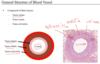
3 layers of the Blood vessel and their components
Tunica intima (closest to the lumen)
-
Endothelium (simple squamous epithelium) connected by tight junctions
- Basal lamina of the endothelium
- Subendothelial layer of loose connective tissue
- contains an INternal elastic lamina (IEL) in some
- Site of atherosclerotic plaque formation
Tunica Media (middle layer)
- Smooth muscle layer arranged in concentric layers
- Gap junctions between smooth muscle cells (SMC)
- Sheets of reticular fibers, elastic fibers, ground substances (proteoglycans) and lamellae are intertwined between layers of (SMC)
- Thickest layer for arteries vs. tunica adventitia for venules and veins
- contains an EXternal elastic lamina (EEL) in some
- Smooth muscle regulates the diameter of blood vessel [more diameter = more blood flow] [less diameter = less blood flow]
Tunica Adventitia/Externa (outermost layer)
-
Dense irregular connective tissue (longitudinal collagen type 1 fibers with elastic fibers and fibroblasts)
- Thickest layer of venules and veins vs. tunica media for arteries
-
Larger vessels’ tunica adventitia contain:
- Nervi vasorum/vascularis- unmyelinated sympathetic nerve fibers (vasoconstrictor- NE)
- Vasa Vasorum- “vessel of the vessel”- blood vessel (arterioles, capillaries, venules) that provide perfusion to larger blood vessels (aorta, SVC, IVC)
Compare and contrast Large arteries, Medium arteries, Small arteries and arterioles
Large Arteries (Elastic arteries)- arteries that branch directly off the heart (aorta and pulmonary arteries)- Conduct blood from heart and with elastic recoil to help move blood forward under steady pressure
- Tunica intima- Thick and IEL is not always discernable
-
Tunica media- contains multiple sheets of elastic laminae; External elastic lamina is not always discernable
- Enables stretching to accomomdate a large volume of blood ejected from the ventricles during systole, which can recoil during diastole to maintain continuous pulsatile blood flow
- Tunica adventitia- Thin; contains vasa vasorum and nervi vasorum
Medium Arteries (Muscular arteries) - branch form large arteries to provide blood flow to specific regions and organs (axillary, radial, femoral, carotid, renal, mesenteric, & coronary arteries)- Distribute blood to all organs and maintain steady blood pressure and flow with vasodilation and constriction
- Tunica intima- prominent internal elastic lamina
-
Tunica media- contains 3-40 layers of smooth muscle with elastic laminae that decrease in number as the artery size decreases
- External elastic lamina present between tunica media and tunica adventitia
- Tunica adventitia- Prominent; can be equal to or less than the thickness of the tunica media
Small Arteries (Distribute blood to arterioles) &
Arterioles (Resist and control blood flow to capillaries)
- Tunica intima- Small arteries contain prominent internal elastic lamina- Arterioles do NOT
- Tunica media- Small arteries contain up 3 to 8 layers of smooth muscle- Arterioles only contain 1-2 layers
- Tunica adventitia- thin layer that blends with surrounding connective tissue for both

Important role of Arterioles
Arterioles regulate blood flow through capillaries via contraction (vasoconstriction) and relaxation (vasodilation) of the smooth muscle layer of their tunica media
Major site of vascular resistance that play large roles in hypertension and shock
Increase blood flow = decrease resistance
Decrease blood flow = increase resistance
Metarterioles
Metarterioles are short vessels that link arterioles to capillary beds with thickened tunica media layer called a precapillary shincter, which regulate capillary blood flow
Contract-relax 5-10cycles/min in a pulsatile manner
When sphincters are closed, blood flow directly to the Thoroughfare channel (lack muscle) which is connected to the postcapillary venules

Capillaries
Capillaries:
- Smallest diameter blood vessels
- Flattened nuclei; Heterochromatic
- Sites of exchange of gases, metabolites, & waste products between the blood and peripheral tissues
- Consist of a single endothelial cell with a surrounding basal lamina layer with tight junction for permeability
- Cytoplasm contains mitochondria and vesicles
- Increased metabolic demand = Increased capillary density
- Classified into 3 categories based on permeability
- Continuous
- Fenestrated
- Discontinuous

3 Histological types of Capillaries
- Continuous capillaries (least permeable):
- Endothelium bound together by occluding (tight) junctions
- Surrounded by continuous basal lamina
- Pinocytic vesicles are found beneath the lamina and basal membrane surfaces to facilitate the exchange of larger molecules between the blood and surrounding tissue
- Found in the CNS (BBB), lung*, *muscle*, & *cortex of thymus
- Fenestrated capillaries:
- Endothelium with numerous fenestrations that facilitate rapid exchange of molecules
- Surrounded by a continuous basal lamina
- Found in the kidney*, *endocrine tissue*, *intestinal villi*, & *choroid plexus
- Discontinuous capillaries:
- Endothelium with large fenestrations and no diaphragms
- Discontinuous gaps exist between endothelium (inter-endothelial gaps)
- Discontinuous and sometimes absent basal lamina
- Reticular fibers present
- Enable easy phagocytosis of blood molecules
- Found in liver*, *bone marrow, & spleen

Pericytes
Pericytes:
- Located along continuous capillaries and postcapillary venules
- Mesenchymal cells with long cytoplasmic processes surrounding the endothelial layer
- Secrete many ECM & form their own basal lamina which fuses with the basement membrane of the endothelial cells
- Cytoskeletal component of myosin, actin, tropomyosin (indicates that pericytes dilate or constrict capillaries helping to regulate blood flow)
- Important for maintaining the BBB in the CNS
- Proliferate and differentiate to form smooth muscle after injury

Diabetic microangiopathy
Diffuse thickening of capillary basal lamina and decrease in metabolic exchange at the vessels (affecting kidneys, retina, skeletal muscles, skin)
Excessive blood sugar that occurs with diabetes (hyperglycemia) can lead to this
Primary site of diapedesis
Post- capillary venules are the primary site of diapedesis
Junction between the endothelial cells of post-capillary venules are the looses of the microvasculature as it facilitates transedothelial migration of leukocytes during inflammation

Compare and contrast Venules, Small and medium veins, Large veins
Venules:
- First vessel of the venous system that drain capillary blood
- Vessel wall is often as thin as a capillary, but a much larger lumen
- Can transition from low permeability to high permeability when stimulated by vasoactive molecules (histamine)
- Post-capillary venules are the primary site of diapedesis
- Tunica intima- very thin; NO internal elastic lamina
- Tunica media is comprised of 1-2 bundles of smooth muscle cells and pericytes with longitudinal collagen fibers; No external elastic lamina
- Tunica adventitia- indistinct to none
Small/Medium veins:
- Veins that drain tissues/organs into the large veins
- Contain one way endothelial line valves formed by the thin tunica intima layer to enable unidirectional flow
- Prevents retrograde flow due to gravity in the lower extermities
- Tunica media consists of 3-5 layers of smooth muscle cells, collagen, and elastic fibers that is perfused by the vasa vasorum
- Tunica adventitia is thicker than the tunica media with some longitudinal smooth muscle
Large vein:
- Thin tunica intima; indistinct internal elastic lamina
- Tunica media- several layers of smooth muscles; indistinct external elastic lamina
-
Tunica adventitia is the thickest layer, up to 4X the thickness of the tunica media
- Prominent bundles of longitudinal smooth muscle
- Contain collagen and elastic fibers, nerves, and vasa vasorum
- Examples: Superior and inferior vena cavae, mesenteric, renal, femoral, portal, and internal jugular veins

Systole and Diastole
Pressure gradient between atria and ventricles- main driver of blood flow into the ventricles
Blood flow is moved through the arteries forcefully during contraction (Systole)
- aortic and pulmonary valves opened
- tricuspid and mitral valves closed
Venricular pressure drops to a low level the ventricles relax (Diastole):
- aortic and pulmonary valves closed
- tricuspid and mitral valves opened
- pressure in atria is greater than pressure in ventricles (Pa>Pv)
- Elastin rebounds passively to maintain arterial pressure
- Aortic and pulmonary valves prevents backflow of blood so the rebound continues the blood flow away from the heart

Difference of pressure requirement in correlation with the functions of arteries and veins
Arteries are distributing system (higher pressure)
Veins are collecting system (higher volume so need lower pressure)
Pressure in artery > pressure in vein that’s why arteries need much stronger wall to stand pressures
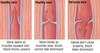
Atherosclerosis
Atherosclerosis
- Disease of elastic arteries (large) and muscular arteries (medium)
- Buildup of plaque within arterial walls that leads to wall hardening and subsequent narrowing of the vessel wall
- Endothelial damage leads to macrophages entering the tunica intima and platelets adhering to the damaged endothelium triggering and inflammatory response
- Causes of endothelial damage- oxidized LDL, hypertension, smoking
- Inflammatory cells stimulate hyperplasia of smooth muscle cells then migrate to the tunica intima
- Macrophages engulf cholesterol to form foam cells that appear as fatty streaks
- Atheroma: Smooth muscle cells and macrophages release cytokines that stimulate production of extracellular matrix components that form a fibrous cap over a necrotic center (cellular debris, cholesterol, foam cells)
- Rupture of fibrous cap and release of the necrotic center components stimulate thrombus formation and subsequent vessel occlusion and ischemia

Aortic Aneurysm
Aortic Aneurym
- Pathologc dilation of the aorta that can occur at any point along the aorta but most commonly seen in the abdomen (AAA-abdominal aortic aneurysm)
- Major risk factors: hypertension, smoking, diabetes, and hyperlipidemia
- Aortic dissection occurs when a break in the tunica intima allows blood to leak into the tunica media layer, resulting in abnormal dilation of the aorta
Identify the type of vessel

Medium artery-
- thicker tunica media (smooth muscle layer), rounder lumen
- Internal elastic lamina in the Tunica Intima is very prominent in medium artery- histological marker of medium or muscular arteries
Identify the type of vessel

Medium vein-
- thinner tunica media (smooth muscle layer), almost stellate-shaped lumen
- very thin tunica intima
Identify the histological marker of this vessel

Internal elastic lamina in the Tunica Intima is very prominent in medium artery- histological marker of medium or muscular arteries

Identify the type of vessel

Arterioles (surrounded by adipocytes)- endothelium and 1 layer of smooth muscle

Identify the type of vessel

Large artery
- Significant tunica intima
-
Tunica media- contains multiple sheets of elastic laminae
- Enables stretching to accommodate a large volume of blood ejected from the ventricles during systole, which can recoil during diastole to maintain continuous pulsatile blood flow
Identify the type of vessel

Large vein
- Very thin intima, very thin media, very large adventitia (hallmark for large vein)
- 2 layers of smooth muscle- 1 in media and 1 in adventitia (bundles of smooth muscle interspersed between the dense irregular connective tissue running at a 90 degree angle to the tunica media- orthogonal)

Identify the type of vessel

Small artery-
- prominent internal elastic lamina, there is not much smooth muscle to qualify for medium artery
Identify the 2 vessels

Venule (left)
Arteriole (right)- with 1 layer of smooth muscle below the endothelium and much more defined lumen