Cardiovascular pathology Flashcards
What kind of tissue is this and what are some key characteristics of this tissue type?

This is the normal appearance of myocardial fibers in longitudinal section. Note the central nuclei and the syncytial arrangement of the fibers, some of which have pale pink intercalated disks.
What is this a cross-section of?

This is a normal coronary artery. The lumen is large, without any narrowing by atheromatous plaque. The muscular arterial wall is of normal proportion.
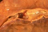
Which valve is this?

This is the tricuspid valve. The leaflets and thin and delicate. Just like the mitral valve, the leaflets have thin chordae tendineae that attach the leaflet margins to the papillary muscles of the ventricular wall below.
What valve is this?

The aortic valve shows three thin and delicate cusps. The coronary artery orifices can be seen just above.The endocardium is smooth, beneath which can be seen a red-brown myocardium. The aorta above the valve displays a smooth intima with no atherosclerosis.
What is in this image?

This is the external appearance of a normal heart.The epicardial surface is smooth and glistening.The amount of epicardial fat is usual.The left anterior descending coronary artery extends down from the aortic root to the apex.
What is shown here?

The coronary artery shown here has narrowing of the lumen due to build up of atherosclerotic plaque.
What is occurring in this coronary artery?

This section of coronary artery demonstrates remote thrombosis with recanalization to leave only two small, narrow channels.
What is occurring in this coronary artery?

There is a severe degree of narrowing in this coronary artery. It is “complex” in that there is a large area of calcification on the lower right, which appears bluish on this H&E stain.
What is shown here?

This distal portion of coronary artery shows significant narrowing. Such distal involvement is typical of severe coronary atherosclerosis, such as can appear with diabetes mellitus or familial hypercholesterolemia. This would make a coronary bypass operation difficult.
What is the arrow pointing at?

There is a pink to red recent thrombosis in this narrowed coronary artery. The open, needle-like spaces in the atheromatous plaque are cholesterol clefts.
This is a magnified atheroma. What are some histological characteristics seen here?

This high magnification of the atheroma shows numerous foam cells and an occasional cholesterol cleft. A few dark blue inflammatory cells (lymphocytes) are scattered within the atheroma.
What is the white arrow pointing at?

A fatty streak in the aorta.
What disease is seen in these aortas? Rank them from least to most severe.

These three aortas demonstrate mild, moderate, and severe atherosclerosis from bottom to top. At the bottom, the mild atherosclerosis shows only scattered lipid plaques. The aorta in the middle shows many more larger plaques. The severe atherosclerosis in the aorta at the top shows extensive ulceration in the plaques.
What is seen here?

This high magnification microscopic view of an aortic atheroma shows prominent foam cells as well as cholesterol clefts.
This cross section shows…

…a large overlying atheroma on the left. Cholesterol clefts are numerous in this atheroma. The surface on the far left shows ulceration and hemorrhage. Despite this ulceration, atheromatous emboli are rare (or at least, complications of them are rare).
What pathological process led to this gross appearance?

This is severe atherosclerosis of the aorta in which the atheromatous plaques have undergone ulceration along with formation of overlying mural thrombus.
What is seen in the vessel on the far right?

This is the left coronary artery from the aortic root on the left. Extending across the middle of the picture to the right is the anterior descending branch. This coronary shows severe atherosclerosis with extensive calcification. At the far right, there is an area of significantnarrowing.

At high magnification, the dark red thrombus is apparent in the lumen of the coronary. The yellow tan plaques of atheroma narrow this coronary significantly, and the thrombus occludes it completely.

A thrombosis of a coronary artery is shown here in cross section. This acute thrombosis diminishes blood flow and leads to ischemia and/or infarction with damage to the myocardial fibers. This can be evidenced clinically by the onset of chest pain–angina.
Coronary atherosclerosis is shown here complicated by hemorrhage into the atheromatous plaque. Such hemorrhage acutely may narrow the arterial lumen.

Cross sections of this anterior descending coronary artery demonstrate marked atherosclerosis with narrowing. This is most pronounced at the left in the more proximal portion of this artery. In general, the worst atherosclerosis is proximal, where arterial blood flow is more turbulent.

Within the lumen of the LAD can be seen a dark red recent coronary thrombosis. The dull red color to the myocardium as seen below the glistening epicardium to the lower right of the thrombus is consistent with underlying myocardial infarction.
This is the left ventricular wall which has been sectioned lengthwise to reveal…….?

This is a large recent myocardial infarction. The center of the infarct contains necrotic muscle that appears yellow-tan. Surrounding this is a zone of red hyperemia. Remaining viable myocardium is reddish- brown.
Location? How old?

Anteroseptal, ~1 week. The center is tan with surrounding hyperemia.
Describe the earliest histological changes associated with MI.

The earliest change histologically seen with acute myocardial infarction in the first day is coagulation/gragmented/contraction band necrosis. The myocardial fibers are beginning to lose cross striations and the nuclei are not clearly visible in most of the cells seen here. Note the many irregular darker pink wavy contraction bands extending across the fibers.

These images are of an MI that is 1-2 days old. The necrotic myocardial fibers have dark red contraction bands extending across them. The myocardial cell nuclei have almost all disappeared. There is beginning acute inflammation. Clinically, such an acute myocardial infarction is marked by changes in the electrocardiogram and by a rise in the MB fraction of creatine kinase. In the second image, there is hemorrhage as well.

How old is this MI? What are the associated histological changes?

This myocardial infarction is about 3 to 4 days old. There is an extensive acute inflammatory cell infiltrate and the myocardial fibers are so necrotic that the outlines of them are only barely visible.
How old is this MI? What are the associated histological changes?

This is an intermediate myocardial infarction of 1 to 2 weeks in age. Note that there are remaining normal myocardial fibers at the top. The neutrophils died off, and macrophages replaced them as main inflammatory cells. There are also numerous capillaries (responsible for the dark red border formed around the infarct after ~1 week).
How old is this MI? What are the associated histological and clinical changes?

At 3 to 4 weeks of age the intermediate myocardial infarction shown involving a papillary muscle at low power have decreasing cellularity along with more prominence of collagen. Note that there are remaining normal red myocardial fibers. Cardiac biomarkers are not positive at this stage and myocardial rupture is unlikely. The degree of cardiac failure depends upon the extent of myocardial loss

The heart is opened to reveal the left ventricular free wall on the right and the septum in the center. There has been a remote myocardial infarction that extensively involved the anterior left ventricular free wall and septum. The white appearance of the endocardial surface indicates the extensive scarring.
How old is this MI? What are the associated histological changes?

The myocardium shown demonstrates pale fibrosis with collagenization following healing of a myocardial infarction. There is minimal cellularity; a few remaining viable red myocardial fibers are present. This stage is reached about 2 months following the initial ischemic event. This collagenous scar is nonfunctional for contraction and will diminish the ejection fraction. Such a scar will not rupture.
When is rupture most likely to occur after an MI?

In cross section, the point of rupture of the myocardium is shown with the arrow. This is most likely to occur in the first week between 3 to 5 days following the initial event, when the myocardium is the softest.

There has been a previous extensive transmural myocardial infarction involving the free wall of the left ventricle. Note that the thickness of the myocardial wall is normal superiorly, but inferiorly is only a thin fibrous wall. The infarction was so extensive that, after healing, the ventricular wall was replaced by a thin band of collagen, forming an aneurysm. Such an aneurysm represents non-contractile tissue that reduces stroke volume and strains the remaining myocardium. The stasis of blood in the aneurysm predisposes to mural thrombosis.
What is shown, and what does this predispose the patient to?

A cross section through the heart reveals a ventricular aneurysm with a very thin wall at the arrow. Note how the aneurysm bulges out. The stasis in this aneurysm allows mural thrombus, which is present here, to form within the aneurysm.

There is a tear (arrow) located 7 cm above the aortic valve and proximal to the great vessels in this aorta with marked atherosclerosis. This is an aortic dissection.

Microscopically, the tear (arrow) in this aorta extends through the media, but blood also dissects along the media (asterisk).
What complication of aortic dissection is pictured?

An aortic dissection may lead to hemopericardium when blood dissects through the media proximally. Such a massive amount of hemorrhage can lead to cardiac tamponade.

Here, the aortic dissection went into the muscular wall. In any case, an aortic dissection is an extreme emergency and can lead to death in a matter of minutes. The blood can dissect up or down the aorta. Blood dissecting up around the great vessels can close off the carotids. Blood can dissect down to the coronaries and shut them off.

The right carotid artery is compressed by blood dissecting upward from a tear with aortic dissection. Blood may also dissect to coronary arteries. Thus patients with aortic dissection may have symptoms of severe chest pain (for distal dissection) or may present with findings that suggest a stroke (with carotid dissection) or myocardial ischemia (with coronary dissection).
What is pictured in this cross section? What are some complications?

This microscopic cross section of the aorta demonstrates a red blood clot that is compressing the aortic lumen. This occurred as a result of aortic dissection in which there was a tear in the intima followed by dissection of blood at high pressure out through the muscular wall to the adventitia. This blood dissecting out can lead to hemopericardium, cardiac tamponade, and hemothorax.
What is a condition that can predispose a patient to this?

This aortic dissection occurred just above the aortic root in a patient with Marfan’s syndrome.
This patient has Marfan syndrome. What pathological changes are seen?

Mitral valve prolapse. The leaftlets of the mitral valve are redundant, and the one on the far left is ballooned upward. The chordae tendineae that hold the leaflets become long and thin.

This is infective endocarditis. The aortic valve demonstrates a large, irregular, reddish tan vegetation. Virulent organisms, such as Staphylococcus aureus, produce an “acute” bacterial endocarditis, while some organisms such as Streptococcus viridans group produce a “subacute” bacterial endocarditis.

The more virulent bacteria causing the acute bacterial form of infective endocarditis can lead to serious destruction, as shown here in the aortic valve. Irregular reddish tan vegetations overlie valve cusps that are being destroyed. Portions of the vegetation can break off and become septic emboli.

In this case, the infective endocarditis shows how the infection tends to spread from the valve surface onto adjacent endocardium. Shown here, vegetations extend onto the endocardial surfaces, and the infection is also extending into to underlying myocardium.

Microscopically, the valve in infective endocarditis demonstrates friable vegetations of fibrin and platelets (pink) mixed with inflammatory cells and bacterial colonies (blue). The friability explains how portions of the vegetation can break off and embolize.

Here is a valve with infective endocarditis. The blue bacterial colonies on the lower left are extending into the pink connective tissue of the valve. Valves are relatively avascular, so high dose antibiotic therapy is needed to eradicate the infection.

Here, infective endocarditis involving the mitral valve has spread into the interventricular septum all the way to the tricuspid valve, producing a fistula (the hole in the center).

Seen here involving the finger at the right are small splinter hemorrhages in a patient with infective endocarditis. These hemorrhages are subungual, linear, dark red streaks. Similar hemorrhages can also appear with trauma.
What is this pathology, and who is predisposed to this disease?

The small pink vegetation on the rightmost cusp margin represents the typical finding with non-bacterial thrombotic endocarditis (or so-called “marantic endocarditis”). This is non-infective. It tends to occur in persons with a hypercoagulable state (Trousseau’s syndrome, a paraneoplastic syndrome associated with malignancies) and in very ill persons.

Here is another marantic vegetation (non-bacterial thrombotic endocarditis) on the leftmost cusp. These vegetations are rarely over 0.5 cm in size. However, they are friable and very prone to embolize.

The valve is seen on the left, and a bland vegetation is seen on the right. It appears pink because it is composed of fibrin and platelets. It displays about as much morphologic variation as a brown paper bag. Such bland vegetations are typical of the non-infective forms of endocarditis.

This patient has systemic lupus erythematosus. Here are flat, pale tan, spreading vegetations over the mitral valve surface and even on the chordae tendineae. These vegetations that can be on any valve or even on endocardial surfaces are consistent with Libman-Sacks endocarditis. These vegetations appear in about 4% of SLE patients and rarely cause problems because they are not large and rarely embolize. Note also the thickened, shortened, and fused chordae tendineae that represent remote rheumatic heart disease.

The small verrucous vegetations seen along the closure line of this mitral valve are associated with acute rheumatic fever. These warty vegetations average only a few millimeters and form along the line of valve closure over areas of endocardial inflammation. Such verrucae are too small to cause serious cardiac problems.
What disease caused this? What valves are most affected by this disease?

Mitral valve as seen from above in the left atrium. The mitral valve demonstrates the typical “fish mouth” shape with chronic rheumatic scarring. Mitral valve is most often affected with rheumatic heart disease, followed by mitral and aortic together, then aortic alone, then mitral, aortic, and tricuspid together.

A window of adherent pericardium has been opened to reveal the surface of the heart. There are thin strands of fibrinous exudate that extend from the epicardial surface to the pericarial sac. This is typical for a fibrinous pericarditis.

This is an example of a fibrinous pericarditis. The surface appears roughened from the normal glistening appearance by the strands of pink-tan fibrin.
What is this pathology, and what is a diagnostic physical finding?

The epicardial surface of the heart shows a shaggy fibrinous exudate. This is an example of fibrinous pericarditis. This appearance has often been called a “bread and butter” pericarditis, but you would have to drop your buttered bread on the carpet to really get this effect. The fibrin often results in the the finding on physical examination of a “friction rub” as the strands of fibrin on epicardium and pericardium rub against each other.

Fibrinous pericarditis. Microscopically, the pericardial surface here shows strands of pink fibrin extending outward. There is underlying inflammation. Eventually, the fibrin can be organized and cleared, though sometimes adhesions may remain.

The pericarditis here not only has fibrin, but also hemorrhage. Thus, this is called a “hemorrhagic pericarditis”. It is really just fibrinous pericarditis with hemorrhage. Without inflammation, blood in the pericardial sac would be called “hemopericardium”.
What is this, and what are some causes?

The surface of the heart with hemorrhagic pericarditis demonstrates a roughened and red appearance. Hemorrhagic pericarditis is most likely to occur with metastatic tumor and with tuberculosis (TB). TB can also lead to a granulomatous pericarditis that may calcify and produce a “constrictive” pericarditis.

This is a purulent pericarditis. Note the yellowish exudate that has pooled in the lower pericardial sac that has been opened here. A bacterial organism is usually implicated in this process, and the infection typically spreads from the lungs.

Microscopically, acute rheumatic carditis is marked by a peculiar form of granulomatous inflammation with so-called “Aschoff nodules” seen best in myocardium. These are centered in interstitium around vessels as shown here. The myocarditis may be severe enough to cause congestive heart failure.

Here is an Aschoff nodule at high magnification. The most characteristic component is the Aschoff giant cell. Several appear here as large cells with two or more nuclei that have prominent nucleoli. Scattered inflammatory cells accompany them and can be mononuclears or occasionally neutrophils.

Another peculiar cell seen with acute rheumatic carditis is the Anitschkow myocyte. This is a long, thin cell with an elongated nucleus.

In time, chronic rheumatic valvulitis may develop by organization of the acute endocardial inflammation along with fibrosis, as shown here affecting the mitral valve. Note the shortened and thickened chordae tendineae.
What drugs can be used to lyse clots in coronary vessels?
tissue plasminogen activator (t-PA) or streptokinase
Causes of arterial thrombosis?
- atherosclerosis
- endothelial injury
- vasculitis
- trauma
What is the temporal sequence of pathologic changes following an acute MI?
- 20-40 mins: irreversible myocardial damage
- 1-3 hrs: wavy fibers
- 4-12 hrs: necrosis
- next 24 hrs: progressive loss of myocardial zone
What are the 3 types of post-MI rupture and what do they result in?
- free wall rupture -> hemopericardium/tamponade
- IV septum rupture -> L to R shunt
- papillary muscle rupture -> acute mitral insufficiency
Major complications of acute MI?
- arrhythmias
- sudden death
- cardiogenic shock
- aneurysm
- mural thrombus
What is the difference between type A and type B aortic dissection?
- Type A: proximal lesion, more common, more dangerous
- Type B: doesn’t involve ascending aorta, less common, better prognosis
How much myocardium must a patient lose before signs and symptoms of cardiogenic shock manifest?
>40% of LV
What pathological changes are seen in athlete’s heart?
Haphazardly arranged hypertrophied septal myocytes
What are some complications of infective endocarditis?
- valve insufficiency
- ring abscess
- suppurative pericarditis
- hemopericardium (rare)
- glomerulonephritis (with hematuria, albuminuria, or renal failure)
- arrhythmia
- systemic embolization
- atrial dilation if valve is insufficient
What lab test is diagnostic of Libman Sacks endocarditis when combined with clinical indications?
ANA

This section of myocardium shows amorphous deposits of pale pink material between myocardial fibers. This is characteristic for amyloid. Amyloidosis is a cause for an “infiltrative” or “restrictive” form of cardiomyopathy. It is a nightmare for anesthesiologists when intractable arrhythmias occur during surgery on such patients.
What causes amyloidosis?
Protein misfolding, causing extracellular fibrils to aggregate and disrupt normal function.

Cardiac amyloidosis. The atrophic myocardial fibers are separated by structureless, pink-staining amyloid.
How do you diagnose amyloidosis?
Congo red staining

What are some complications of amyloidosis?
- heart failure
- restrictive cardiomyopathy
- arrhythmias
What does a healed (3+ months) MI look like histologically?
- no dead leukocytes remain
- what was once myocardium is now scar tissue
- no inflammation remains
- fibrosis
Name a risk factor for dilated cardiomyopathy.
History of alcoholism.
What is the “classic” clinical presentation of SLE?
The triad of fever, joint pain, and rash in a woman of childbearing age
What is a lab test that can be done to test for fibrous pericarditis, and why?
Urea nitrogen, because uremia is the most common systemic disorder associated with pericarditis
What are the components of a thrombus?
Fibrin, platelets, and red cells.
What causes arterial thrombosis? What causes venous thrombosis?
Arterial thrombosis is caused by endothelial damage (atherosclerosis, vasculitis, etc). Venous thrombosis is caused by blood stasis. Both are affected by hypercoagulable states (antithrombin or protein C deficiency)
At what time after infarction are patients at the greatest risk for rupture of an acute MI?
4-7 days.
What happens in an aortic dissection?
Intimal tear (often in proximal aorta) -> blood enters aortic media and splits it apart -> possible rupture into pericardium -> hemopericardium & death
What inherited disorder commonly causes aortic dissection? What is the molecular basis of this disorder?
Marfan syndrome. Autosomal dominant disorder resulting from a mutation in fibrillin (a glycoprotein secreted by fibroblasts, which allows elastic fiber deposition in the ECM).
What is the difference between type A and type B aortic dissection?
Type A: proximal, common, dangerous.
Type B: do not involve ascending aorta, better prognosis.
What is the most common site for bacterial endocarditis in IV drug users?
Tricuspid valve.
Name 3 general factors that predispose to endocarditis.
- Abnormal flow (hemodynamically abnormal valves, congenital shunts, etc)
- bacteremia (instrumentation, IV drugs, surgery)
- abnormal immune response (neutropenia, immunodeficiency, immunosuppression)
What are the most common causative organisms of infective endocarditis?
Mostly gram positive bacteria. In previously abnormal hearts, most often streptococci (S viridans). Staphylococci are also common.
How can infective endocarditis lead to shock?
- septicemia
- bacterial release of endotoxins trigger TNF release
- TNF promotes release of inflammatory mediators (IL-1, IL-6, IL-8, NO)
- in high quantities, these mediators develop shock through systemic vasodilation, impaired contractility, and widespread endothelial injury
What is non-bacterial thrombotic endocarditis? Compare it with infective endocarditis.
In some chronically ill patients (ie cancer), there is hypercoagulatbility of the blood, forming vegetations on normal valves. Valves are not distorted or destroyed, but there is still risk of systemic embolism. Infective endocarditis occurs on previously damaged valves (ie RHD) or on normal valves if the organisms are very virulent. In IE, valvular damage is either present before infection or caused by IE itself.
What is the clinical presentation of nonbacterial thrombotic endocarditis?
- chronic illness
- malignancies
- trauma to valves
What are the pathologic manifestations of acute RF?
- Endocardium: vegetations (often on mitral valve)
- Myocardium: myocarditis with widely disseminated Aschoff bodies (inflammatory lesions)
- Epicardium: fibrinous/serofibrinous pericarditis, sometimes resulting in constrictive pericarditis
What is the etiology of RHD?
Type II hypersensitivity reaction induced by GABHS. After initial GABHS infection, antibodies to streptococcal M proteins develop. These antibodies then cross-react with cardiac glycoprotein antigens.
Describe an Aschoff body.
- a form of granuloma, with central necrosis in early stages of ARF
- contains activated macrophages (Anitschkow cells), some of which converge into multinucleated Aschoff giant cells
- also contains lymphocytes and plasma cells
- over time, fibrosis of affected regions leads to chronic rheumatic heart disease, in which Aschoff bodies are no longer seen.
Is the cardiac dysfunction in patients with ARF caused by valve dysfunction or myocardial damage?
Myocardial damage. Valvular damage becomes an issue years later, with valvular scarring and deformity. This most commonly affects mitral and aortic valves, with mitral stenosis being the most characteristic finding. Stenosis can lead to atrial fibrillation, thromboembolism, and if severe, heart failure.
