Cardiovascular Medicine Flashcards
Helen Taussig

Cardiovascular Health - Part 1
Cardiovascular Health - Part 2

Cardiovascular Health - Part 3

Cardiovascular symptoms- Chest pain
Character
Radiation
Precipitants
Relieving factors

Cardiovascular symptoms- Chest pain
Associations
Pleuritic pain
Chest pain and acutely unwell

Cardiovascular symptoms
Dyspnoea
Palpitations
Syncope
25% of non-cardiac chest pain
Tietze’s syndrome

Chest pain from patient’s perspective

Chest pain from patient’s perspective: Dialogue transformed symptoms

ECG—a methodical approach:
Reading an ECG >>> Rate & Rhythm

ECG—a methodical approach:
Reading an ECG >>> Axis, P wave, PR interval, QRS figure

ECG—a methodical approach:
Reading an ECG >>> QRS complex, QT interval, ST segment, T wave, J wave

ECG—a methodical approach:
Schematic diagram of a normal ECG trace

ECG—a methodical approach
Calculating the heart rate

ECG—a methodical approach:
Determining the ECG axis

ECG—abnormalities:
Sinus bradycardia
Sinus trachycardia
AF
Heart block

ECG—abnormalities
ST elevation
ST depression
T inversion
Myocardial infarction

ECG—abnormalities:
Pulmonary embolism
Metabolic abnormalities

ECG—abnormalities
Diagnosis?

ECG—abnormalities
Location, location, location &
ECG territories

ECG—abnormalities
ECG territories & posterior MI

ECG—additional points
Where to place the chest leads

ECG—additional points
QRS complex: the long and the short

ECG—additional points
RBBB
LBBB
Bifascicular block
Trifascicular block

ECG—additional points
Causes of low voltage QRS complex
Only follow >>> causes of low-voltage QRS complex

ECG— Diagnosis


ECG— Diagnosis


ECG— Diagnosis


ECG— Diagnosis


ECG— Diagnosis


ECG— Diagnosis


ECG— Diagnosis

Cardiac imaging
Intro &
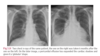
Chest X-ray

Cardiac imaging
Echocardiography &
Cardiac CT

Cardiac imaging
Cardiac MR & Nuclear imaging

Cardiac imaging
changes in the second image > ?

Cardiac imaging
Cardiac CT demostrating > ?
Cardiac MR demonstrating > ?


Echocardiography
Intro

Echocardiography
Types of scan (name only)

Echocardiography
M-mode (motion mode): > ?
Two-dimensional (real time): > ?

Echocardiography
3D echocardiography > ?
Doppler and colour-flow echocardiography > ?
Tissue Doppler imaging: > ?

Echocardiography
Transoesophageal echocardiography (TOE) > ?
Stress echocardiography > ?

Echocardiography
Uses of echocardiography (names)
Only the names

Echocardiography
Use in >>> “Quantification of global LV function”
Echocardiography
Use in >>>
‘estimating right heart haemodynamics’
‘valve disease’
‘congenital heart disease’

Echocardiography
Use in >>>
‘Endocarditis’
‘Pericardial effusion’
‘HCM’

Structures seen in this picture


Echocardiography


Cardiac catheterization
function and indications

Cardiac catheterization
Pre-procedure & post-procedure checks

Cardiac catheterization:
Complications

Cardiac catheterization:
Mortality and intra-cardiac electrophysiology

Coronary artery anatomy

Coronary artery angiography > image
what do the two images show (b & c)?


CVS drugs
Antiplatelet drugs
Anticoagulants

CVS drugs
Beta blockers
ACE inhibitors

CVS drugs
Diuretics
Vasodilators

CVS drugs
Calcium antagonists

CVS drugs
Digoxin

CVS drugs:
Na channel blockers
Amiodarone
Ivabradine

CVS drugs
Statins
Anti-anginal
Anti-HTN
Drugs acting on AV node

Angina pectoris:
Features
Precipitants
Causes

Angina Pectoris:
Types
Tests
Only focus on types and tests

Angina pectoris:
Management

Angina pectoris:
Management > Revascularization

Investigations of IHD
NICE:
Recommended investigations when considering stable angina

CT angiogram data:
white arrow points towards?


Vasospastic/ Prinzmetal angina:
Risks & Triggers
Treatment

Acute Coronary Syndrome (ACS):
Definitions &
Risk factors

Acute Coronary Syndrome (ACS):
Incidence &
Diagnosis

Acute Coronary Syndrome (ACS):
Symptoms &
Signs

Acute Coronary Syndrome (ACS):
Tests

Acute Coronary Syndrome (ACS):
Differentials & mortality

Sequential ECG changes following acute MI

Graph of cardiac enzymes

Troponin:
Introduction &
Rise of troponin > cardiac cause

Troponin:
Rise of troponin > non-cardiac cause

ACS Management:
First step

ACS Management:
Acute STEMI >>> Initial treatment flowchart

ACS Management:
Acute STEMI >>> Initial treatment details

ACS Management:
Acute STEMI > Reperfusion therapy >
criteria
PCI
thrombolysis

ACS Management:
ACS without ST elevation >>> assessment and investigations
ACS management:
ACS without ST elevation >>> Acute/initial management
>>> Flow chart

ACS management:
ACS without ST elevation >>> Acute/initial management
>>> details

ACS management:
ACS without ST elevation >>> prognosis

ACS management:
ACS without ST elevation >>> further measures
ACS management:
what is GRACE ?

ACS management
After immediate actions >> focuses on ?

ACS management:
After initial management >>>
- Symptom control ?
- Modifying CVS risk factors ?

ACS management:
After initial treatment >>>
- Optimize cardioprotective medications ?
- Revascularization ?

ACS management:
After initial treatment >>>
- Discharge and General advice

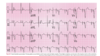
ECG Diagnosis ?


Complications of MI >>> names
Cardiac arrest
Cardiogenic shock
Left ventricular failure
Bradyarrythmias
Tachyarrythmias
Right ventricular failure/infarction
Pericarditis
Systemic embolism
Cardiac temponade
Mitral regurgitation
Ventricular septal defect (VSD)
Late malignant ventricular arrythmias
Dressler’s syndrome
Left ventricular aneurysm
Bradyarrythmias & their management

Tachyarrythmias & their management

Some MI complications and their management:
Right ventricular failure/infarction
Pericarditis
Systemic embolism
Cardiac temponade
Mitral regurgitation
Ventricular septal defect (VSD)

Some MI complications and their management:
Late malignant ventricular arrythmias
Dressler’s syndrome
Left ventricular aneurysm

ACLS algorithm of cardiac arrest: 2015

Cardiogenic shock:
Intro & causes

Cardiogenic shock:
management flow chart

Cardiogenic shock:
management details

PICCO & LIDCO ?

CABG indications

CABG vs PCI

Procedures of CABG

After CABG

Cardiac temponade:
Causes
Signs
Management

Severe pulmonary oedema:
Causes & differentials

Severe pulmonary oedema:
Symptoms, Signs, & Investigations

Severe pulmonary oedema:
monitoring progress &
once stable & improving

Management of acute heart failure/ severe pulmonary oedema/ heart failure (not chronic)

Arrhythmias:
Introduction &
Causes

Arrhythmias
presentation
history
tests

Arrhythmias:
different arrhythmias, bradycardia, and management

Continuous ECG monitoring:
Telemetry
Exercise ECG
Holter monitors

Continuous ECG monitoring:
Loop recorders
Pace makers & ICDs

Sick sinus syndrome

Narrow complex tachycardia:
Definition & Differentials

Narrow complex tachycardia:
Principles of management

Narrow complex tachycardia:
Differential diagnosis from ‘emergency section’

Management of Narrow complex tachycardia/ SVT > Flow chart

Management of ‘irregular’ Narrow complex tachycardia

Management of narrow complex tachycardia (in brief)

Adenosine in narrow complex tachycardia

Narrow complex tachycardia:
specifics in emergency management

Narrow complex tachycardia:
WPW syndrome (in brief in the emergency section)

Holiday heart syndrome

Normal conduction through heart

Regular rhythm tachycardia:
Sinus tachycardia
Focal atrial tachycardia
Atrial flutter

AVRT
AVNRT
Junctional tachycardia
Bundle branch block
VT

Broad complex tachycardia:
Definition and Principles of management

Broad complex tachycardia:
Differential diagnosis

Broad complex tachycardia:
Differentiating VT from SVT with aberrancy

Ventricular extrasystoles (ectopics)

ECG diagnosis


ECG diagnosis


ECG diagnosis


Broad complex tachycardia:
Differentials in ‘emergency section’

Broad complex tachycardia:
Management (in brief text)

Borad complex tachycardia:
After correction of VT ?

Broad complex tachycardias:
VF
SVT with aberrant conduction
Ventricular extrasystoles (ectopics)
Torsades de pointes

Broad complex tachycardia:
management (flow chart)

Bradycardia:
Intro
Symptoms
Rhythm

Bradycardia:
Causes

Bradycardia:
Management (in flow chart)

Bradycardia:
Management (in text)

