Cardiovascular Examination Flashcards
State the 7 stages of your cardio examination
- Introduction
- General inspection
- Hands & arms
- Head & neck
- Chest
- Back
- Legs
Describe what you must do in the introduction stage of your cardio examination
- Name
- Role
- Explain & gain informed consent
- Check patients full details (Name & DOB)
- Offer chaperone
- Check for any pain- particulary in right shoulder
- Wash hands
Describe what you must do in the general inspection stage of your cardio examination
- Position & expose pt
- Inspect surroundings: cardiac monitor, ECG, oxygen, GTN spray, nitrate infusion, pacing wires, warfarin yellow book
- Inspect pt: comfortable at rest?, SOB, pallor, body habitus, scars
Describe what you must do in the hands & arms stage of your cardio examination
- Check for tremor
- Inspect hands: clubbing, tar staining, splinter haemorrhages, Osler’s nodes, Janeway lesions, peripheral cyanosis, xanthoma, radial artery/vein harvest scar
- Capillary refill
- Radial pulse (rate, rhythm)
- Radio-radial delay
- Offer radio-femoral delay
- Collapsing pulse
- Brachial pulse (volume & character) **NOTE: could just do carotid
- Request bp in each arm
Describe what you must do in the head & neck stage of the cardio examination
- Inspect eyes: malar flush, conjunctival pallor, xanthelasma, corneal arcus
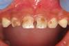
- Inspect mouth: central cyanosis, dental hygiene, high arched palate
- Inspect neck: JVP (may do hepatojugular reflux to confirm)
- Auscultate for carotid bruit
- Carotid pulse bilaterally (volume & pulse)
Describe what you must do in the chest stage of your cardio examination
- Inspection: scars, deformity (pectus excavatum or carinatum)
- Palpate for pacemaker
- Thrills & heaves
- Palpate apex beat
- Ausculate chest
- Ausculate (radiation)
- Ausculate (manoeuvres)
Describe what you must do in the back stage of your cardio examination
- Inspect sacral oedema
- Auscultate lung bases
Describe what you must do in the legs section of your cardio examination
- Inspect: skin changes associated with peripheral vascular disease, vein harvest scar, pitting oedema
- Check for pitting oedema
What position should you have your pt in for your cardio examination?
Led at 45 degrees with chest exposed
What 3 categories can we broadly split cardiac conditions into?
- Arrhythmias
- Heart failure
- Ischaemia
What is normal capillary refill time?
< 3 secs
Why do some clinicians advocate auscultation prior to palpation of carotid?
Argue you should check for atherosclerotic disease prior to palpation as if disease if present palpation may produce atheromatous emboli and subsequent stroke
Describe how you would palpate for the apex beat
- Start lateral
- Work medially
Describe how you check for thrills
- Fingers either side of sternum at around level of sternal angle in 2nd ICS
- Push in quite firmly
- Should feel pulse
- If thrill is present will feel “vibration” as a thrill is a palpable murmur
Describe how you check for heaves
- Place hands either side of sternum and see if feel movement with your hands
- Place hand horizontally across chest (heel of hand around midline)
*Some people may like to check for apex beat after checking for heaves as it requires same hand positioning
Describe where you should auscultate on the chest
“Four basic points”
- Over apex (mitral)
- 4th ICS left (tricuspid)
- 2nd ICS left sternal edge(pulmonary)
- 2nd ICS right sternal edge (aortic)
*Palpate carotid pulse whilst auscultating
Auscultate for radiation?
- Auscultate left axilla on expiration for mitral regurg/stenosis
- Auscultate carotids with bell on expiration on expiration for aortic stenosis/regurg
Auscultation manoeuvres
- Ask pt to roll to left and auscultate with bell on expiration to check for mitral stenosis
- Ask pt to lean forwards and auscultate over tricuspid area for aortic regurg
State at least 3 things/appropriate next steps that you could suggest at the end of your cardio examination
- ECG
- Peripheral vascular examination
- BP
- Focused history
- Observations
- Abdominal examination (for hepatomegaly if tricuspid regug suspected and for hepatosplenomegaly if infective endocarditis suspected, for ascites if HF suspected)
- Fundoscopy
Why would you like to do a fundoscopy following your cardio examination?
To check for:
- Roth spots in infective endocarditis
- Hypertensive retinopathy
- Diabetic retinopathy

What does the image show?

Roth spots which are seen in infective endocarditis; retinal haemorrhages with white or pale centres
Clubbing is a non-specific sign of systemic disease; what cardiovascular diseases can it be indicative of?
- Infection: endocarditis
- Malignancy: mitral myxoma
- Congenital: cyanotic heart disease
What does this image show?
What pathology can they indicate?

- Splinter haemorrhages (capillary nail bed microemboli)
- Pathology:
- Infective endocarditis
- Local inflammation e.g. psoriasis, lichen planus
- Trauma
What does this image show?
Where are these found?
Are they tender?
What pathology can they indicate?

- Osler nodes
- Red-purple, slightly raised, tender lumps often found on fingers and toes
- Considred pathognomonic of subacute infective endocarditis
What does this image show?
Where are they found?
Are they tender?
What pathology do they indicate?
- Janeway lesions
- Red, non-tender found commonly on palms and soles. Disappear after
What 3 signs in hands are widely considered pathognomonic for subacute infective endocarditis?
- Osler’s nodes
- Janeway lesions
- Roth’s spots