Cardiovascular Flashcards
RCA supplies what areas of the heart?
The right atrium, most of the right ventricle, and a variable portion of the inferior portion of the left ventricle.
The right atrium receives blood from where?
The superior and inferior vena cava, and the coronary sinus (drains from the coronary vessels)
Describe atrial kick.
AV node delay conduction and enables atrial contraction to prime the ventricle.
What is the point of maximal impulse?
Where the apex of the heart is positioned anteriorly and inferiorly at the left 5th intercostal space.
What structures is the heart surrounded by?
It is bound anteriorly by the sternum and costal cartilages of the 3rd-5th ribs and inferiorly by the diaphragm.
What are the AV valves? What heart sound do they produce?
tricuspid and mitral valves. S1 is the closing of AV valves.
What are the semilunar valves? What sound do they produce?
The pulmonic and aortic valves. S2 is the closing of semilunar valves.
What produces turbulent flow in the heart? What sound is produced?
Stenotic lesions or regurgitant valves. A murmur is produced.
Describe a heart “click”?
a short, high pitched sound
What is a heart “rub”?
scratchy, creaking, high pitched sound. Could be associated with pericarditis.
What is the left ventricle comprised of?
Most of the apex and the lower-left lateral border.
The anterior surface of the heart is almost entirely made up of which structure?
The right ventricle.
What is the pericardium? What does it consist of?
It is a fibrous double-walled sac that surrounds the heart and roots of the great vessels. Consists of a visceral (epicardium) and outer parietal portion.
What are the visceral and parietal pericardium separated by? What is in this space and how much?
The pericardial space. 10-25 mL of serous fluid which provides lubrication.
What is the base of the pericardium attached to?
It is fused with the central tendon of the diaphragm.
What separates the atria from the ventricles?
The coronary sulcus.
What separates the left from the right ventricle?
The anterior and posterior interventricular sulcus.
What vessel travels within the sulcus?
The RCA.
What does the circumflex artery arise from?
The LCA and it travels in the coronary sulcus.
Where does the LAD course through?
It runs along the anterior interventricular sulcus, then travels over the interventricular septum and continues into the posterior interventricular sulcus.
Which two arteries entirely supply myocardial oxygen supply?
The RCA and LCA.
It is the fixation point for cardiac musculature and plays an important role in the structure, function, and efficiency of the heart. It helps to provide stability and protect against dysrhythmias. It helps to increase the electrical mechanical efficiency of the heart and helps to serve as an insulator.
Annulus fibrosis.
How thick is the muscle wall of the right atrium? The RA is made up of what two parts?
~ 2 mm. An anterior, thin-walled trabeculated portion and a posterior, smooth-walled portion.
What is the fossa ovalis cordis?
Remnant of the fetal foramen ovale.
How thick is the muscle wall of the right ventricle?
~4-5 mm
What prevents the eversion of the tricuspid valve, preventing regurgitation?
The chordae tendineae and papillary muscles. The chordae tendineae are attached to the cusps of the tricuspid valve and papillary muscle have attachments to the ventricular walls and chordae tendineae.
By how much does the LA increase the LVEDV?
20-30% aka the atrial kick. Compromised patients rely on this kick to maintain CO.
How thick is the muscle wall of the left atrium?
~ 3 mm and the atrial wall is smooth.
3 Layers of cardiac muscle
- Epicardium- composed of mesothelium, connective tissue, and fat. 2. Myocardium - consists of two layers that provide strength during contraction 3. Endocardium - endothelium and a layer of connective tissue
What causes opening and closing of cardiac valves?
Pressure gradients
Tricuspid valve. When do you start seeing the signs and symptoms of insufficiency?
AV valve. 3 leaflets of unequal size: anterior, septal, and posterior that are attached to cordae tendineae and papillary muscles. Normal valve area is 7 cm2. Insufficiency becomes evident when it is 1.5 cm2
Mitral Valve. When do you start seeing signs and symptoms of insufficiency?
AV valve. Two leaflets: anteromedial and posterolateral attached to chordae tendineae and papillary muscles. Normal valve area is 4-6 cm2. Insufficiency seen when reduced by 50%.
Semilunar Valves
Pulmonic and aortic valve. Each have 3 cusps with cusps of the aortic being slightly thicker due to being subjected to higher pressures.
How many cusps does the pulmonic and aortic valve have? What lies above the aortic valve and what role does it play?
3 cusps. The sinus of Valsalva, and it helps to maintain blood flow.
Normal aortic valve area.
1-3 cm2
What is right dominant circulation and what percentage of the population is right dominant?
When the RCA gives rise to the PDA which supplies the superior posterior interventricular septum and inferior wall.. 50% of people have a right dominant circulation.
What is left dominant circulation and what percentage of the population is left dominant?
When the circumflex artery (which branches of the LCA) gives rise to the PDA. 10-15%.
What does the LCA supply? What does it branch into?
The left atrium and most of the interventricular septum and left ventricle (septal, anterior, and lateral walls). It bifurcates into the circumflex which supplies the lateral ventricular wall and the LAD which supplies the septum and anterior wall.
After perfusing the myocardium blood returns to the right atrium via…
the coronary sinus and the anterior cardiac veins.
Thebesian veins
Where a small amount of blood returns directly into the chambers of the heart.
Arterial supply to SA node.
From RCA in 60% of individuals. LAD in 40%.
Arterial supply to AV node.
RCA in 85-90% of the population or CX in 10-15%.
Bundle of His blood supply.
Dual blood supply from PDA and LAD.
Blood supply to anterior papillary muscle of mitral valve?
Dual blood supply from LAD and CX.
Blood supply to posterior papillary muscle of mitral valve.
PDA only, therefore much more vulnerable to ischemic dysfunction.
3 major systems of venous drainage in the heart.
coronary sinus, anterior cardiac veins, and Thebesian veins.
What is coronary perfusion pressure determined by?
It is determined by the difference between aortic pressure and ventricular pressure. Arterial diastolic pressure - LVEDP. Decreases in aortic pressure or increases in LVEDP can reduce coronary perfusion pressure.
How much is coronary blood flow at rest?
250 mL/min
How much oxygen does the myocardium usually extract from arterial blood?
65%
What is the normal ventricular cell resting membrane potential? An action potential raised the membrane potential to what?
-80 to -90 mV. + 20 mV.
What does the SA node consist of?
P cells (pacemaker cells) and intermediate/transitional cells that conduct impulses within and away from the SA node.
3 major internodal tracts. What is the impulse time from SA to AV node?
Anterior: Sends fiber to the LA and travels down through the atrial septum to the AV node. Middle: curves behind SVC before descending to AV node. Posterior: Continues along terminal crest to enter atrial septum and then passes to AV node. Impulse time is 0.04 sec
What is the preferential channel for conduction of the action potential from the atria to the ventricles?
The AV bundle (bundle of His)
What contributes to diastolic ventricular filling?
Most diastolic ventricular filling occurs passively. Contraction of the atria contributes 20-30% of filling.
Describe systole.
Isovolumetric contraction. Period of rapid ejection (first 1/3 of systole) and a period of reduced ejection (last 2/3 of systole).
Describe the three CVP waveforms.
A wave: atrial contraction C wave: ventricular contraction V: pressure build-up from venous return before AV valve opens again.
Four factors that affect cardiac performance directly.
- preload 2. afterload 3. heart rate 4. myocardial contractility Preload and afterload depend on both the heart and vascular system. HR and contractility are characteristics of cardiac tissue and are influenced by neural and humoral mechanisms.
Frank-Starling Law
Cardiac muscle increases its strength of contraction when it is stretched.
Laplace’s Law
Wall tension is related to the product of intraventricular pressure and internal radius and inversely to the wall thickness. Useful in understanding formation of aneurysms and blood vessel distensibility.
Factors affecting ventricular preload.
venous return, blood volume, distribution of blood volume, heart rhythm, and heart rate.
Afterload
The force the left ventricle must overcome during systole. AKA ventricular wall tension during systole or arterial impedance to ejection.
Normal intrinsic HR formula
118 beats/min - 0.57 x age
Stroke volume
Volume of blood ejected during systole and depends on myocardial contractility.
Two major factors that affect contractility.
Preload and alterations in sympathetic activation of the ventricles.
Formulas for: cardiac output cardiac index ejection fraction systemic vascular resistance
CO = HR x SV. Normal 4-8 L/min.
CI = CO/BSA. Normal 2-4.
EF= (SV/EDV) x 100. Normal 60-70%
SVR = (MAP - CVP)/CO x 80. Normal 800-1200 mmHG. Relationship between CO, preload, and afterload.
3 contraction abnormalities.
hypokinesis: decreased contraction
akinesis: failure to contract
dyskinesis: paradoxic bulging
What effect does HTN have on afterload and myocardial oxygen demand? What effect does this have on the heart?
It increases both afterload and myocardial oxygen demand. This can result in concentric left ventricular hypertrophy and altered diastolic dysfunction.
Postoperatively, what is a primary antihypertensive consideration?
Adequate control of pain
What effect does HTN have on cerebral blood flow?
Cerebral autoregulation is altered
Normal amount of fluid in pericardial space.
20-25 mL of clear fluid.
Clinical presentation of acute pericarditis
Chest pain with sudden onset. Fever with pericardial friction rub. Diffuse ST-segment elevation but an absence of elevated cardiac enzyme levels.
What is the fundamental hemodynamic abnormality in chronic constrictive pericarditis?
Abnormal diastolic filling. SV and CO are then decreased which leads to pulmonary congestion, peripheral congestion, and over time systolic dysfunction will also occur.
Clinical presentation of cardiac tamponade
Becks triad: JVD, hypotension, distant/muffled heart sounds. Pulsus paradoxus. EKG will show decreased voltage in all leads in QRS complex.
What are the two most important preoperative risk factors for CV complications?
History of recent MI and evidence of CHF
CV contraindications to elective noncardiac surgery?
Recent MI < 1 month, severe aortic or mitral valve stenosis, and uncompensated heart failure.
What population has a high risk of SILENT ischemia?
Diabetics
What paralytics would not be indicated in a patient with ischemic heart disease?
Pancuronium because of its vagolytic effects.
What signs are most important to recognize in the preoperative evaluation of a patient with valvular disease?
Signs of CHF. S3 gallop and rales with left sided failure. JVD, hepatosplenomegaly, hepatojugular reflux, and pedal edema with right sided failure.
What additional lab tests would be indicated int he patient with valvular disease?
LFTs and check for adequate reversal of warfarin or heparin anticoagulation with PTT and PT/INR studies. CXR may be useful to assess for cardiac size and pulmonary congestion. EKG is generally nonspecific.
Prophylactic antibiotics recommended for patients with valvular disease
Amp and gent
When do symptoms of mitral stenosis generally develop?
After 20-30 years and when normal valve orifice is reduced from 4-6 cm2 to less than 2 cm 2.
In mitral stenosis, blood flow is dependent on…
HR, CO, and atrial kick
In mitral stenosis, the left atrium is often dilated. This promotes the onset of…
SVT and afib, which can lead to the formation of thrombi and embolic events.
Normal atrial systole is responsible for what % of ventricular filling?
20-30%
What are the principle goals in the anesthetic management of patients with mitral stenosis?
Maintaining sinus rhythm, avoiding tachycardia and large increases in CO, hypovolemia, and fluid overload.
Acute mitral regurgitation is often the result of…
MI, papillary muscle dysfunction or rupture of chordae tendinea, infective endocarditis, or chest trauma.
What is the most common cause of obstruction in left ventricular outflow?
Aortic stenosis
What is considered critical aortic stenosis?
When valve area is reduced to 0.5-0.7 cm2.
What is the principle derangement in mitral regurgitation? How does the LV compensate?
A reduction in forward stroke volume. The LV compensates by dilating and increasing EDV.
Patients with chronic MVR eventually develop…
ECCENTRIC LV hypertrophy and progressive impairment in contractility.
Factors that exacerbate MVR
slow HR and acute increased in afterload. HR should ideally be kept between 80-100 bpm. A decrease in SVR increases forward flow and decreases regurgitant volume.
Factors that exacerbate mitral valve prolapse
maneuvers that decrease ventricular volume (preload). Hypovolemia, reduced afterload, and increased ventricular emptying should be avoided.
Mitral valve prolapse increases the risk of…
ventricular arrhythmias intraoperatively and infective endocarditis.
Triad of advanced aortic stenosis
HF (DOE), angine, and syncope
Prominent feature of aortic stenosis
Decrease in left ventricular compliance as a result of hypertrophy (concentric). Filling is impaired.
What are critical priorities in the anesthetic management of patients with aortic stenosis?
Maintenance of NSR, HR, vascular resistance, and intravascular volume.
Causes of acute aortic regurgitation
infective endocarditis, trauma, aortic dissection
Consequences of chronic aortic regurgitation
LV dilation and and development of eccentric hypertrophy. Eventually will manifest as CHF.
Manifestations of acute aortic regurgitation
Does not allow for compensatory dilation/hypertrophy of LV. Sudden onset of pulmonary edema and hypotension.
Anesthetic management of aortic regurgitation
HR toward upper limits of normal 80-100 bpm. Avoiding excessive myocardial depression and overzealous fluid replacement.
Factors that worsen LV outflow in hypertrophic cardiomyopathy
Enhanced contractility, decreased ventricular volume, decreased LV afterload.
Anesthetic management of hypertrophic cardiomyopathy
Maintaining adequate intravascular volume, avoiding vasodilatation, and reducing myocardial contractility through the use of beta blockers.
What is absent in a transplanted heart?
Autonomic influences due to denervation. Vagal influences absent, causing a higher resting HR 100-120 bpm. HR-related responses to various meds administered intraop will be absent and MIs are almost always silent due to denervation.
Preferred meds to increase HR in transplanted heart are…
isuprel and epinephrine should be immediately available
Anesthetic considerations for heart transplant
Maintenance of normal or high cardiac preload desirable (graft is preload dependent). Indirect vasopressors are less effective because of absence of catecholamine stores.
2 types of arteries
conducting (elastic) and distributing (muscular)
3 layers of blood vessel walls
tunica intima, tunica media, and tunica externa (adventitia)
Venous drainage of the brain
From the sagittal, transverse, and sigmoid sinuses into the internal jugular vein

Venous drainage of the superficial structures of the face and head
via the external jugular vein
Drainage of the superficial veins of the upper extremity
into the axillary vein, cephalic vein laterally, and basilica vein medially

Axillary vein drainage
Into subclavian vein, then brachiocephalic vein, then SVC
Venous drainage of the thorax
from branches of SVC, IVC, and the azygous and hemiazygous systems. The azygous and hemiazygous systems provide an alternative blood return route if a major obstruction of the IVC occurs; vessels include intercostal vessels, bronchial veins, and pericardial veins.

Venous circulation of lower extremities
superficial and deep venous drainage from the legs >>> deep femoral and femoral veins >>> external iliac arteries
Venous circulation of the pelvis
Blood from pelvis drains into internal iliac veins
Venous circulation of common iliac vessels
Common iliac vessels receive blood from internal and external iliac veins and drain into IVC. Common iliac vessels are joined by branches from the abdomen and hepatic portal system.
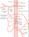
What are the branches of the ascending aorta?
The right and left coronary arteries and three major branches: the brachiocephalic or innominate artery (splits into right common carotid and right SC artery), the left common carotid artery, and the left subclavian artery. The right and left common carotid arteries then branch into the internal and external carotid arteries.
The external carotid arteries supply blood to what area?
To the face and neck. Major branches include the superior thyroid artery, the lingual artery, the facial artery, the posterior auricular artery, the maxillary artery, the transverse facial artery, the middle temporal artery, and the superficial temporal artery.
The internal carotid arteries supply blood to what area(s)?
They supply 80% of blood to the brain via the circle of Willis, and to the eyes via the ophthalmic arteries.
The circle of Willis receives blood from what areas?
The internal carotid arteries and the vertebral branches of the subclavian artery.

What are the branches of the subclavian artery before entering the upper extremity?
The vertebral arteries; the thyrocervical trunk, which supplies blood to the thyroid gland and other structures of the neck; the internal thoracic artery, which supplies blood to the anterior chest; and the costocervical trunk, which supplies blood to the first two intercostal spaces and the muscles of the neck.
Circulation in the upper extremity coming from the subclavian artery
Continues as the axillary artery which branches into the highest thoracic, subscapular, and anterior/posterior circumflex humeral arteries.

Brachial artery circulation
It begins at the terminal end of the axillary artery and continues to neck of radius where it divides into radial and ulnar arteries.
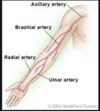
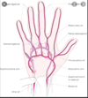
Radial and ulnar artery circulation
Radial artery forms deep palmar arch and ulnar artery forms superficial palmar arch.
Branches of the descending thoracic aorta
- Lower 9 posterior intercostal arteries.
- Subcostal arteries
- Pericardial arteries
- Esophageal arteries
- Bronchial arteries

Branches of the abdominal aorta
- Inferior phrenic arteries (first branches)
- Celiac trunk
- Right and left renal arteries
- Superior mesenteric artery
- Inferior mesenteric artery

Branches of the celiac trunk
- Splenic artery
- Left gastric artery
- Gastroduodenal artery
- Hepatic artery
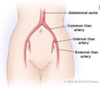
Where does the abdominal aorta terminate?
At the common iliac arteries in the pelvis. The common iliacs divide into internal and external iliac arteries.
Femoral artery circulation
Descends from the external iliac artery as the femoral and deep femoral artery. Then becomes the popliteal artery behind the knee, then divides into the anterior and posterior tibial arteries.

What do the anterior/posterior tibial arteries supply?
The anterior continues as the dorsal artery of the foot and the posterior supplies blood to the plantar arches.

Movement of fluid volume is determined by
- capillary pressure
- interstitial fluid pressure
- plasma colloid osmotic pressure
- interstitial fluid colloid osmotic pressure
Diffusion of substances in the microcirculation is determined by
- lipid and water solubility
- size of molecule
- concentration gradient
What are 2 major theories of capillary blood flow?
The vasodilator theory and the oxygen demand theory.
Theories indicate an ACTIVE microcirculatory process that responds to tissue metabolic needs.
What is the purpose of autoregulation?
It maintains constant blood flow through the capillary bed in response to fluctuations in MAP.
What is a good example of vascular growth that can occur to provide collateral circulation?
Microcirculation.
How does collateral circulation occur?
Substances are released from ischemic tissues, rapidly growing tissues, and tissues with high metabolic rates. The dissolution of the basement membrane occurs with eventual cords developing from cells in vessels. The cells in these cords divided and fold over into a tube. Tubes then connect with other tubes to form a vascular network.
What is the most common cause of peripheral vascular disease?
Atherosclerosis
What are 3 pathophysiologic processes that affect arteries?
- Plaque formation (obstructs lumen)
- Thrombosis (results in acute ischemia)
- Aneurysm formation (from weakening in the arterial wall)
Symptoms associated with peripheral vascular disease?
Claudication, skin ulcerations, gangrene, and impotence.
2 major risk factors in the pathogenesis of atherosclerosis or PVD?
Cigarette smoking and diabetes
What determines the extent of disability from PVD?
The development of collateral blood flow.
As the disease progresses, supply no longer meets demand and limb ischemia becomes symptomatic.
Treatment for PVD
Pharmacological:
- ASA
- Plavix
Surgical:
- transluminal angioplasty
- endarterectomy
- thrombectomy
- multiple bypass procedures
What must be done to the artery in multiple bypass procedures? What medication is administered for this?
Temporary occlusion of the operative artery occurs. Heparin (5k-7k units) is usually administered.
What should be considered in the preop evaluation in patients with PVD?
That there may concurrent CAD, pulmonary, renal, neurologic, and endocrine dysfunction.
Anesthetic selection in patients with PVD
Depends on type of procedure and presence of coexisting disease.
- Local and IV conscious sedation
- Regional (especially if surgery is limited to lower extremities, usually results in more CV stability)
- General anesthesia
Postoperative considerations in patients with PVD
Pain management and postoperative monitoring for MI, respiratory failure, and postop bleeding.
Surgery on the aorta is complicated by what 2 factors?
- Need to cross-clamp the aorta
- Potential for large intraoperative blood loss
What factors affect blood flow in the vascular tree?
The autonomic nervous system, local and metabolic control mechanisms, endothelium-derived factors (vasodilators/vasoconstrictors), and circulating hormones.
Arterial BP is directly related to _____ and inversely proportional to ______.
stroke volume, compliance of the arterial tree
Immediate control of BP
Primarily a function of the ANS reflexes and peripheral baroreceptors (bifurcation of common carotid arteries and aortic arch). BP changes are sensed centrally in the brainstem.
Intermediate control of BP
Activation of RAAS. Angiotensin II is a potent arterial vasoconstrictor.
Long-term control of BP
Slower renal mechanisms such as secretion of aldosterone and sodium regulation.
What are the CV effects of aortic cross-clamping?
Acutely increases LV afterload and could cause severe HTN, myocardial ischemia, LV failure, or aortic valve regurgitation.
Aortic cross-clamping may affect perfusion to what areas?
It may compromise perfusion to the spinal cord and kidneys, producing paraplegia and renal failure.
What type of patients may not be able to tolerate aortic cross-clamping as well? Describe the positioning of the cross-clamp in relation to the severity of its effects.
Patients with precipitating LV dysfunction and pulmonary disease. The more distally the clamp is applied, the less it will affect perfusion to other areas.
Indications for aortic surgery
- Aortic dissection
- Aneurysms
- Occlusive disease
- Trauma
- Coarctation (congenital narrowing of aorta in reference to the ductus arteriosis): preductal diagnosed in childhood and postductal may not be found until adulthood.
Specific site lesions for aortic surgery
Ascending aorta and aortic arch (both require bypass). Distal to the left subclavian artery and above the diaphragm known as the thoracic aorta and below the diaphragm known as the abdominal aorta.
Variations in wall integrity that may contribute to aortic dissection are related primarily to what?
A degenerative process called medial cystic necrosis often seen in Marfan’s disease.
What is the most common factor contributing to the progression of aortic dissection?
HTN. Goal is to maintain SBP 90-120 mmHg.
What is the most serious complication of aortic dissection?
Aneurysm rupture
Treatment of dissecting aortic lesions:
Proximal dissections almost always require surgery.
Distal dissections may be managed medically initially to decrease HTN and slow down the progression of dissection, but eventual surgical tx will ensure long term survival.
Aortic aneurysms commonly involve what area? The majority are caused by what? What may be other causes of aneurysm formation?
Abdominal aortic aneurysms are the most common.
Atherosclerosis.
Medial cystic necrosis, RA, spondyloarthropathies, and trauma.
Syphilitic aneurysms typically involve ascending aorta.
Complications of aortic aneurysms
- Aortic regurgitation
- Tracheal or bronchial compression or deviation
- hemoptysis
- superior vena cava syndrome
- rupture and exsanguination GREATEST DANGER
Super vena cava syndrome
severe airway obstruction, deviation of the airway, problems breathing, neck and head engorgement.
Pseudoaneurysm formation
Rupture of INTIMA and MEDIAL layers of the artery
What is the normal size of the aorta? When are elective resections generally performed?
2-3 cm in width. Surgery when greater than 4 cm.
Surgical treatment for occlusive disease of the aorta
- aorto-bifemoral bypass with synthetic graft (not with CPB)
- possible proximal thromboendarterectomy
Preoperative evaluation in the patient undergoing vascular surgery should focus on
Cardiac, renal, and neurologic function
For vascular surgery on the aorta, when would a double-lumen ETT be needed?
For one-lung ventilation to expose the descending thoracic aorta.
Key points for surgery on the ascending aorta
Uses median sternotomy and CPB.
Intraoperative complications:
- Aortic regurgitation
- Long aortic cross-clamp times
- Large intraoperative blood loss
The left radial artery used to monitor ABP due to clamping of the innominate artery.
Control of BP with Nipride/Cardene.
Medications that reduce surgical bleeding and have an antifibrinolytic effect?
- aminocaproic acid (Amicar)
- tranxenemic acid (TXA)
- Aprotin - renal failure
Key points for surgery involving the aortic arch
Usually performed through median sternotomy with deep hypothermic circulatory arrest FOLLOWING CBP
Achieving optimal cerebral protection by:
- systemic and topical hypothermia
- thiopental infusion (not anymore)
- methylprednisolone or dexamethasone
- mannitol
- phenytoin
Long rewarming periods contribute to intraoperative blood loss.
Patient will also need higher doses of meds with rewarming.
Surgical approach on the descending thoracic aorta
Left thoracotomy approach if limited only to thoracic.
Thoracoabdominal incision if abdominal aorta also involved.
One lung ventilation.
Aortic cross-clamping above and below the lesion
How does aortic cross-clamping affect blood pressure? Where should ABP be monitored?
Acute HTN develops above the clamp and hypotension below the clamp.
In the right radial artery due to clamping of left subclavian artery.
What may happen following the release of cross-clamping?
What are some anesthetic considerations?
Hemodynamic instability or release hypotension due to:
- An abrupt decrease in afterload
- Bleeding
- Release of vasodilating acid metabolites
Anesthetic considerations:
- Decrease anesthetic depth
- Volume loading
- Partial or slow release of cross-clamp
Use of vasopressors, NaHCO3 to treat metabolic acidosis, and/or CaCl (following transfusion) may be necessary
Major neurological complications of thoracic aorta clamping
Spinal cord ischemia and paraplegia.
The classic deficit is that of anterior spinal artery syndrome.
Anterior spinal artery syndrome
Loss of motor function and pinprick sensation but preservation of vibration and proprioception.
Blood supply to the spinal cord
From the vertebral arteries and the thoracic and abdominal aorta:
- One anterior and two posterior arteries descend along the cord.
- Intercostal arteries
- Artery of Adamkiewicz (almost always on the left side of spinal cord)

What does the artery of Adamkiewicz supply?
The lower thoracic and lumbar spinal cord. However, anatomical origin varies.
Protective measures against paraplegia with thoracic aortic cross-clamping
- Monitoring SSEPs
- Use of temporary heparin-coated shunt or partial CPB with hypothermia
- methylprednisolone
- mild hypothermia
- mannitol (decrease CF production)
- drainage of CSF
Spinal cord perfusion pressure
MAP - CSF pressure
Caution: Excessive BP reduction can reduce spinal cord perfusion.
Increased incidence of renal failure following aortic surgery is associated with:
- emergency procedures
- prolonged cross-clamping periods
- prolonged hypotension
Especially in patients with pre-existing renal disease
Surgery on the abdominal aorta
- anterior transperitoneal or anterolateral approach
- cross clamp applied supraceliac, suprarenal, or infrarenal
- Heparinization prior to occlusion is necessary
- intra-arterial blood pressure monitoring on either arm
- renal prophylaxis with mannitol, esp with preexisting renal disease
- epidural anesthesia in conjunction with general anesthesia. Make sure you have coags before and start heparin after epidural is done (risk for epidural hematoma). Check coags again before discontinuing epidural.
Measures to decrease the incidence of renal failure secondary to aortic cross-clamping
- Infusion of mannitol (0.5 g/kg prior to cross-clamping
- low (renal) dose dopamine
- fenoldopam infusions (very renal specific, but you will have systemic and coronary vasodilation)
- Maintain adequate cardiac function: preload, contractility, and systemic perfusion pressure.
Postoperative considerations for aortic surgery
Maintaining hemodynamic stability and monitoring for postoperative bleeding. Increased maintenance fluids required. May need to remain intubated.
Endovascular aortic aneurysm repair
Involves deployment of an endovascular stent graft within the aortic lumen. The graft restricts blood flow to the portion of the aorta in which the aneurysm exists.
Advantages of endovascular aortic aneurysm repair
- improved hemodynamic stability
- decreased incidence of thrombo-embolic events
- reduced stress response
- less renal dysfunction
- improved post-operative comfort.
What is the most common site of atherosclerosis leading to TIA or stroke?
The internal carotid artery
Complications of endovascular aortic aneurysm repair
- endo leak between graft and aneurysm
- migration or rupture of the graft
- infection
- iliac artery rupture
- lower extremity ischemia
Stroke vs. TIAs
Stroke is a neurological deficit that lasts > 24 hr and you will have a pathological focal infarction. TIA could be related to an embolic event or low flow state.
Indications for carotid endarterectomy
significant stenotic lesions:
severe carotid stenosis 60-70% occlusion or
as little as 30% if patient has had preexisting stroke.
Preoperative considerations for carotid artery surgery
- Manage uncontrolled preop HTN (if not you will increase incidence of postop risks)
- Manage uncontrolled hyperglycemia (will increase cerebral ischemia injury)
- Medications should be taken as usual, except for diuretics
Blood pressure management in carotid artery surgery
- MAP maintained at or slightly above normal
- Intraoperative HTN is common
- Hypotension should be immediately treated, and pronounced or sustained reflex bradycardia treated with atropine. Stimulation of baroreceptors can also cause reflex response, surgeon can also use lidocaine to blunt response and bradycardia.
Heparin and Protamine administration for carotid artery surgery
Heparin will be administered prior to clamping. 5-7k units and wait 3 minutes.
Protamine should be administered before closure. For every 100 units of heparin, you give 1 mg of protamine.
3 significant side effects of protamine
- systemic arterial hypotension
- pulmonary HTN
- allergic reaction
CEA under regional anesthesia
Technique?
Advantages?
Disadvantages?
- Superficial and deep cervical plexus blocks (C2-C4 nerves)
- The patient may be assessed and examined intraoperatively for LOC, speech, and contralateral handgrip.
- Requires full cooperation of the patient and airway is not secured.
Indirect methods for monitoring cerebral perfusion during carotid cross-clamping when regional anesthesia is not used
Shunts are routinely used
Carotid stump pressures, EEG, and SSEPs have been used to determine the need for a shunt.
Changes lasting more than ten minutes may be associated with a new postoperative neurological deficit.
What is the disadvantage of inserting a shunt during a CEA?
If there is a lot of plaque, it could dislodge the calcification creating an embolic event.
