Cardiology Flashcards
What is the definition of orthostatic hypotension?
Orthostatic hypotension can be diagnosed when there is a drop in SBP of at least 20 mmHg or a drop in DBP of at least 10 mmHg after 3 minutes of standing.
What scroing system is used in assessment of suspected obstructive sleep apnoea?
Epworth Sleepiness Scale
What scoring system is uses to determine whether and AF patient needs anticoagulation=
The CHA2DS2-VASc
What is the most common cause of aortic stenosis in:
a) patients <65 years old
b) >65 years old
Patients <65 years old: Bicuspid aortic valve
Patients >65 years old: calcification
What ECG changes migh you expect in a hypothermic patient provided their electorlytes are all normal?
- Bradycardia
- J-wave (small hump at end of QRS complex see image)
- First degree heart block
- Long QT interval
- Atrial and ventricular arrhythmias

Describe the murmur you would expect to hear with mitral regurgitation.
- Pansystolic murmur
- The murmur is best heard at apex (5ht ICS mid-clavicular line).
- It radiates to the axilla.
- Soft S1 due to incomplete closure of mitral valve
- Severe MR: Widely split S2 as pressure in pulmonary circulation is high
List the causes of mitral regurgitation
- Post MI: papillary muscle rupture/chordae tendiniae rupture
- Mitral valve prolapse: Occurs when the leaflets of the mitral valve are slightly deformed.
- Infective endocarditis: the infective vegitations prevent the valve form closing properly
- Rheumatic fever
- Congenital. E.g. Collagen Disorders are predisposed to mitral regurgitation
Name the symptoms a patient with mitral regurgitation might complain of.
- Often asymptomatic
- Symptoms of heart failure
- Arrhythmias
- Breathlessness/cough
- Fatigue
What do the heartsounds S3 and S4 indicate?
S3: Commonly heard in heart failure. Indicates rapid ventiruclar filling, usually associated with volume overload as part of congestive cardiac failure.
S4: less commonly heard in heart failure. Thought to be due to contraction of the atria against a stiff ventricle.
What is the target blood pressure in DM?
If end organ damage (e.g. renal disease, retinopathy): < 130/80
Otherwise: < 140/80
What is the first line investigation for stable chest pain (i.e. on exertion exclusively) of suspected coronary artery aetilogy?
CT coronary angiography.
Then, do non-invasive cardiac imaging (look for reversible myocardial ischaemia) and after this, invasive coronary angiography.
(Examples of non-invasive functional cardiac imaging: Myocardial perfusions scintigraphy with single photon emission CT, MPS with SPECT; Stress echo; Contrast cardiac MRI)
What is the name given to abnormally large drop in BP during inspiration?
In what condition might you see this?
This is called puslus paradoxus. (a drop in BP > 10 mmHg with inspiration). Mechanism: there is normal increased venous return to the righ heart during inspiration. As the right ventricle cannot dilate in constrictive pericarditis or cardiac tamponade, the pressure is exerted onto the septum, so the left ventricle fills less and decrease in stroke volume, therefore lower BP during inspiration.
What is Kussmaul’s Sign?
In what condition might you see this?
Kussmaul’s Sign is the abnormal rise in JVP with inspiration. This typcially occurs in constrictive pericarditis, and very rarely in Cardiac Tamponade.
Mechanism of Kussmaul’s Sign: usually, during inspiration, the intrathoracic pressure decreases. The decrease in right atrial pressure, as well as the increased abdominal pressure, leads to increased venous return. A normal heart accommodates by increasing filling of the right ventricle and increasing heart rate. In constrictive pericarditis, the hear cannot dilate and therefore the increased venous pressure backs up to the JVP.
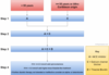
Describe the diagnostic pathway for hypertension.

Describe the management of primary hypertension in primary care.
Conservative: lifestyle advie (low sodium diet aiming for < 6g btu ideally <3g/day. Reduce caffeine intake. Smoking cessation, exercise, diet etc.)
Medical: see flowchart
If a patient with warfarin and on target INR for AF (2-3) suffers from a haemorrhagic stroke, what do you do with regards to the warfarin?
In any patient on warfarin with INR 2-3 that suffers from major bleed (such as intracranial haemorrhage):
Stop the warfarin, give 5mg of Vit K IV and give prothrombin complex concentrate IV. (if PCC nto available, give FFP).
What is the treatment protocol for a patient on warfarin that suffers from a major bleed?
In any patient on warfarin with INR 2-3 that suffers from major bleed (such as intracranial haemorrhage or GI haemorrhage):
Stop the warfarin, give 5mgof Vit K IV and give prothrombin complex concentrate IV. (if PCC nto available, give FFP).
What is the treatment protocol for a patient on warfarin that has an INR of >8.0 and suffers from a minor bleed?
If INR >8.0 and minor bleed:
- Stop warfarin
- 1-3 mg IV Vitamin K
- Repeat INR after 24 hours; if still high -> repeat vitamin K
- Restart warfarin when INR < 5.0
What is the treatment protocol for a patient on warfarin that has an INR of >8.0 with no bleed?
If INR >8.0 and no bleed:
- Stop warfarin
- 1-5 g Vitamin K PO
- Repeat INR after 24 hours; if still high -> repeat vitamin K
- Restart warfarin when INR < 5.0
What is the treatment protocol for a patient on warfarin that has an INR of 5.0-8.0 with minor bleeding?
When INR 5.0-8.0 and minot bleeding:
- Stop warfarin
- 1-3mg Vitamin K IV
- Restart warfarin when INR < 5.0
What is the treatment protocol for a patient on warfarin that has an INR of 5.0-8.0 with no bleed?
If INR 5.0-8.0 with no bleed:
- Withold 1-2 doses of warfarin
- Re-titrate maintenance dose subsequently (likely needs to be reduced)
What are the side effects of ACE-inhibitors?
- Cough (15% of patients, due to increased bradykinin levles)
- Angioedema: after up to a year of starting treatment
- Hyperkalaemia (if ≥6mmol/L should cessate ACEi in patient with CKD)
- First-dose hypotension (more common if also taking diuretics)
What are causes of RBBB?
- Normal variant - more common with increasing age
- Right ventricular hypertrophy
- Myocardial infarction - new onset RBBb should be concerning
- Atrial septal defect
- Pulmonary embolism
- Cardiomyopathy/myocarditis
What are the features of RBBB on ECG?
Broad QRS (>120 ms)
rSR pattern in V1-3 (“M” shaped QRS)
Wide, slurred S wave in latearl leads (aVL, V5-6; “W” shaped)


