Cardio Flashcards
(57 cards)
Cardiac Causes of Clubbing
- Atrial Myxoma
- Bacterial Infective Endocarditis
- Congenital Cyanotic Heart Disease
Causes of collapsing pulse
- AR
- Thyrotoxicosis
- Pregnancy
- Anaemia
Causes of absent radial pulse
- Dead
- Trauma
- Thrombosis or embolism
- Coarctation of the aorta
- Takayasu’s Arteritis
Causes of impalpable apex beat
- COPD
- Obesity
- Pericardial effusion
- Dextrocardia
What are the features of Pulmonary HTN
- ↑JVP
- Left parasternal heave
- Loud P2 + PSM of TR
- Pulsatile hepatomegaly
- Ascites and peripheral oedema
What are the four heart sounds
- S1: mitral valve closure
- S2: aortic valve closure
- S3: rapid ventricular filling of dilated left ventricle
- S4: atrial contraction against stiff ventricle
What investigations would you want to do at the end of a cardio examination?
- ECG
- Blood
- FBC: anaemia exacerbates cardiac symptoms
- U+E: renovascular disease
- NT-proBNP: heart failure
- Fasting lipids and glucose: cardiac risk
- Trops
- Imaging
- CXR
- Echo
- Cardiac catheterisation
Features of Aortic Stenosis
- Crescendo decrescendo ESM
- Right 2nd ICS
- Radiates to carotids
- Sitting forward in end-expiration
- May be an ejection click in bicuspid valve disease
Features of severe disease:
- Low-volume pulse
- Slow-rising (anacrotic)
- Narrow pulse pressure (<30mmHg)
- Aortic thrill
- Heaving apex
- Reversed splitting of S2
- Soft aortic component of S2
- 4th HS
- Pulmonary HTN
Ddx of AS
- Aortic sclerosis: no radiation, normal pulse character
- MR
- HOCM:
- valsalva ↑s murmur
- squatting ↓s murmur
- Right-sided: PS
- Supra-valvular aortic stenosis (William’s syndrome)
Causes of AS
Common:
- Age-related senile calcification
- Bicuspid aortic valve and other congenital causes
- Rheumatic heart disease
Rare:
- Infective endocarditis
- Hyperuricaemia
- Alkaptonuria
- Paget’s disease
Rx of AS

Clinical signs of severe MR
- LVF
- AF
- Soft first HS
- 3rd and 4th HS
- Displaced apex beat
- Precordial thrill
- Mid-diastolic flow murmur
- Widely split second HS
Murmur in MR
Blowing PSM
Apex
Left lateral position in end-expiration
Radiates to the axilla
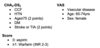
DDx for MR
Right-sided: TR
AS
VSD
Causes of MR
Chronic:
- Functional: LV dilatation (e.g. 2O to HTN or idiopathic)
- Annular calcification → contraction
- Rheumatic heart disease
- Mitral valve prolapse
- Connective tissue disorders: Marfan’s, Ehler’s danlos, osteogenesis imperfecta
- Cardiomyopathies
- SLE (Libman-Sachs endocarditis)
- Papillary muscle dysfunction (ischaemia)
Acute
- Rupture of chordae tendinae
- Infective endocarditis
- Trauma
What might you see on ECG and CXR for a patient with MR?
ECG
- AF
- P mitrale (LA hypertrophy)
CXR:
- LA (double right heart border) and LV hypertrophy
- Splaying of the carina (LA dilatation)
- Left atrial appendage
- Mitral valve calcification
- Pulmonary oedema
Echo features for severe MR
Jet width >0.6cm
Systolic pulmonary flow reversal
Regurgitant volume >60ml
Specific Mx of MR
AF: rate control and anticoagulation
Emboli: anticoagulant
↓ afterload
ACEi or β-B (esp. carvedilol)
Diuretics
Valve replacement
Murmur in AR
High-pitched early diastolic murmur
LLSE (3rd left IC parasternal)
Sitting forward in end-expiration
Additional Murmurs:
Ejection systolic flow murmur
Austin-Flint murmur (rumbling MDM @ apex secondary to regurgitant jet fluttering the anterior mitral valve)
Signs of AR
Eponymous Signs
Quincke’s: capillary pulsation in nail beds
Corrigan’s: visible vigorous carotid pulsation
De Musset’s: head nodding
Traube’s: pistol-shot sound over femorals
Duroziez’s
Systolic murmur over the femoral artery ̄c proximal compression.
Diastolic murmur ̄c distal compression
Mueller’s: systolic pulsations of the uvula
Rosenbach’s: systolic pulsations of the liver
Causes of AR
Chronic
- Bicuspid aortic valve
- HTN
- Rheumatic heart disease
- Autoimmune: Ank spond, RA, SLE
- Connective tissue: Marfan’s, Ehler’s Danlos
- Aortitis: Takayasu, syphilis, Reiter’s syndrome
- Perimembranous VSD
Acute
- IE
- Type A Aortic dissection
Indications of valve replacement for AR
Symptomatic: NYHA >2
LV dysfunction
Pulse pressure >100mmHg
ECG changes: T inversion in lateral leads
LV enlargement on CXR or EF <50%
Mitral Stenosis Murmur
Opening snap
Rumbling MDM
Apex - tapping apex
Left lateral position in end-expiration
With the Bell
Radiates to the axilla
Loud first HS
Pre-systolic accentuation if pt. in sinus rhythm
Atrial contraction
Graham Steell murmur (EDM secondary to PR)
ECG Features of Mitral Stenosis
P-mitrale
AF