Cardiac Electrophysiology- I Cardiac Muscle Flashcards
What are cardiac myocytes & what are their specialized functions?
- Cardiac myocytes
- all electrically active with specialized function
- Specialized function
- Contraction: force generation (working myocytes)
- Conduction: conduction pathways and working myocytes
- Automaticity: pacemaker function & conduction pathways in pathology

Expression of what traits that produce myocyte-specific & tissue-specific characteristics & what are those characteristics?
How are they expressed?
- Traits
- cardiac ion channels
- gap junctions
- contractile proteins
- Characteristics
- Distribution of channels
- Distribution of gap junctions
- Distribution of force-generating capacity
- Current density
- Action potential morphology
- They are expressed in a heterogeneous continuum across the heart
What are the factors affecting conduction?
What are the characterisic of each of these factors in Rapid & Slow conduction areas of the heart?
- Factors
- Internal resistance
- Cell-to-cell resistance
- Rate of rise of AP
- AP amplitude
- Rapid Conduction
- Large cells
- many gap junction
- AP - fast rate-of-rise
- AP - greater amplitude
- carried by Na+ channels
- Slow Conduction
- small cells
- fewer gap junctions
- AP - slow rate-of-rise
- AP - lesser amplitude
- carried by Ca2+ channels
Describe the progression of electrical syncytium in the heart
Describe the progression of the runctional syncytium in the heart
- Electrical Syncytium (conduction system)
- Atria –> AV Node –> His bundle-Purkinje cells –> Ventircle
- Functional Syncytium (force generation– working cells)
- atrial contraction –> ventricular contraction

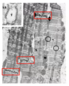
What are features of ventricular myocytes?
Shape?
Prominant organelles?
- Ventricular myocytes
- single central nucleus
- broad sheets of branching cells
- lots of mitochondria (20%)
- Striated muscle
- Z-lines
- myofibrils in parallel
- Sarcoplasmic reticulum
- well developed t-tubule system
- triads and diads (circles)
- Inset
- t-tubule, triad, lateral sacs
- Intercalated disc: end to end transmission
What is the function of intercalated discs?
Identify the features of intercalated discs shown in the provided image & describe their functions

- Intercalated cell-to-cell electro-mechanical function : functional syncytium
- Fascia Adherens
- cell-cell connection transmits force
- Desmosomes
- “press studs” cytoskeleton attachment
- Gap junctions
- electrical connection
- normal heart rhythm depends on coupling cardiac myocytes via gap junctions– dependent on:
- type & amount connexin expressed
- size & distribution of GJ plaques
- proportionof of connexin subtypes assembled
- distribution related to chamber & how chamber conducts
- gating and connexin type
- Fascia Adherens

Describe the specific features of atrial working myocytes
Shape?
prominent organelles?
Rapid, impulse transmission end-to-end and side-to-side
- bundles of 2-3 cells
- elliptical shape
- generally, no branching
- Intercalated discs
- horizontally oriented intercalated discs
- occassional end-end intercalated discs
- can get side-to-side & end-to-end transmission and this particular structure sets up arrhythmias
- series of desmosome & gap junctions
- SR, but abscence of t-tubules
- part of what makes conduction slow

What is myocardial connective tissue composed of?
What functions does it provide?
- Composition
- collagen-elastin matrix
- connects myocytes nerve and capillary networks embedded in meshwork
- Provides
- structure
- collagen struts
- support
- passive elastic component
- prevents overstretching of the heart
- force transmission
- may “hold” vessels open during contraction to counter surround pressure
- structure

How is the cardiac action potential different from skeletal action potential?
- Skeletal muscle
- force of contraction is much longer and happens after the action potential
- important for force production through spatial & temporal summation of action potentials
- Cardiac muscle
- Cardiac has a different shape and is longer
- contraction begins with the action potential, & the duration of the contraction and the action potential are similar
- prevents temporal summation & can not have tympany


- a. large cells
- c. many gap junctions
- f. Na+ channels
- g. large action potential amplitude

longer

b. the duration of contraction is roughly the same as the duration of the AP
Cardiac action potentials consist of which membrane boltage-gated, time-dependent currents?
Which electrogenic transporters carry current?
Who are the important players in action potentials?
- Membrane coltage-gated, time-dependent currents
- sodium current (INa)
- funny current (If)
- Calcium current (ICa)
- Potassium current (IK)
- IK1
- It01
- electrogenic transporters carry current
- Na+ - Ca2+ exchanger (INCX)
- Na+ - K+ ATPase (INa-K)
- Sarcoplasmic Reticulum
- SERCA (uptake Calcium)
- inhibited by phospholambam
- when phosphorylated, it is inhibited itself
- inhibited by phospholambam
- RYR (release Calcium)
- SERCA (uptake Calcium)
- Adenylate Cyclase
- converts ATP to cAMP which activates PKA, which phosphorylates
- phospholambam
- RYR
- L type Calcium channels
- Potassium Channels
- converts ATP to cAMP which activates PKA, which phosphorylates

What stage in indicated by the provided photo?
What is the determinig factor of this stage?

- Phase 4: Resting Potential
- flat line
- determined by stable potassium conductance
- IK1 - inward rectifying K+ (maintains the membrane potential at -90 mV)
Describe what occurs in phase 0-3 of a cardiac action potential
- Phase 0: Sodium - Rapid Depolarizaion
- INa - Na+ channels
- gNa (sodium conductance)- rapid increase
- CaL - L type calcium channels are open
- INCX - Na-Ca Exchanger (bring some sodium into the cell)
- Phase 2: CaL closing
- Phase 3: INCX reverses - Calcium removal

Describe the state of the sodium channel during each of the 4 phases of a cardiac action potential
- In Phase 4, the sodium channel is activatable
- As we enter Phase 0, and membrane potential approaches 0, the sodium gates will be deactivated, so sodium can no longer be conducted at the end of Phas 0
- Phase 1 & 2 the sodium gate & the channel is inactivated & inactivatable

What are the important repolarizing currents & in what phases do they occur?
-
Repolarizing currents
- transient outward (It0)
- Rapid repolarization of Phase 1
- other potassium channels begin to open as It0 closed
- Plateau phase (2) is due to the fact that the calcium that is entering the cell is roughly proportional to the charge caused by the potassium leaving the cell
- End of phase 2, beginning phase 3, calcium channels close & potassium channels open, and we repolarize the cell and go back to phase 4
- transient outward (It0)

What is an imporant characteristic about IK1 at resting membrane potential
- Around resting membrane potential, IK1 can bring potassium into the cell
- As a rectifier, it may go negative (inward) or positive (outward), but in the end you have a very stable baseline

c. opening of Na+ channels

d. movement of K+ in and Ca2+ out

b. Ca2+ close

d. IK1 channels
What is the clinical importance of the cardiac refractory period?
any change in heart rate is due to a change in refractory period
and most if not all arrhythmias are due to a change in refractory period
What is a refractory period & during which cardiac phases is the absolute refractory period?
Relative Refractory period?
Super normal period?
- Period of:
- abolished excitability
- reduced ability to resopnd to a stimulus
- absolute refractory (phase 1 to end of phase 2)
- sodium channels are closed & cannot be activated
- Relative refractory (phase 3)
- sodium channels are able to be activated
- with a strong enough impulse, you can get an action potential, but not a very good one – it will have a slower rate of rise & lower amplitude
- can be disturbing, enough to excite other cells
- Super normal period (phase 4)
- membrane is back at rest and any impulse can generate an action potential


