Cardiac Flashcards

1
Q

What is ischemic heart disease

A

characterized by an imbalance between metabolic oxygen demand and myocardial oxygen supply

  • Metabolic oxygen DEMAND that EXCEEDS oxygen SUPPLY
  • Most commonly caused by
    • Narrowing of coronaries →
      • Atherosclerosis of the coronary arteries
  • Can also be caused by:
    • Severe hypertension or tachycardia
      • extra demand on heart/O2 supply
    • Coronary artery vasospasm (or plaques that lead to vasospasm)
      • Supply reduced
    • Severe hypotension (decreased supply to heart)
      • Reduced supply
    • Hypoxia
    • Anemia
      • Reducing O2 carrying capacity
    • Severe AI or AS
      • Aortic insufficiency (regurgitation)/Stenosis
2
Q

Incidence of ischemic heart disease?

A
  • Incidence
    • Present in 10 million American adults
    • 1.5 million suffer from MI
      • 1/3 mortality
    • Major cause of perioperative morbidity and mortality
    • Overall incidence in surgical pts: 30% of surgical patients
3
Q

Risk factors for ischemic heart disease?

A

MAJOR

  • **increasing age
  • **male gender
    • (men > 60 yo → assume they have some sort of atherosclerosis)
  • Hypercholesteremia (increased LDL → more plaques)
  • Diabetes
  • Hypertension (increased demand)
  • Cigarette Smoking
    • Vascular disease → narrowing
  • Positive family hx (genetic predisposition

Other:

  • Obesity
  • (probably have hyperlipidemia, hypercholesteremia)
  • Cerebral Vascular Disease
  • Peripheral vascular disease
  • (for this and CVD, if they have atherosclerosis in one part of the body they probably have it in another part)
  • Menopause
  • Risk equalize with men
  • Use of high estrogen contraceptives
  • Sedentary lifestyle
  • Psychosocial characteristics (high stress)
4
Q

What happens when ischemic chest pain present (flowchart)?

A
  • Angina is one of the biggest sign of ischemic heart disease
    • New or changed angina = Acute Coronary Syndrome
      • Acute onset of S/D issue

GET 12 lead EKG!!!

  • No ST segment elevation and negative troponin/ CK-MB = unstable angina
    • Signals an impending MI
  • If no ST segment elevation and positive troponin/ CK-MB= NSTEMI (myocardial infarction)
  • ST segment elevation and positive troponin/ CK-MB= STEMI (ST elevated myocardial infarction)

Gold Standard- Troponin/CK-MB test

  • get a 12 lead and follow this algorithm and get a cardiology consult
  • ST elevation not always present in MI
  • **if they tell you that their chest pain has changed –> DO NOT do the case
5
Q

Pathogenesis of unstable plaques and thrombus formation?

A

MI→ formation of cardiac plaque that formed over time

  • Body detects and grows collateral vessels over time → typically enough supplemental O2
  • These are not the issue (acutely)

Atherosclerotic plaque

  • Why is unstable angina not good?
    • Because at any moment that person can have an MI – very high risk
  • What is an unstable plaque?
    • An atherosclerotic plaque with lipids inside and a thin fibrous cap
  • The type of plaque is important
    • Vulnerable plaques= rich lipid cores and thin fibrous caps

Athersclerotic plaque with lipid core and thin fibrous cap (typical culprit for MI)

  • Causes shear force → anything causes blood to come through quicker or constriction (excited/stressed)
  • Inflammation
  • Apoptosis
  • Macrophage- derived degradative enzymes
    • (^) Responses to lipid rich core → thin fibrous cap gets sheared off and allows for rupture of plaque
    • Increased inflammation with release of multiple cytokines, platelet activation and adherence → production of thrombin and vasoconstrictors (vasospasms)
    • Thrombus formation over lesion plus vasoconstriction (release of thromboxane A2) of vessel → Acute decrease in coronary blood flow → lack of O2/blood arriving to myocardium →
    • UNSTABLE ANGINA OR MYOCARDIAL INFARCTION
6
Q

What does this picture show?

A

Angiography

  • Plaque formation at baseline (A)
  • Transient total occlusion (mental stress) (B)
  • Give CCB or nitrate → allows for vasodilation of constricted vessel (C)
    • Opens at the same diameter for blood to go through

*ACUTE OCCULUSION- complete occlusion distal/downstream to plaque upon exercise/stress

If meds don’t work → need stent or bypass placed

Why do you want to avoid phenylephrine in patients with a history of an MI? (pictures of the vessels)

  • It can cause coronary artery vasospasm
  • Also remember when surgery occurs you are causing stress
  • If an occlusion occurs, administration of nitrates and nifedipine can reopen the artery to same diameter as baseline
7
Q

What is the role of stress in trigerrign cardiac clinical events?

A

Acute mental stress –> Central and autonomic nervous system activity

  • Physiologic responses
    • Increased catecholamines
    • Increased HR
    • Increased BP
    • Decreased plasma volume
    • Increased coronary constriction
    • Increased platelet activity
  • Cardiac Effects
    • Increased electrical instability
      • (due to incr catecholamines, HR and BP)
    • Increased demand (d/t incr catecholamines, HR and BP)
    • Decreased supply (d/t decr plasma volume and increased coronary constriction)
      • ­ S/D = really what leads to ischemia, plaque rupture, and ventricular F/T
  • Pathologic Result
    • VF/VT (d/t electrical instability)
    • Ischemia (d/t increased demand and decreased supply)
    • Plaque rupture (d/t decreased supply and plaque rupture also leads to decreased supply)
    • Coronary thrombosis (d/t increased plt activity)
      • Bottom 3 is what we see in ER → leads to MI
  • Cardiac Event
    • Cardiac sudden death ( from VF/VT)
    • Myocardial infarction (d/t ischemia, plaque rupture, coronary thrombosis)
  • Background factors Are (has cardiac effects)
    • Atherosclerosis
    • Prior MI
    • Poor LV function
8
Q

What are some EKG changes that can be indicative of ischemia or infarction

A
  • ST segment depression (chronic/previous MI)
  • T wave inversion
  • ST segment elevation (ACUTE MI/ischemia)
9
Q

What is stable angina?

A
  • Baseline CAD, ischemic disease
  • No change in precipitating factors for at least 60 days (2 months)
    • Frequency or duration of pain
      • Associated with fixed narrowing (usually 75% or greater)
      • O2 demand may be normal under basal conditions
      • Tx:
        • Vasodilators (Nitrates or CCB)
        • Relieved by rest, reducing demand
10
Q

What is unstable angina?

A
  • Caused by less than normal activity
  • Lasts for prolonged period
  • Occurring more frequently
  • Signals impending myocardial infarction
  • Imminent danger of CV event
    • Crescendo- starts slow and then becomes unbearable (worse and worse)
    • Increased frequency, duration, etc
      • New precipitating thing changes → unable to walk up steps acutely
    • Can cause irreversible infarction
    • Acute plaque changes
    • Usually also partial thrombosis (shear forces that dislodged plaque)
  • Tx: Medically manage, stent placement, cardiac bypass
11
Q

What is prinzmetal angina?

A
  • At rest
  • Coronary spasm- usually caused by this instead of atherosclerosis
    • Occurrence:
      • In a plaque area or normal vessel
      • Associated with other vasospastic diseases (Raynaud’s Disease)
        • Tx: medically manage, Nitrates, CCB
          • *avoid phenylephrine (vasospasm)
          • Maybe have nitro in the room
12
Q

What is myocardial infarction?

A
  • Necrosis caused by ischemia
  • Within 20-30 minutes of ischemia
    • Typically begin in the subendocardial regions (in between epi and endocardial layer)
  • Full damage seen 3-6 hours after ischemia occurs
  • Size depends on:
    • Proximity of lesion
    • Collateral circulation (if no collateral circ, damage can be very great because heart hasn’t time to develop collateral)
      • CKMB/troponin usually increase right away and are even higher at 3-6 hours
      • Numbers correspond to size of the infarct
13
Q

Complications of MI?

A
  1. Papillary muscle dysfunction
    • Causes valvular disease
  2. External rupture of infarct
    • day 4-7 most common – cardiac tamponade → followed by death
      • Cardiac Tamponade- pericardium sac filled with blood and heart cant pump properly
  3. Mural thrombi – lead to stroke
  4. Acute pericarditis (acute infection of the necrosed area): day 2-4 most common
    • Pericardium filled with blood and leads to infection
  5. Ventricular aneurysm – fibrous outpouching of the ventricle- anteroapical region most common → cardiomyopathy
  6. Arrythmias
  7. LVF +/- pulmonary edema
  8. Cardiogenic shock (rare ~10%) → MODS
  9. Rupture of wall, septum, papillary muscle
  10. Thromboembolism
    • MI: mortality dependent on SIZE and LOCATION of thrombi/plaque
14
Q

What are various sensitivities of blood tests used to detect MIs?

A

(Graph)Rate and extent of rise of cardiac troponin and myocardial creatine kinase (CK-MB) levels after a typical acute MI

  • Troponin level: rise QUICLKY and ACUTELY
    • GOLD STANDARD→ can determine the presence and extent of injury (degree of myocardium damage)
    • Cardiac microinfarctions: can raise the troponin levels without increasing the CK-MB levels
      • *Microinfarction: infarction of smaller areas and vessels
  • CK-MB level: elevated but not as good as troponin rate or rise and extent of rise
    • More sensitive to larger vessels of heart
15
Q

What areas of the heart are infarcted with occlusion of RCA? LCA? LAD? LCX?

A
  • Occlusions of the coronary arteries
    • RCA= posterior, inferior MI (small portion of LV)
    • LCA= massive anterolateral MI (big portion of LV)
    • LAD= anteroseptal MI (portion of LV and RV)
    • LCX= Lateral MI

All leads some LV dysfunction → large vessels

Don’t pump properly → poor systemic outcomes

16
Q

What is defnition of vascular hyertension? Incidence? Categories?

A
  • Defined:
    • Sustained diastolic of 90mmHg
    • Sustained systolic of 140mmHg
      • Average of three different measurements at 3 different times
  • Incidence
    • 30% of general public (~1/3)
  • Categories
    • Normal < 120/80
    • Prehypertension 120-139/80-89
    • Stage 1 140-159/90-99
    • Stage 2 >160/100
17
Q

Importance of controlling HTN?

A
  • HTN is the most important risk factor in
    • CAD
    • CVA
    • Cardiac Hypertrophy- LV tries to pump against HTN → grows larger, vessels constrict → CAD
    • Renal Failure
    • Aortic Dissection/Aneurysm
18
Q

What are some causes of HTN?

A
  • 90-95% of HTN is idiopathic apparently primary (Essential HTN)- don’t know reason why it occurs
  • 5-10% : mostly secondary d/t renal disease
    • We know what the risk factors are for it (atherosclerosis, stress, pain, life style, smoking, renal, etc) but we don’t actually know what causes it except for organic things like pheochromocytoma
19
Q

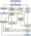
What are some effects of HTN on the heart?

A
  • Hypertension causes increased myocardial wall tension which can lead to:
    • Increased myocardial oxygen demand (need to feed a bigger muscle)
      • Leads to coronary insufficiency
      • Which can lead to heart failure
    • Left ventricular hypertrophy (which leads to increased o2 demand)
      • Which leads to heart failure
    • Infarction dysrhythmias can lead to coronary artery insufficiency and heart failure
20
Q

What are types of HTN?

A
  • Systolic and Diastolic
    • Essential hypertension – 95%
    • Secondary hypertension – 5%
      • Renal
      • Endocrine
      • Cardiovascular
      • Neurologic
21
Q

How is blood pressure normally regulated?

A

BP= Cardiac Output x Peripheral Resistance

  • Cardiac Output
    • Determined by blood volume
      • (stroke volume)
    • Cardiac factors
      • (how well is myocardium functioning → such as HR- frank starling curves)
    • CO = SV x HR
  • Peripheral Resistance
    • Humoral Factors (constrictors/dilators)
    • Neural Factors (stress response, vagal response)
    • Local (released locally, thromboxane vs NO produced)
22
Q

What is the pathogenesis of essential (primary ) HTN?

A

Risk factors

  • Genetic
    • Polygenic and heterogenous
    • Polymorphisms at several gene loci
  • Environmental
    • *Stress
    • Obesity
    • Smoking
    • Salt consumption
    • Sedentary lifestyle
23
Q

Pathophys of HTN?

A
  • Increased SNS activity in response to stress
  • Overproduction of sodium retaining hormones
  • Overproduction of vasoconstrictors
  • Underproduction of vasodilators (NO, prostaglandins)
  • Increase Na intake
  • Inadequate intake of Ca and K
  • DM – changes to vasculature that occur
  • Obesity- plague formation and vasocontraction that occurs in disease
24
Q

Effect of sodium retention on BP?

A
  • Salt/Water Retention: The final common pathway in the pathophysiology of essential hypertension
    • PROBLEM: BV (going back to pic)
  • TX:
    • Sodium restriction lowers BP
    • Diuretics - act as effective antihypertensives by promoting sodium excretion/H2O
25
Q

What are some pathogenetic factors and clinical prsentation of HTN heart disease?

A
  • Top brown box → LVH (BLUE)
    • BIGGEST MANIFESTATION OF HTN
    • Boxes on side: contribute to the LVH
  • LVH:
    • Microangiopathy→ blood supply increase in response to increase in demand on heart → MI
      • reduced coronary reserve
    • Impaired Contractility
      • As grows larger, not pump as efficient → impaired contractility (→ HF)
    • Reduced LV filling
      • Heart becomes stiff while growing larger
      • During Diastole (when heart fills) becomes stiff after systole and wont fill properly
    • Atrial Fibrillation/Ventricular dysrhythmia
      • Heart enlarges, pushing against itself
      • Ex: Push on→ Atria = a. fib or Purkinje fibers= vent. dysrhythmia
        • → sudden death, cardiac emboli result
  • Most important thing is – is HTN well controlled?
26
Q

Pathogenesis of secondary HTN?

A

(5% population)

  • Oral contraceptives (high estrogen)
  • Renal parenchymal disease
  • Renin-secreting tumors
  • Primary aldosteronism
  • Cushings Syndrome
  • Pheochromocytoma
    • Endocrine disorders… discuss in endocrine
27
Q

Treatments for HTN?

A
  • Drug therapy (possibly a combination of 2 for stage 2 hypertension-usually a diuretic and something else)
    • Diuretics- offload volume. (1st line)
    • Ca channel blockers
    • Ace inhibitors – helpful for renal prob (DM)
    • Beta blockers- decreases workload of heart
    • Angiotensin receptor blockers (similar to ACEI)
  • Non drug Therapy
    • Lifestyle changes: weight reduction, smoking cessation, increased physical activity
    • Sodium restriction, diets
    • Reduction of ETOH
    • Relaxation techniques
28
Q

What is peripheral vascular diseaes? What causes it? Prevalence?

A

Compromised blood flow to the extremities

  • Ankle-brachial index of less than 0.9
    • Perfusion to upper and lower extremity not match (index < 0.9) = PVD
    • Ratio of SBP in ankle vs SBP in brachial arteria?
  • What causes it?
    • Atherosclerosis- most often (PVD and CAD go hand in hand)
    • Arterial embolism
    • Vasculitis
  • Prevalence
    • 2 million Americans are symptomatic (have pain bc PVD)
    • Associated with aging
    • Exceeds 70% in people over the age of 75
    • Increases with risk factors
      • Way to avoid PVD= EXERCISE
29
Q

Risk factors for atherosclerosis?

A
  • Diabetes
  • Advanced age
  • Male
  • Obesity and physical inactivity
  • Sedentary lifestyle
  • Essential HTN
  • Smoking
  • Dyslipidemia
  • Family hx
30
Q

Atherosclerosis/PVD signs and symptoms?

A
  • The most common symptom = intermittent claudication
    • “Angina of the legs”
    • Claudication- the pain starts with walking or exercise and stops with rest
  • Other symptoms may include
    • Cool/cold feet to touch
    • Pain while lying flat and relieved by a sitting position
    • Loss of pulses in legs or feet, pale color when legs are raised up
    • Dependent rubor (redness in a dependent position)
    • Shiny skin, loss of hair
    • Thickened toenail- fungal infections (not good profusion to feet)
  • The most SEVERE symptom- Critical limb ischemia(“rest pain”)
    • Lack of oxygen to the limb/leg at rest- associated with non-healing ulcers and gangrene
31
Q

Treatment of atherosclerosis?

A
  • Identify and treat the risk factors
    • Exercise, lifestyle changes, weight loss
    • Smoking cessation
  • Lipid lowering therapy (statin drugs)- help with CAD too
  • Vitamin C and E & folate supplements help BV and cause local vasodilation
  • Antiplatelet therapy- prevent thrombus formation
  • Revascularization procedures
    • Typically for Critical Limb ischemia
  • Amputation- when vasculature is too poor and can’t fix it medically/surgically
32
Q

Summary of difference between stenosis and regurg?

A

Stenosis= pressure problem

  • Narrowing of leaflets of outlet → portion of heart that needs to pump through valve needs to generate enough pressure to have outflow through stenotic valve

Regurgitation/insufficiency= volume problem

  • Leaflets don’t work properly (when needing to close, doesn’t close properly or over flaps)
    • BACKFLOW- back to area of heart that just pumped it out → volume issue
33
Q

Pathophysiology of mitral stenosis?

A
    1. Almost always due to fusion of the mitral valve leaflets at the commissures during the healing process of acute rheumatic fever
      * Inflammatory process (rheumatic fever) → involvement of commissures and papillary muscles
      • Rheumatic Fever- risk for ALL valvular disorders
  • 2. When the mitral valve area/orifice < 1 cm2
    • Normal: 4-6cm
    • Stenotic: < 1-1.5cm (become symptomatic)
    • → A mean left atrial pressure of about 25mmHG is necessary to maintain an adequate resting cardiac output (normal is 8-10mmHG)
      • Atrial pressures need to get high to overcome stenotic valve= pressure problem!
  • 3. Left atrial enlargement predisposes to atrial fibrillation
    • Increased pressures in the LA required to move blood through tiny mitral opening which causes LA enlargement
    • LV enlargement → changes morphology of SA node (predispose of a.fib)
  • a. Stasis of blood in the distended LA predisposes to the formation of thrombi
    • Mitral stenosis may be receiving chronic anticoagulant therapy. Thrombi can cause increase R/F MI or CVA.
    • pooling of blood can also increase r/f infection

Symptoms:

  • Dyspnea on exertion when CO is increased
  • Severe MS →leads to CHF

mitral stenosis is heart on left of pic!!

34
Q

Effects of mitral stenosis on heart?

A
  • Decrease of LA emptying–> Increase LA preload (pressure)
    • Leads to back up in system (P. Edema) → → RV failure
    • Decrease LA force of contraction as heart gets enlarged (frank starling not fx prop as getting larger) → decrease delivery of blood → decrease O2 supply → LV failure
  • Eventual Tx: mitral valve stenosis–> mitral valve replacement
  • Mitral stenosis also leads to “fish mouth” appearance on valve
35
Q

Pathophysiology of Aortic Stenosis?

A
  • Pathophysiology
    • Isolated non-rheumatic aortic stenosis usually results from progressive calcification and stenosis of a congenitally abnormal bicuspid valve
    • Rheumatic fever-aortic stenosis
      • due to rheumatic fever
      • Almost always occurs in association with mitral valve stenosis
  • Hemodynamically significant Aortic stenosis
    • Associated w/:
    • Transvalvular pressure gradient > 50mmHG and
    • Aortic valve orifice area < 1cm
      • Normal is 2.5-3.5cm
        • Heart needs to generate enough pressure to get from LV to aorta (pressure gradient massive)
  • Two factors associated with the development of aortic stenosis
    • Aging- degeneration and calcification of the aortic leaflets- incidence is increasing as population is aging
    • Presence of bicuspid aortic valve- which is common congenital valve abnormality
      • b/c of abnormal shape, calcification and fibrosis more likely
36
Q

What are the triad of symptoms that occur with aortic stenosis?

A
  1. Angina pectoris (chest pain and ischemia) often in the absence of ischemic heart disease
    • Bc LV enlarged so much→ less O2 (demand more)
    • Heart cant relax properly → not filling well→ supply reduced
  2. Dyspnea on exertion
    • Any increase in demand will cause difficulty breathing
  3. Syncope
    • Pressure gradient high and blood flow reduced in aorta→ faint (not enough O2, BF to head)
      • Severe reduction in SV, BP
37
Q

Aortic stenosis effects on heart?

A

LV outflow obstruction from stenotic valve leads to:

  • Increase SBP (to get blood through valve)
  • Increase LVET (LV exit time)- heart tries to compensate by forcing blood out in first portion of contraction (become reliant on atrial kick)
  • Increase DBP (cant relax during diastole)
  • Decrease Ao pressure (not enough blood coming through the aorta)- Increase in LV SBP and decrease Ao pressure creates gradient
    • Increase myocardial O2 consumption and decrease diastolic time (when coronaries fill properly to get BF to heart itself)
      • Decrease supply!!! → myocardial ischemia → LV failure
      • Heart no longer efficient in pumping
38
Q

Pathophysiology of mitral regurgitation?

A

Mitral Regurgitation = volume issue

Pathophysiology

  • Mitral regurg is usually due to rheumatic fever and is almost always associated with mitral stenosis
    • Book said it is not usually due to rheumatic fever
    • Can be associated with ischemic heart disease or papillary muscle dysfunction
  • Left atrial volume overload by retrograde flow of a portion of the left ventricular stroke volume into the left atrium is the principal pathophysiologic change produced by mitral regurgitation
    • This causes LA volume overload and pulmonary congestion
    • The amount that regurgitates depends on
      • Size of mitral valve orifice
      • Heart rate, which determines the duration of ventricular ejection
      • The pressure gradient across the mitral valve
    • Pharmacological changes in SVR have a major impact on the regurgitation fraction
39
Q

What is seen on central line with mitral regurgitation?

A
  • Regurgitant flow is responsible for exaggerated V wave present on the recording of PAOP
    • The size of the V wave correlates with the magnitude of the regurgitant flow
  • This pic shows a normal wave (about 12) – an exaggerated wave will go to about 20-25 with regurgitation
    • A wave- p wave (atrial contraction
    • C wave- back pressure
    • V wave- SV (ventricular side)
      • Regurgitation – when heart goes to pump blood, blood comes back into LA (exaggerated)
40
Q

Causes, pathophys of aortic regurgitation?

A

Pathophysiology

  • Acute
    • infective endocarditis- infectious vegetation (dental infection, drugs)
    • trauma
    • dissection of thoracic aneurysm
      • morbidity/mortality rate worse for acute causes of AR → heart did not have time to compensate for damages
  • Chronic
    • prior rheumatic fever
    • persistent systemic hypertension – afterload increased → ventricle becomes enlarged. Papillary muscles grow outward → wont work as well (regurg)
  • The basic hemodynamic problem in aortic Regurgitation:
    • Regurgitation of part of the ejected stroke volume from aorta back into the LV
    • Decrease in forward left ventricular stroke volume
  • Heart starts enlarging -Heart dilated → cant make good SV
  • Problems with the aortic root
    • Idiopathic aortic root dilation
    • Aortic dissection *acute
    • Marfan syndrome
    • Ankylosing spondylitis
41
Q

Cardiac effects of aortic regurgitation?

A
  • Decreased CO d/t regurgitation of part of the SV back into the LV during diastole
  • Increased SV by increasing LVET ( increase time for LV ejection, this causes decrease in diastolic time of heart= decrase O2 available to heart)
  • Decrease in effective stroke volume
  • Causes combined pressure and volume overload on LV

(summary- increase demand/decrease supply)

42
Q

What does LV dysfunction result in? Types of hypertrophy?

A
  • LV dysfunction- results in progressive remodeling (myocytes themselves change) of the ventricular chamber
    • Leads to:
      • Dilation
      • Low Ejection Fraction (EF)

Can get either:

  • Eccentric Cardiac Hypertrophy: chamber size increase from increase volume → thinning membrane and dilation
    • Chamber enlarged – muscle dilated
  • Concentric Hypertrophy- thickening of heart muscle because pressure problem. Muscle grows inward into chamber (chamber gets small)
    • Chamber smaller- muscle hypertrophied
43
Q

Examples of causes for left ventricular dysfunction?

Effects of low EF?

A
  • Ex: CAD/HTN → Blood supply not adequate (→ remodeling to myocytes bc reduction of O2 delivery) → areas that’s happening…
    • Dyskinesic
    • Akinesic
      • Pump not working properly because pump not getting enough O2
  • Chronic ETOH use – direct effect on myocytes causing cardiomyopathy

Low enough EF → dysrhythmias

  • → Baroreceptors (on *carotid and aorta)→ carotid baroreceptors → detect drop in SV and EF → increase HR → tachyarrhythmias (increasing size of heart)→ change morphology of SA node → other rhythms
  • As heart enlarges (impacts purkinjie fibers) → more arrythm
44
Q

What happens to the heart shape in dilated cardiomyopathy?

Hypertrophic?

Restrictive CMP?

A
  • Normal vs cardiomyopathy
    • B. In the dilated type of cardiomyopathy
      • the heart has a globular shape and the largest circumference of the left ventricle is not at its base but midway between apex and base
        • Eccentric Hypertrophy
    • C. In the hypertrophic type
      • the wall of the left ventricle is greatly thickened; the left ventricular cavity is small but the left atrium might be dilated because of poor diastolic relaxation of the ventricle.
      • This kind is not very common
        • Concentric Hypertrophy
    • D. restrictive type (amyloid)
      • the left ventricular cavity is normal size, but again the left atrium is dilated because of the reduced diastolic compliance of the ventricle.
      • Usually due to mitral stenosis or regurg/stenosis together

Any time have B,C,D → heart cant relax properly during diastole (not good diastolic filling)

** Diastolic Filling pressures go up → changes in valves anatomically and LA dilation from backpressure

45
Q

Cuases of dilated CMP?

A

Inflammatory and non inflammatory

46
Q

What are symptoms of inflammatory dilated CMP? Progresses to?

A

Inflammatory myocarditis early symptoms

  • Fatigue
  • Dyspnea
  • Palpitations – carotid baroreceptors detect drop in BV → increase HR to compensate
    • These are vague symptoms which can be dangerous because heart issues aren’t the first things you would consider
    • Inflammation- some infection or maybe massive MI and some of muscle dies and causes infection
  • Progress to:
    • CHF- bc compensatory mechanisms
      • Venoconstriction–> increase preload to heart
      • Constriction on arterial side (from kidneys detecting drop in CO) → increase afterload.
        • Increase volume coming back to heart but cant pump forward (congestive HF)
    • Pulsus alternans (BP/SV that varies with each pulse)
    • Tachycardia- baroreceptors
    • Pulmonary edema (increase flow to heart and not good enough forward flow–>things start to back up)
      • Complete recovery usually ensues with treatment by antibiotics
      • Since symptoms are so vague at first- might have it for a while and remodeling could have occurred
47
Q

Cuase for noninflammatory CMP?

A
  • Usually due to
    • Toxicity (ethanol)→ direct toxicity to myocytes
    • Idiopathic process
    • Degenerative process-aging
    • Infiltrative process- that infection that happened caused remodeling
    • Post myocardial infarct- remodeling
      • Turn Akinesic (don’t move at all) or dyskinesic (not pump as efficiently)
48
Q

What are dilated cardiomyopatheis characterized by?

A
  • Elevated filling pressures
    • Look at Diastolic function
    • heart is not compliant, frank starling curve is off
  • Failure of contractile strength
    • problems with myocytes and its dilated already so it cant stretch much more (frank starling curve failure)
  • Marked inverse relationship between arterial impendence stroke volume
    • Ex: RAAS → vasoconstriction → increase preload but impedes SV bc arterial vasoconstriction
    • Higher impendence, lower stroke volume
49
Q

Clniical picture of CHF associated with dilated CMP?

A
  • Forward failure (decrease CO)
    • Fatigue – not getting enough blood
    • Hypotension- no stroke volume
    • Oliguria- kidneys are reacting
      • Reduced renal perfusion caused the activations of RAAS
      • Increases the effective circulating blood volume through sodium and water retention
50
Q

Left sided heart failure vs right sided HF?

A

Left Sided Heart Failure

  • Elevated filling pressures required by the failing heart
  • Secondary mitral regurg caused by dilation of the ventricles (leaflets cant close because its so large)
    • Orthopnea- changes in breathing with position changes
    • Pulmonary edema
    • Paroxysmal nocturnal dyspnea (3-4 pillows at night)

Right Sided HF

  • Hepatomegaly- back up in portal system (nice store for blood but don’t want it all there)
  • JVD
  • Peripheral edema
51
Q

What does dilated CMP do to heart structure? Myocardial tissue?

A

Dilated cardiomyopathy (DCM).

  • A: Four-chamber dilatation and hypertrophy are evident. There is a small mural thrombus (arrow) at the apex of the left ventricle (on the right in this apical four-chamber view). There was no coronary artery disease.
  • B: Histology of typical DCM demonstrating variable myocyte hypertrophy and interstitial fibrosis (collagen is highlighted as blue in this Masson trichrome stain)
52
Q

What are various other names for hypertrophic CMP?

A

It has been called many other names

  • Idiopathic hypertrophic subaortic stenosis (IHSS) – most common name
    • Anatomical changes bc valves stuck together
  • Asymmetric septal hypertrophy
  • Hypertrophic obstructive cardiomyopathy
    • Concentric hypertrophy and pushes papillary muscle and leaflets together
  • Muscular subaortic stenosis
    • (subaortic- right below aorta-septum gets large)
53
Q

Clinical features of hypertrophic CMP?

A
  • Usually younger 15-16 year old kids
  • AUTOSOMAL DOMINANT TRAIT
    • Main defect is contractile elements of the heart – then there is an increase in the density of the calcium channels
  • Majority of patients are asymptomatic- until increased demand placed on heart
  • 50% initially present with sudden death or cardiac arrest
    • Symptoms (if they have them): mimic aortic stenosis
      • dyspnea, angina (d/t high demand of O2 and the inability to relax), syncope
    • 75% ventricular dysrhythmias (purkinjie don’t fx prop)
    • 25% SVT- changed morphology of SA node
    • 5-10% atrial fibrillation
  • Annual mortality 3-8% (post op 1-3%)
  • Most patients symptomatic in second-third decades
    • Sudden death patients: The most frequent finding at autopsy in a young previously healthy athletes
54
Q

Pathophysiology of hypertrophic CMP?

A
  • Asymmetric myocardial hypertrophy
  • Diastolic dysfunction
  • Disproportionate enlargement of the intrerventricular septum; usually in the top portion right below the aortic valve (where it gets enlarged and muscular)
    • Cant relax during diastole → high filling pressures and diastolic dyfx
  • Rapid LV ejection (80% during early systole) – normal is 20%
    • Reduced LV ejection time**
    • SV comes from atrial kick
    • Sudden death→ running and increasing HR → tachyarrythmias→ lose atria kick and portion of SV → SV DROPS
  • Often sub-aortic pressure gradient
  • Most patients have some degree of mitral regurgitation
    • Vasodilators worsen the MR while vasoconstrictors attenuate obstruction and decrease the MR
      • Ex: MR: increase rate and give vasodilators
  • Poor diastolic compliance, atrial contribution may be as high as 70%
    • **Phenylephrine, volume volume volume, lots of gas to decrease contractility
  • Imbalances in myocardial o2 requirements- heart is so muscular
55
Q

When is obsturction worsened in HOCM?

A
  • Dynamic obstruction is worsened by
    • Decreased preload (decreased ventricular volume)- diastolic dysfunction worsened
      • also decrease in volume makes stenosis worse
    • Decreased afterload (they already get rid of blood so fast you don’t want to make it any faster)
    • Increased contractility- exercise (→ reduction in ventricular volume → increasing prox of mitral valve leafs)
      • All these produce a reduction in ventricular volume thus increasing the proximity of the anterior MV leaflet to the IVS → mitral valve stenosis worse (DEATH)
56
Q

What are ways to improve systolic function in HOCM?

A
  • Factors that impair contractility but improve systolic function (opposite than everything else)
    • Volume loading
    • Vasoconstriction – increase afterload to slow heart down allowing the heart to fill in diastole
    • Myocardial depression- slow it down!
57
Q

Treatments for hypertrophic CMP? Sx management? Echo findings?

A
  • Beta blockers
    • Blunt sympathetically mediated increases in subaortic stenosis
    • Decrease tachyarrythmias
  • Calcium channel blockers
    • Improve diastolic relaxation
  • Surgical management
    • Myomectomy- surgically remove parts of enlarged ventricle
  • Echo findings- usually diagnose with this
    • Thickened IVS (base to apex)
    • Poor septal motion
    • Anterior displacement of the mitral valve

PIC: Concentric Hypertrophy: Enlarged heart muscle with small chamber size, muscle growing into chamber. During diastole → cant adequate fill

58
Q

Frank starling law of the heart? Effects during heart failure?

A

Frank Starling Curve:

Y axis: CO

X axis: end diastolic pressure (pump fx of heart-) How we measure SV

Increase EDP → increase SV → increase CO

HF:

As heart trying to generate pressure → cant get out even with increasing pressures. CO decreases as EDP increase. (not normal- should be letting CO increase)

Drugs:

  • Nitrates
  • Diuretics → decrease arterial side compensation (constriction→ dilate)
    • Does not impact CO
  • Potent vasodilator (phentolamine) with inotrope (Digitalis) → increasing contractility → more normal
  • Nitroprusside & dopamine → closer to normal
59
Q

What are some common congenital heart defects?

A
  • Patent ductus arteriosis
  • Atrial septal defect
  • Ventricular septal defect
  • Tetrology of Fallot
  • Coartation of the Aorta
  • Pulmonary stenosis with right-to-left shunt
60
Q

What are left to right shunt congenital heart defects?

A

Left to Right Shunt (noncyanotic because its been oxygenated)

  • Blood flows from L heart to R heart- depending on size of defect, may not be able to detect right away. No necessarily corrected with sx. May not lead to cyanosis right away
    • ASD
    • PDA- normally closes 24 hrs after birth
    • VSD
      • With VSD the shunt is left to right, and the pressures are the same in both ventricles. Pressure hypertrophy of the right ventricle and volume hypertrophy of the left ventricle are generally present.
      • → Oxygenated blood mixes with unoxygenated blood (mixing) but oxygenated blood goes to system
  • Sometimes leads to tardive (delayed) cyanosis, but does not cause cyanosis from the onset
    • Can compensate for a little while so it takes a bit (can grow and wait for closure or surgery)
  • Left to right shunt is more common than R–> L shunt
    • Blood from left –> right
      • Left atrium to right atrium
      • Aorta to pulmonary artery
      • Left ventricle to right ventricle
    • Blood that makes it to systemic circulation is oxygenated
    • Increase in blood flow to pulmonic system
61
Q

What is an atrial septal defect?

A

ASD: FORAMEN OVALE not closing properly or completely (1st day of life)

  • Allows for blood to flow from L atrium to right atrium
  • Oxygenated blood goes back to the lungs and increases blood flow through the lungs
  • Can lead to pulm HTN
  • Bigger the defect, the bigger the problem

Mixed blood ends up going through the PA

Blood that enters systemically is still oxygenated blood

  • Most common of the cardiac malformations diagnosed in adulthood
    • Normally: Atrial Septum forms between 4-6 weeks of embryonic life
      • *Pregnancy ETOH ingestion associated with ASD!
62
Q

Clinical features of ASD?

A
  • CLINICAL FEATURES
    • Eventually lead to pulm HTN (overtime)
    • Can cause reversal of R to L shunt → cause cyanosis and CHF (the pressure in R atrium can be greater than L and it flips)
    • Can also be associated with mitral insufficiency – just because of the anatomy of the heart
63
Q

What is a ventricular septal defect?

A
  • Most common heart defect at birth- more severe than ASD
    • Normally: Develops between 4-8 weeks gestation
      • Many close in childhood spontaneously
      • Develops at the beginning of pregnancy (ETOH ingestion)
  • VSD: Can cause severe L to R shunts, with pulm HTN and CHF as well as infective endocarditis
    • Surgery is needed for larger VSDs- optimize and then surgery
      • Sometimes they’ll watch these kids and they can close spont
  • VSDs become severe because pressure increase rapidly
  • The bigger the hole, the bigger the problem, not enough blood to the body (blood is properly oxygenated, just not enough in systemic circulation)
  • Kid is pink but lethargic from decrease in blood flow to body
  • Increased pulmonary blood flow à pulm htn
  • Often closes on its own
    • Failure to thrive is symptom to note
64
Q

What is eisenmenger syndrome?

A
  • Development of severe pulmonary HTN (leading to shunt reversal) as a result of L to Right intracardiac shunt- becomes predominantly R to Left
  • Remodeling of the pulmonary arteries and veins due to prolonged volume and pressure overload
  • Common cause of Eisenmenger syndrome is an unrestricted and unrepaired VSD, half of which will end up in this syndrome
  • More blood to lungs à increased pulm pressure (norm is 20mmHg)
    • Right-left shunt may occur 20-30 years later
    • Pressure in right ventricle grows to match left side and becomes Rà shunt
      • Throwing clots to systemic circulation
    • People with chromosomal disorders have more risk of congenital heart disease
65
Q

What is a patent ductus arteriosus?

A
  • Arterial channel that courses between the pulmonary artery and the aorta- allowing for bypass of unoxygenated lungs in utero
  • Constricts and closes at birth due to:
    • Increased O2 level
    • decreased pulmonary resistance
    • decrease in PGE2 (prostaglandin, vasodilation causes PDA to close at birth)
      • If anomaly → remains open (patent)
  • If it doesn’t close, leads to high pressure L to R shunt
    • Between aorta and pulm artery- supposed to go to system but goes back to pulm artery
    • Possible R ventricular hypertrophy
  • Leads to: Pulm HTN, Cyanosis, and CHF with bigger lesions, infective endocarditis can occur
66
Q

What are right to left shunts?

A
  • Cyanotic at BIRTH
    • Poorly oxygenated blood from the right side of the heart is introduced directly into arterial circulation via the left heart
  • Examples:
    • Tetralogy of fallot
    • Transposition of the great vessels (aorta on R side and pulmonic on L side)
      • Normally pressure is lower in right side but with R to L shunt blood goes from right side to left side
      • Mixture of oxygenated and deoxygenated blood- kid is blue
67
Q

What is tetralogy of fallot?

A

example right to left shunt

  • Most common cause of cyanotic congenital heart disease
    • Risks: >40 yo moms, ETOH in pregnancy, DM, children with down syndrome
  • Caused by abnormal division of the truncus arteriosus into a pulmonary trunk and aortic root
    • Main defect: Anterior deviation of pulmonary outflow septum → narrowing of ventricular outflow tract → prevention of BF via aorta because pulmonary artery valve tends to be constricted
  • Has 4 components
    • VSD- if they didn’t have this they wouldn’t survive
    • Dextrapoased aortic root that overrides the VSD
    • RV outflow obstruction
    • RV hypertrophy
  • ​VSD- needs to be there for child to survive
  • Narrowing of pulmonary passageway

Misaligned aorta- has to be misaligned or wont get O2 to systemic system

68
Q

Clinical signs and manifestations of tetralogy of fallot?

A
  • Clinical signs
    • R to left shunt
    • Decreased blood flow to the lungs as well
    • Increased blood flow to the aorta
      • Extent of shunting really depends on the degree of outflow obstruction
        • (bc main defect is deviation of pulm outflow and narrowing of pulm outflow)
  • Manifestations
    • Can be avoided by surgical repairs and correction
    • Due to chronic cyanosis
      • Erythrocytosis
      • Increased blood viscosity
      • Digital clubbing
      • Infective endocarditis
      • Systemic emboli (d/t increased RBC)
      • Brain abscesses
69
Q

What is transposition of great vessels?

A
  • Cyanotic Right to left shunt
  • Aorta rises from R ventricle
  • PA rises from L ventricle
    • Must be associated with ASD, VSD, or PDA for the pt to survive in extrauterine life (to allow for some oxygenated blood to go to system)
  • Clinical features
    • Cyanosis
  • This is where half of everything is reversed- this is much worse
  • From vena cava –> R atrium –> right ventricle –> aorta
  • From pulmonary vein –> L atrium –> left ventricle –> pulm artery
    • Get two independent circuits of blood flow
    • One pumping to body, and the other pumping to lungs
  • Not a big deal in utero but not consistent with life
    • Sometimes fixed in utero because it is a huge deal after birth
    • A little bit of blood will trickle through ductus arteriosus (will give a few hours of life)
70
Q

What is coarctation of the aorta?

A
  • Abnormal narrowing of aorta
    • More common in males than females
  • Can be preductal or post ductal- named due to where the narrowing occurs in relation to ductus arteriosus
    • Post ductal (distal to the ductus arteriosus) is the more common type
  • Preductal COA
    • Infantile
    • Weak femoral pulses
    • Cyanosis of lower extrems (mottled)
    • CHF
    • Need surgical correction to survive!
      • Pink hands, blue feet= R to L shunt
  • Post ductal COA
    • Older children/ young adults
    • Collaterals have developed
    • Decreased perfusion to the kidneys
      • Activation of RAS
    • High pressures in upper extrem and low pressures in lower
    • Intermittent claudication- exercise with leg pain
  • High pressure above (head, arms), low pressure below (everything else)
  • Blood has normal oxygenation, kid is pink
    • Decreased or absent pedal pulses
  • Problem: left ventricle hypertrophies à high BP in upper body, low BP in lower body
    • Might be seen in older patients where nothing is bringing BP down
  • Kidneys do long term control of blood pressure
    • Renin- trying to increase BP to where it needs to be
  • BP in ankle very low due to coarctation
  • Upstream of coarctation extremely high
  • Strong radial and carotid pulses